Case Report: BRCA1 and BRCA2 loss in a young man with primary cutaneous extraskeletal osteosarcoma
Wen-Feng Luo, Yu-Hang Hou, Yu-Teng Huang, Jun-Dong Lai, Hui-Shan Jiang, Wei-Liang Wang

TL;DR
A 28-year-old man with a rare skin cancer had reduced BRCA1 and BRCA2 genes, the first such case reported, highlighting the need for detailed testing.
Contribution
First reported case of primary cutaneous extraskeletal osteosarcoma with decreased copy number of BRCA1 and BRCA2.
Findings
Patient had primary cutaneous extraskeletal osteosarcoma with reduced BRCA1 and BRCA2 copy numbers.
No metastasis was found, and the patient remained recurrence-free after treatment.
BRCA1 and BRCA2 may be linked to worse outcomes in this rare cancer.
Abstract
Extraskeletal osteosarcoma is an uncommon and high-grade soft tissue malignancy. The incidence is even lower when the skin is the primary site. To the best of our knowledge, the primary cutaneous osteosarcoma has fewer than 30 reported cases worldwide, which with decreased copy number ofBRCA1 and BRCA2 has never been reported before. A 28-year-old man was hospitalized for a skin mass on the left shoulder. The histological examination showed a large number of tumor giant cells and fibroblasts, and nuclear division was easy to see. Immunohistochemistry showed positive for CK, EMA, S100, CD34, CK7, Bcl-2, ACTin, and NSE, and negative for Vim, SATB2, CD99, SMA (focal), and Ki67 was about 40%. Shoulder joint CT and PET-CT showed that no metastasis presented. Germline testing showed decreased copy number ofBRCA1 and BRCA2. The diagnosis was cutaneous extraskeletal osteosarcomas of the left…
Genes, proteins, chemicals, diseases, species, mutations and cell lines named across the full text — each resolved to its canonical identifier and authoritative record.
Click any figure to enlarge with its caption.
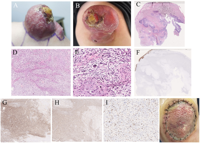 Figure 1
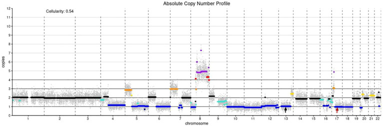 Figure 2
Peer Reviews
No public reviews on file for this paper yet. If you reviewed it on a platform where reviews are public (OpenReview, ICLR, NeurIPS, ICML), you can paste yours below so the community can read it here.
Videos
No videos yet. Explain this paper in a talk, walkthrough, or lecture? Add one.
Taxonomy
TopicsSarcoma Diagnosis and Treatment · Cancer Genomics and Diagnostics · Virus-based gene therapy research
Introduction
1
Extraskeletal osteosarcoma can present as a skin tumor without involvement of deep soft tissues or internal organs. Its clinical presentation is variable and can manifest as subcutaneous nodules or exogenous masses. Due to the lack of specific features, preoperative diagnosis can be challenging. Now we summarize the diagnosis and treatment process of a 28-year-old male with osteosarcoma of the left shoulder, in order to share our experience and lessons learned and improve understanding of this disease.
Case description
2
A 28-year-old man presented with an exogenous mass on the left shoulder for 3 months, that was about 3×3×4 (mm) in initial and quickly increased in size over the past time. He had a history of keloids but denied any history of local trauma, radiotherapy, or chemotherapy. Family history was negative for relevant malignancies. Physical examination showed a mass of about 6×6.5×2 (cm) on the left shoulder. (Figures 1A, B). The ultrasound examination revealed a hypoechoic solid mass in the subcutaneous tissue that had irregular bordersand uneven internal echogenicity. His results on complete blood cell count and differential count, four coagulation tests, liver and kidney function tests, electrolyte profiles and abdominal ultrasound were negative for disease. The lesion was completely excised. The histological examination showed numerous tumor giant cells and fibroblasts, and nuclear division was easily seen. There is also abundant osteoid, chondroid elements, with delicate lace-like bone or wide bone trabeculae. (Figures 1C–E). Immunohistochemistry showed positive for CK, EMA, S100, CD34, CK7, Bcl-2, ACTin, and NSE, and negative for Vim, SATB2, CD99, SMA(focal), and Ki67 was about 40% (Figures 1F–I). Shoulder joint CT and PET-CT showed that the tumor localized on the skin without involvement of the shoulder joint or humerus, and no metastasis presented. A diagnosis of cutaneous extraskeletal osteosarcomas of the left shoulder was made. Due to the early onset of cancer, the patient conducted germline testing that revealed BRCA1, BRCA2 and RB1 losses, and amplification of the MYC gene (Figure 2). Considering a high possibility of recurrence, the patient underwent an enlarged resection (Figure 1J), followed by local radiotherapy four cycles. No recurrence or metastasis occurred on a 1-year of follow-up.
Clinical and histopathological features. (A) A 66.52 (cm) hemispherical mass was presented on the left shoulder. (B) scattered scales were visible in the surrounding area, with ulcerated surfaces and yellow crusts on top. (C) A large irregular mass was seen in the subcutaneous soft tissue,with erosion in the superficial regions (HE staining, ×4). (D) Pleomorphic chondrocytes were visible, with a cell-rich periphery of lobules mixed with areas of osteoblastic activity (HE staining, ×50). (E) The tumor cells exhibited pleomorphism with various shapes including spindle-shaped, triangular, and oval. A large number of tumor giant cells and fibroblasts were seen, and nuclear division was easily observed (HE staining, ×200). (F) CK (-). (G) CD99 (+). (H) Vim (+). (I) Ki67 (40%+). (J) There was a circular incision on the left shoulder measuring approximately 10cm in diameter. And the incision edges were aligned and sutured in place, with mild redness, swelling, and oozing.
Absolute copy number profile: The color code in the plot is as follows (in parentheses the call in the data frame): black - not called (0); gold - subclonal gain (0.5); turquoise - subclonal loss (-0.5);dark orange - single copy gain (1);blue - single copy loss (-1); red - double copy gain (2); dark blue - double copy or full loss(-2); purple - amplification (3); Simple annotation of copy numbers of coding genes using devtools:BRCA1 CN= 1; BRCA2 CN= 1; MYC CN=4; black arrow: BRCA2, red arrow: BRCA1..
Discussion
3
Extraskeletal osteosarcoma (EOS), first reported by Wilson in 1941, is a rare malignant soft tissue sarcoma of mesenchymal origin. It accounts for about 1% of all soft tissue sarcomas and about 4% of osteosarcomas, and produces tumor-like bone or cartilage material without obvious attachment to bone or periosteum (1).
Etiopathogenesis remains poorly understood, potential etiologies are proposed to be sun exposure, previous trauma or radiation, burn scars, malignant melanoma, Paget disease of the bone, and germline abnormalities (2). EOS is commonly found in deep tissues such as limbs, trunk, and retroperitoneum, and rarely in solid organs like liver and breast. PC-EOS is exceptionally rarer. The clinical presentation lacks specificity, it may be with or without pain, and may present with ulceration or bleeding (2, 3). Its size range from small nodules to large exophytic masses with slow-growing, however, rapid growing could occur sometimes. Imaging performance lacks specificity, but calcification and ossification are important manifestations. It has been reported that approximately 50% of cases of extraskeletal osteosarcoma exhibit calcification or ossification (4), with eccentric and mature ossification being more common (5). Therefore, when the suspected disease is found clinically, the CT examination should be improved as much as possible to clarify the calcification or ossification in the lesion. Unlike osteosarcoma, PC-EOS primarily affects the elderly. However, its histological features are similar to osteosarcoma, mainly consisting of spindle cells, bone or osteoid-like material, and cartilage tissue (4). Clinical differential diagnoses included squamous cell carcinoma, basal cell carcinoma, Merkel cell carcinoma, dedifferentiated malignant melanoma, simple cyst, neurofibroma, adnexal tumor, lipoma, traumatic myositis ossificans, and metastasis or cutaneous extension of a tumor originating from bone or deep soft tissue (3). Wide surgical excision is the optimal treatment, and postoperative radiotherapy can improve survival rates and delay recurrence (1). Due to the paucity of reported cases, reliable and specific survival data for PC-EOS are scare (3).
BRCA1 and BRCA2 genes were discovered in 1994 and 1995, respectively, that their encoding proteins involved in tumor suppression, regulating cell replication, DNA damage repair, and normal cell growth in the human body. Pathogenic germline variants in the BRCA1/2 genes lead to an increased lifetime risk of breast, ovarian and further less frequently present cancers in women and an increased lifetime risk of breast, prostate and other tumors in men (6). It has been shown that mutations, genomic instability and loss of heterozygosity resulting in BRCA1/2 inactivation occur in 91% and 78% of osteosarcoma, respectively (7). And BRCA1 and BRCA2 are driver genes for osteosarcoma (8). Loss of the BRCA pathway accelerates p53-associated tumor development, possibly without altering the fundamental tumorigenic processes (9). TP53 and RB1 losses and CDKN2A loss are associated with a worse outcome and local recurrence in EOS (10). Therefore, we considered that BRCA1 and BRCA2 genes may play an important role in the occurrence, development, and prognosis of EOS, which has not been fully described in the literature. Thus, it may not have been fully recognized.
The patient is a 28-year-old young man with a rapidly growing mass on the left shoulder, involving the dermis, and with BRCA1 and BRCA2 deletion. He denied any history of local trauma. The onset of the disease may be related to deletion in the BRCA1 and BRCA2 genes, which led to the inability of cells to effectively repair DNA damage, resulting in genomic instability that promoted the occurrence and progression of osteosarcoma. However, the study is based on a single case, and family members have no history of related cancers and refused to conduct germline testing, which limits the generalizability of the findings.
Conclusions
4
Herein, we describe the case of a 28-year-old man diagnosed with the primary cutaneous osteosarcoma with decreased copy number of BRCA1 and BRCA2, based on histological morphology, immunohistochemical examination, and germline testing. This study is the first to report such a case. Although primary cutaneous osteosarcoma is rare, it has been reported in over 20 cases worldwide. Most patients presented with purplish-red solitary exophytic nodules that are firm in texture, with ulcerated surfaces and commonly occurring on the scalp and extremities (2, 11). Therefore, the possibility of this disease should be considered in the rapid growth of exogenous skin lesions, and early identification and treatment are essential for the prognosis of patients.
The reference list from the paper itself. Each links out to its DOI / PubMed record.
- 1Kattepur AK Gulia A Jones RL Rastogi S. Extraskeletal osteosarcomas: current update. Future Oncol. (2021) 17:825–35. doi: 10.2217/fon-2020-0802 33533642 · doi ↗ · pubmed ↗
- 2Jerew KS Mehregan DR. Primary cutaneous extraskeletal osteosarcoma of the pretibial leg: A case report and summary of the literature. J Cutan Pathol. (2022) 49:549–56. doi: 10.1111/cup.14194 34967022 · doi ↗ · pubmed ↗
- 3Habeeb O Weigelt MA Goldblum JR Ko JS Habermehl G Rubin BP. Primary cutaneous extraskeletal osteosarcoma: a series of 16 cases. Pathology. (2023) 55:315–23. doi: 10.1016/j.pathol.2022.10.002 36567163 · doi ↗ · pubmed ↗
- 4Wang XC Zhang L Lin JB Huang XY Liang JH Zhong JP. Imaging diagnosis and differential diagnosis of extraskeletal osteosarcoma. BMC Cancer. (2024) 24:11. doi: 10.1186/s 12885-023-11731-3 38166700 PMC 10763387 · doi ↗ · pubmed ↗
- 5Roller LA Chebib I Bredella MA Chang CY. Clinical, radiological, and pathological features of extraskeletal osteosarcoma. Skeletal Radiol. (2018) 47:1213–20. doi: 10.1007/s 00256-018-2908-6 29502131 · doi ↗ · pubmed ↗
- 6Scherz A Stoll S Rothlisberger B Rabaglio M. A New de novo BRCA 1 Mutation in a Young Breast Cancer Patient: A Case Report. Appl Clin Genet. (2023) 16:83–7. doi: 10.2147/TACG.S 405120 PMC 1018488937197323 · doi ↗ · pubmed ↗
- 7Gaeta R Morelli M Lessi F Mazzanti CM Menicagli M Capanna R. Identification of new potential prognostic and predictive markers in high-grade osteosarcoma using whole exome sequencing. Int J Mol Sci. (2023) 24:10086. doi: 10.3390/ijms 241210086 37373240 PMC 10298506 · doi ↗ · pubmed ↗
- 8Kovac M Blattmann C Ribi S Smida J Mueller NS Engert F. Exome sequencing of osteosarcoma reveals mutation signatures reminiscent of BRCA deficiency. Nat Commun. (2015) 6:8940. doi: 10.1038/ncomms 9940 26632267 PMC 4686819 · doi ↗ · pubmed ↗