A multiplex PCR method to determine the sex of fetal rat tissues
Cristine Camp, Paige Drotos, Adrian Courville, Miranda Reed, Rachel West

TL;DR
This paper introduces a PCR method to determine the sex of fetal rat tissues using genetic markers, aiding research on prenatal sex differences.
Contribution
A novel multiplex PCR protocol using DDX3X and DDX3Y to determine fetal rat sex from small tissue samples.
Findings
The multiplex PCR protocol successfully detects DDX3X in females and both DDX3X and DDX3Y in males.
The method works on fetal tail snips and placentas, enabling early sex determination for prenatal research.
Adult male rat testis confirmed the presence of both DDX3X and DDX3Y, validating the protocol.
Abstract
Fetal and placental sex influence a variety of developmental processes during prenatal life; including metabolism, growth, and the response to in utero insults. Additionally, the National Institute of Health’s requirement that sex as a biological variable be included into proposal design necessitates the development of tools to investigate sex during embryonic and fetal life. Rodent models are insightful models in the study of sexual dimorphism due to large litter sizes, short gestation period, and frequency of use as an animal model. In this methods paper, we demonstrate a multiplex PCR method to determine sex in fetal rat tail snips and placentas. We designed primers for X-chromosome and Y-chromosome homologs, DDX3X and DDX3Y, and developed a single-step PCR protocol that can determine the presence of both genes in one reaction. We performed PCR on fetal tail snips and placentas to…
Genes, proteins, chemicals, diseases, species, mutations and cell lines named across the full text — each resolved to its canonical identifier and authoritative record.
Click any figure to enlarge with its caption.
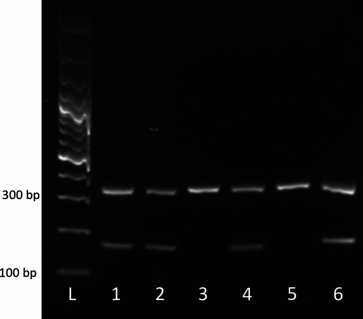 Figure 1
 Figure 2
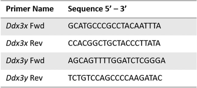 Figure 3
- —NIH/NIDA
- —Auburn University Startup Funds
Peer Reviews
No public reviews on file for this paper yet. If you reviewed it on a platform where reviews are public (OpenReview, ICLR, NeurIPS, ICML), you can paste yours below so the community can read it here.
Videos
No videos yet. Explain this paper in a talk, walkthrough, or lecture? Add one.
Taxonomy
TopicsSex and Gender in Healthcare · Reproductive System and Pregnancy · Genetic and Clinical Aspects of Sex Determination and Chromosomal Abnormalities
Introduction
There is a growing body of work demonstrating that biological sex is a powerful influence on the development and health of humans and animals. These findings have catalyzed funding agencies to account for sex as a biological variable in grant proposals and research projects [1]. As more emphasis has been placed on better understanding sexual dimorphism, we have become increasingly aware that sex influences prenatal growth and development as well [2–4]. As there are significant ethical and scientific limitations that prevent researching the molecular and physiological events that contribute to human pregnancy, the use of rodent models is common. Humans and rodents both have a hemochorial class of placenta, defined by its invasive nature and intimate relationship with the maternal blood supply [5]. Compared to the mouse, the rat placenta has significantly deeper invasion into the placenta and trophoblast-led uterine spiral artery remodeling [6], making it an important animal model for pregnancy related research.
Determining the sex of postnatal rat pups is straightforward as sex can be identified visually by assessing the anogenital distance in pups [7]. However, this external landmark cannot be assessed in rat fetuses. Furthermore, while there are several published PCR assays to determine genetic sex in mice [8–10], there are few protocols for the rat. This creates the need for a method to identify the genetic sex of rat embryos, fetuses, and extraembryonic tissues. We have developed a single step PCR protocol to amplify the X chromosome and Y chromosome homologs, the genes Ddx3x and Ddx3y. Amplification of the X-linked Ddx3x serves as an internal control that can account for the female genome while amplification of the Y-linked Ddx3y indicates the presence of the Y chromosome. We tested our protocol in both fetal and placental tissues and were able to successfully amplify Ddx3x in male and female fetal tail snips and placentas and Ddx3y in only male fetal tail snips and placentas.
Materials
ReagentConcentrationCompany/catalog numberCommentsDNA Isolation Lysis Buffer Nonidet P-40 Substitute0.3%Amresco, M158-100 mL Potassium Chloride (KCl)50 mMVWR, BDH9258-500G Tris10 mMVWR, 97,061–794pH to 8.3 Tween200.3%Fisher Scientific, BPBP337500Proteinase K1 mg/mLBoston BioProducts, P-1460PCR Reaction Ddx3x Fwd Primer10 µM Ddx3x Rev Primer10 µM Ddx3y Fwd Primer10 µM Ddx3y Rev Primer10 µM Molecular biology grade waterVWR, VWRL0201-0500 2X Phusion U Green supermixFisher Scientific, F-564 1X TE BufferInvitrogen, AM9849pH 8.0Gel Electrophoresis FluorStainSMOBio, DS1000 GeneRuler 100 bp Plus DNA LadderThermo Scientific, SM0323 RA AgaroseAmresco RA, N605-500G TAE BufferVWR, K915-1.6 L50x Tritrack 6X Loading DyeThermo Scientific, R1161Gel Imaging ChemiDocBio-Rad
Methods
Primer design
We obtained genomic sequences for Ddx3x (NC_086039.1) and Ddx3y (NC_086040.1) on NCBI Gene. FASTA sequences were used to design primer sequences using the Primer3web software (primer3.ut.ee). Amplicon size ranges were chosen between 150 and 250 base pairs for Ddx3y and 250–350 base pairs for Ddx3x. The sequences for Ddx3x and Ddx3y are as follows; Ddx3x Forward primer: 5′ – GCATGCCCGCCTACAATTTA – 3′, Ddx3x Reverse primer: 5′ – CCACGGCTGCTACCCTTATA – 3′, Ddx3y Forward primer: 5′ – AGCAGTTTTGGATCTCGGGA – 3′, Ddx3y Reverse primer: 5′ – TCTGTCCAGCCCCAAGATAC – 3′. Sequences can also be seen in Table 1.Table 1Ddx3x and Ddx3y primersPrimer sequences used to amplify Ddx3x and Ddx3y
DNA isolation
- Pre-heat two heating blocks, one to 55 °C and one to 98 °C.
- Mix 96 µL of Lysis Buffer with 4 µL Proteinase K in a microcentrifuge tube.
- Add tail snip or placental tissue to microcentrifuge tube containing Lysis Buffer + Proteinase K.
- Vortex briefly and place tube into 55 °C heat block for 1 h.
- After 1 h, move microcentrifuge tube to 98 °C heat block and incubate for 15 min to inactivate Proteinase K.
- After 15 min, spin microcentrifuge tube at 2,000 × g for 3 min at room temperature to separate undigested debris.
- Move supernatant to a fresh microcentrifuge tube.
- At this point, sample can be frozen at −20 °C for long term storage or 4 °C for short term storage.
Multiplex PCR
- Dilute DNA to 20–50 ng/uL using molecular biology grade water.
- Make primer mix by adding 10 µM Ddx3x forward and reverse primers and 10 µM Ddx3y forward and reverse primers at a 1:1 ratio.
- Primers should have previously been reconstituted to 100 µM stock in 1X TE Buffer and then diluted to a 10 µM working solution using molecular biology grade water.
- For each PCR reaction add:
- 10 µL of 2X Phusion U Green Supermix
- 10 µL molecular biology grade water
- 1 µL of primer mix
- 1 µL of diluted DNA sample
- Briefly vortex and centrifuge PCR tubes.
- Place tubes into thermal cycler and use the following PCR protocol:
- 6.Dilute PCR product 1:5 in molecular biology grade water before moving to gel electrophoresis.
Agarose gel electrophoresis
- Make a 3% gel using 3 g of RA agarose and 100 mL of 1X TAE buffer.
- Add 10 µL DNA fluorostain dye.
- Load 5 µL 100 bp ladder.
- Mix 10 µL diluted PCR product with 2 µL loading dye.
- Load 10 µL of the mixture into each well of gel.
- Run gel at 4 °C at 50 V until sufficient separation is achieved.
- Immediately image gel.
Results
Presence of Ddx3x and Ddx3y in male and female fetal tail snips and placentas
The presence of a Y chromosome was examined using primers for the Y-linked gene Ddx3y. As an internal control, primers for Ddx3x were used with each sample. After performing PCR, we ran the amplified DNA on an agarose gel using electrophoresis. The male fetal tail snip and placentas had both X-chromosome specific and Y-chromosome specific amplicons as evidenced by the presence of two bands, one for Ddx3x (amplicon size 320 bp) and one for Ddx3y (amplicon size 185 bp) (Fig. 1). The female fetal tail snips and placentas had the Ddx3x X-chromosome specific amplicon, indicating that the Y chromosome is not present in these samples (Fig. 1).Fig. 1PCR products of rat placental tissues. L – 100 bp ladder, 1 – Adult testis, 2 – Adult testis, 3 – female placenta, 4 – male placenta, 5 – Female fetal tail snip, 6 – Male fetal tail snip. Amplicon at 320 bp is Ddx3x. Amplicon at 180 bp is Ddx3y
Troubleshooting
Failure to amplify both Ddx3x and Ddx3y bands in male samples
A problem we encountered early in the development of this protocol is the presence of only the Ddx3y amplicon of male samples in the multiplex PCR protocol. However, when PCR was performed for each gene independently, amplicons for both Ddx3x and Ddx3y would appear in their individual lanes. To overcome this, we tested different annealing temperatures and found that 58 °C was the ideal annealing temperature to detect the presence of both amplicons using a multiplex PCR reaction.
Additionally, we tried several different master mix recipes, concentrations of primers, and concentrations of template for the single-step PCR reaction. Multiplex PCR requires a delicate balance of magnesium chloride, deoxynucleotide triphosphates (dNTPs), buffers, primers, template DNA, and DNA polymerases [11]. We were most successful using the commercially available high-fidelity Phusion U Multiplex PCR master mix provided by Thermo Scientific.
Discussion
In this methods paper, we provide a simple single-step multiplex PCR protocol to determine genetic sex in fetal rat tissues. Historically, determination of genetic sex by PCR has been performed by amplifying the Y-chromosome specific gene, Sry. However, solely amplifying for the presence of Sry leaves the user without a proper internal control. By amplifying both Ddx3x and Ddx3y, we have created a protocol that tests for the presence of the Y-chromosome while using the X-chromosome as an internal control, ensuring the user that their method is sound by removing the possibility of a false negative. Additionally, by including placental and adult male testis tissue, we have demonstrated that both fetal and adult tissues that are not traditionally used for genotyping can be successfully used to amplify Ddx3x and Ddx3y. This is useful for researchers who have tissues frozen but are unsure of the sex and would like to carry out experiments considering sex as a biological variable.
We acknowledge that there is at least one other publication that provides a protocol to determine genetic sex in rat tissues [12]. However, in our hands, we were unable to amplify both genes in one reaction. In this paper, we selected two different X- and Y- chromosome homologs and designed new primers. Our proposed method provides an alternative approach to determine genetic sex in rat tissues using a multiplex PCR method.
Conclusion
To summarize, we have developed a simple, cost-effective method to determine the genetic sex of rat fetal and placental tissues using multiplex PCR.