Unveiling four new taxa and Nigrosynnemanatarajanensis comb. nov. in Stachybotryaceae (Hypocreales) from monocotyledon plants in Guangdong Province, China

Abstract
Genes, proteins, chemicals, diseases, species, mutations and cell lines named across the full text — each resolved to its canonical identifier and authoritative record.
Click any figure to enlarge with its caption.
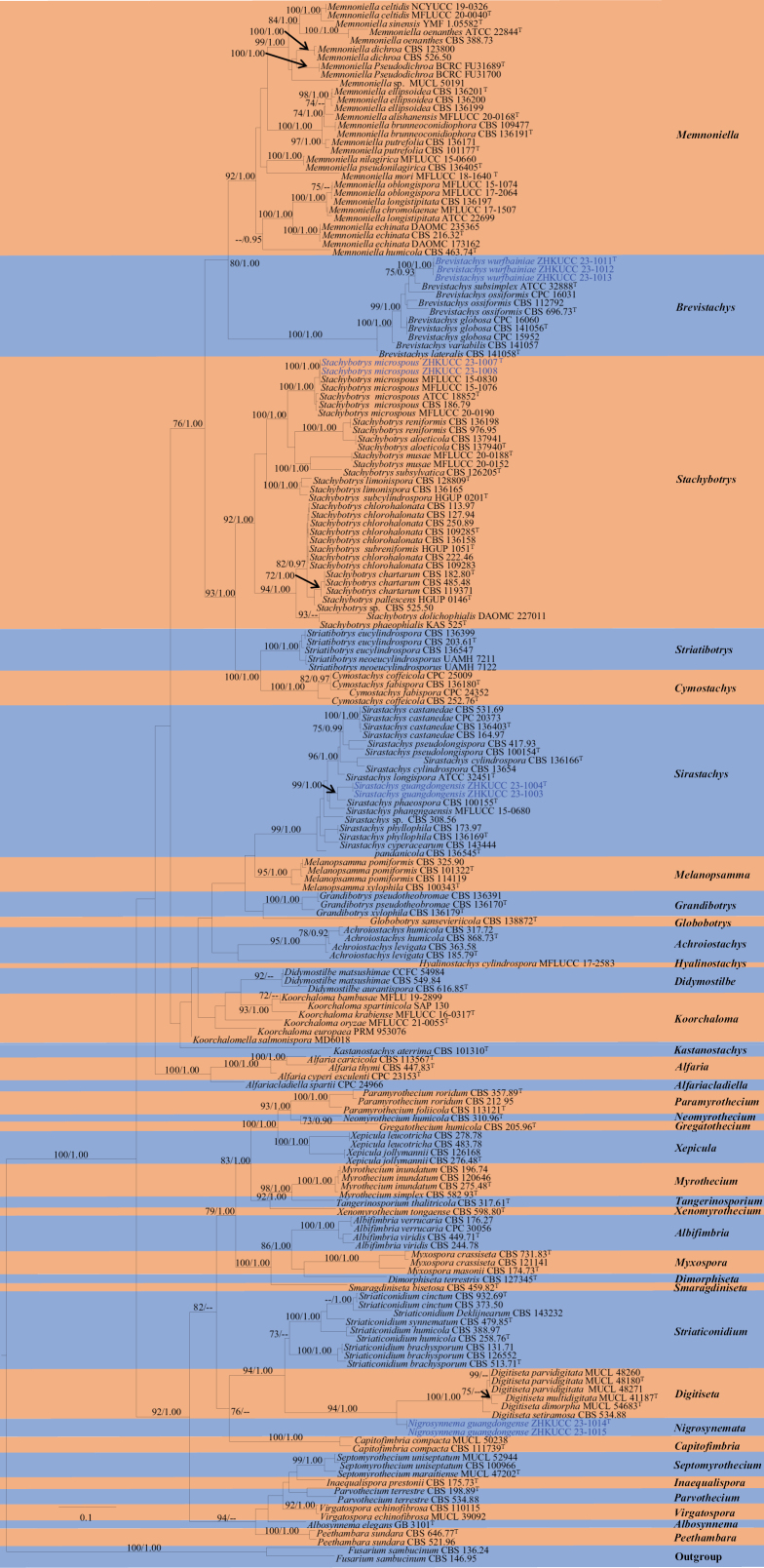 Figure 1
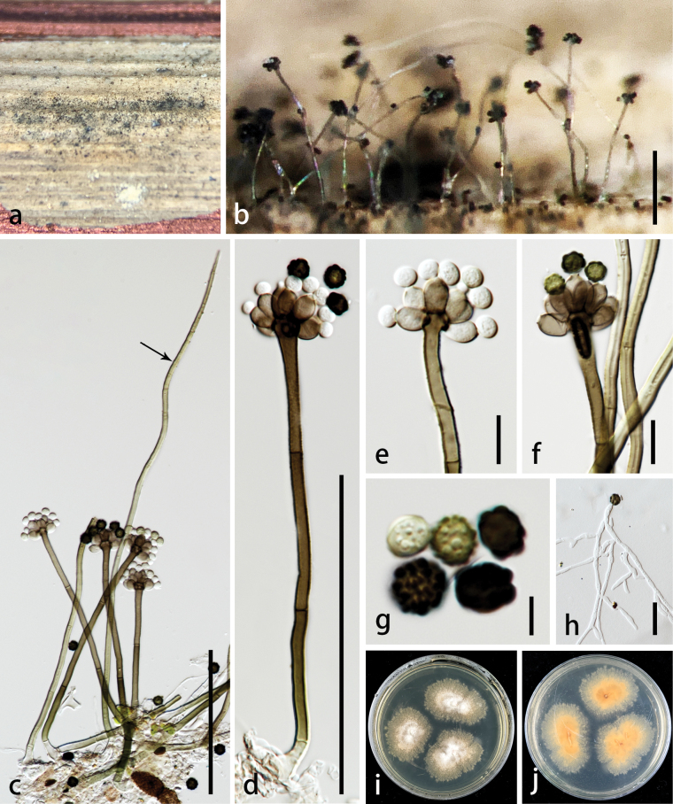 Figure 2
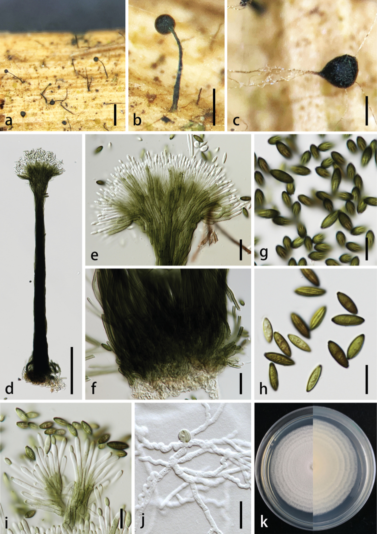 Figure 3
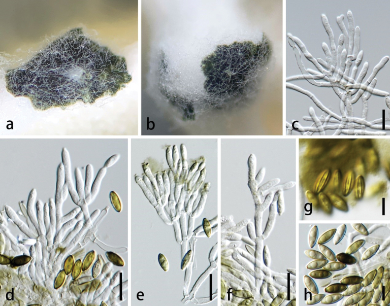 Figure 4
 Figure 5
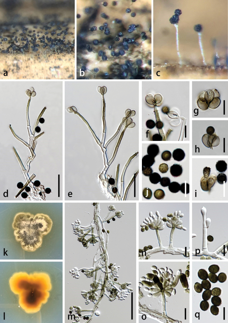 Figure 6
| Taxa | Culture collection numbers | GenBank accession numbers | |||||
|---|---|---|---|---|---|---|---|
|
|
|
|
|
|
| ||
|
| CBS 317.72 |
|
|
|
|
|
|
|
| CBS 868.73T |
|
|
|
|
|
|
|
| CBS 185.79T |
|
|
|
|
|
|
|
| CBS 363.58 |
|
|
|
|
|
|
|
| GB 3101T | - | - |
| - | - | - |
|
| CPC 30056 |
|
|
|
|
|
|
|
| CBS 176.27 |
|
|
|
|
|
|
|
| CBS 244.78 | - |
|
|
|
|
|
|
| CBS 449.71T |
|
|
|
|
|
|
|
| CBS 113567T |
|
|
|
|
|
|
|
| CPC 23153T | - |
|
| - | - | - |
|
| CBS 447.83T |
|
|
| - |
|
|
|
| CPC 24966 | - |
|
| - | - | - |
|
| CBS 141056T |
|
|
| - |
|
|
|
| CPC 15952 |
|
|
| - |
|
|
|
| CPC 16060 | - |
|
| - |
|
|
|
| CBS 141058T |
|
|
|
|
|
|
|
| CBS 696.73T | - |
|
| - | - |
|
|
| CBS 112792 |
|
|
|
|
|
|
|
| CPC 16031 |
|
|
| - |
|
|
|
| ATCC 32888T | - |
| - | - | - | - |
|
| CBS 141057 |
|
|
|
|
|
|
|
|
|
|
|
|
|
| |
|
|
|
|
|
|
|
| |
|
|
|
|
|
|
|
| |
|
| CBS 111739T |
|
|
|
|
|
|
|
| MUCL 50238 | - |
|
|
| - |
|
|
| CBS 252.76T |
|
|
|
|
|
|
|
| CPC 25009 | - |
| - | - | - | - |
|
| CBS 136180T |
|
|
|
|
|
|
|
| CPC 24352 | - |
| - |
|
| - |
|
| CBS 616.85T | - | - |
| - | - | - |
|
| CBS 549.84 | - | - |
| - | - | - |
|
| CCFC 54984 | - | - |
| - | - | - |
|
| MUCL 54683T | - |
|
|
|
|
|
|
| MUCL 41187T | - |
|
|
|
|
|
|
| MUCL 48180T | - |
|
|
|
|
|
|
| MUCL 48271 | - |
|
|
|
|
|
|
| MUCL 48260 | - |
|
|
|
|
|
|
| CBS 534.88 | - |
| - | - |
| - |
|
| CBS 127345T |
|
|
|
|
|
|
|
| CBS 146.95 | - | - |
|
| - | - |
|
| CBS 136.24 | - | - |
| - | - | - |
|
| CBS 138872T | - |
|
| - |
|
|
|
| CBS 136170T | - |
|
|
|
|
|
|
| CBS 136391 | - |
|
|
|
|
|
|
| CBS 136179T |
|
|
|
|
| |
|
| CBS 205.96T |
|
|
|
|
|
|
|
| MFLUCC 17-2583 | - |
|
| - | - | - |
|
| CBS 175.73T |
|
|
|
|
|
|
|
| CBS 101310T | - | - |
|
| - | - |
|
| MFLU 19-2899 | - |
|
|
| - | - |
|
| PRM 953076 | - |
| - | - | - | - |
|
| MFLUCC 16-0317T | - |
|
|
| - | - |
|
| MFLUCC 21-0055T | - |
|
|
| - | - |
|
| SAP 130 | - |
| - | - | - | - |
|
| MD6018 | - | - |
| - | - | - |
|
| CBS 325.90 |
|
|
|
|
|
|
|
| CBS 101322T |
|
|
|
| - | - |
|
| CBS 114119 |
|
|
|
|
|
|
|
| CBS 100343T |
|
|
|
|
| - |
|
| MFLUCC 20-0168T | - |
| - | - | - |
|
|
| CBS 109477 | - |
|
|
|
|
|
|
| CBS 136191T |
|
|
|
|
|
|
|
| MFLUCC 20-0040T | - |
| - | - | - |
|
|
| NCYUCC 19-0326 | - |
| - | - | - |
|
|
| MFLUCC 17-1507 | - |
|
| - | - | - |
|
| CBS 526.50 |
|
|
|
|
| - |
|
| CBS 123800 |
|
|
|
|
| - |
|
| CBS 216.32T |
|
|
|
|
|
|
|
| DAOMC 173162 |
|
|
|
|
|
|
|
| DAOMC 235365 |
|
|
|
|
|
|
|
| CBS 136199 |
|
|
|
|
|
|
|
| CBS 136200 |
|
|
|
|
|
|
|
| CBS 136201T |
|
|
|
|
|
|
|
| CBS 463.74T |
|
|
|
|
| |
|
| CBS 136197 |
|
|
|
|
|
|
|
| ATCC 22699 | - |
| - | - | - | - |
|
| MFLUCC 18-1640T | - |
| - | - | - |
|
|
| MFLUCC 15-0660 | - |
| - |
| - | - |
|
| MFLUCC 17-2064 | - |
| - |
| - | - |
|
| MFLUCC 15-1074 |
|
| - |
|
| |
|
| ATCC 22844T | - |
| - | - | - | - |
|
| CBS 388.73 | - |
|
|
|
| |
|
| BCRC FU31689T | - |
| - |
|
|
|
|
| BCRC FU31700 | - |
| - |
|
|
|
|
| CBS 136405T |
|
|
|
|
|
|
|
| CBS 101177T | - |
|
|
|
|
|
|
| CBS 136171 |
|
|
|
|
|
|
|
| YMF 1.05582T | - |
| - |
|
|
|
| MUCL 50191 |
|
|
|
|
|
| |
|
| CBS 196.74 |
|
|
| - |
|
|
|
| CBS 275.48T |
|
|
| - |
|
|
|
| CBS 120646 |
|
|
| - |
|
|
|
| CBS 582.93T |
|
|
| - |
|
|
|
| CBS 731.83T |
|
|
|
|
|
|
|
| CBS 121141 |
|
|
|
|
|
|
|
| CBS 174.73T |
|
|
|
|
|
|
|
| CBS 310.96T |
|
|
|
|
| - |
|
|
|
|
|
|
|
| |
|
|
|
|
|
|
|
| |
|
| CBS 113121T |
|
|
| - |
|
|
|
| CBS 212.95 |
|
|
|
|
|
|
|
| CBS 357.89T |
|
|
|
|
|
|
|
| CBS 198.89T |
|
|
|
|
|
|
|
| CBS 534.88 |
|
|
|
|
|
|
|
| CBS 521.96 | - |
|
|
|
|
|
|
| CBS 646.77T | - |
|
|
|
|
|
|
| MUCL 47202T | - | - |
|
| - | - |
|
| CBS 100966 | - |
|
|
| - |
|
|
| MUCL 52944 | - | - |
|
| - | - |
|
| CBS 164.97 |
|
|
|
|
|
|
|
| CBS 531.69 |
|
|
|
|
|
|
|
| CBS 136403T |
|
|
|
|
|
|
|
| CPC 20373 |
|
|
|
|
|
|
|
| CBS 136166T |
|
|
|
| - |
|
|
| CBS 13654 |
|
|
|
|
|
|
|
| CBS 143444 | - |
|
| - | - | - |
|
|
|
|
|
|
|
| |
|
|
|
|
|
|
|
| |
|
| ATCC 32451T | - |
| - | - | - | |
|
| CBS 136545T | - |
|
| - | - |
|
|
| CBS 100155T |
|
|
|
|
|
|
|
| MFLUCC 15-0680 | - |
|
|
| - | - |
|
| CBS 173.97 |
|
|
|
|
|
|
|
| CBS 136169T |
|
|
|
|
|
|
|
| CBS 417.93 |
|
|
|
|
|
|
|
| CBS 100154T |
|
|
|
| - |
|
| CBS 308.56 |
|
|
|
|
|
| |
|
| CBS 459.82T |
|
|
|
|
|
|
|
| CBS 137940T |
|
|
|
| - |
|
|
| CBS 137941 |
|
|
|
| - |
|
|
| CBS 182.80T |
|
|
|
|
|
|
|
| CBS 119371 |
|
|
|
|
|
|
|
| CBS 485.48 |
|
|
|
|
|
|
|
| CBS 113.97 |
|
|
|
|
|
|
|
| CBS 127.94 |
|
|
|
|
|
|
|
| CBS 222.46 |
|
|
|
|
|
|
|
| CBS 250.89 |
|
|
|
|
|
|
|
| CBS 109283 |
|
|
|
|
|
|
|
| CBS 109285T |
|
|
|
|
|
|
|
| CBS 136158 |
|
|
|
|
|
|
|
| DAOMC 227011 |
|
|
|
| - |
|
|
| CBS 128809T |
|
|
|
|
|
|
|
| CBS 136165 |
|
|
|
|
|
|
|
| CBS 186.79 |
|
|
|
|
|
|
|
| ATCC 18852T | - |
| - | - | - | - |
|
| MFLUCC 15-0830 |
|
| - |
| - |
|
|
| MFLUCC 15-1076 |
|
| - |
| - |
|
|
| MFLUCC 20-0190 | - |
| - | - |
|
|
|
|
|
|
|
|
|
| |
|
|
|
|
|
|
|
| |
|
| MFLUCC 20-0188T |
|
| - |
| - |
|
|
| MFLUCC 20-0152 |
|
| - |
| - |
|
|
| HGUP 0146T |
|
| - | - | - | |
|
| KAS 525T |
|
|
|
|
|
|
|
| CBS 976.95 |
|
|
|
|
|
|
|
| CBS 136198 | - |
|
| - |
| - |
|
| HGUP 0201T | - |
| - | - | - | - |
|
| HGUP 1051T | - |
| - | - | - | - |
|
| CBS 126205T |
|
|
|
|
|
|
| CBS 525.50 |
|
|
|
|
| ||
|
| CBS 203.61T |
|
|
|
|
|
|
|
| CBS 136399 | - |
|
|
|
|
|
|
| CBS 136547 |
|
|
|
|
|
|
|
| UAMH 7211 | - |
|
|
|
|
|
|
| UAMH 7122 | - |
|
|
|
|
|
|
| CBS 131.71 |
|
|
|
|
|
|
|
| CBS 513.71T |
|
|
|
|
|
|
|
| CBS 126552 |
|
|
|
|
|
|
|
| CBS 373.50 |
|
|
|
| - |
|
|
| CBS 932.69T |
|
|
|
| - |
|
|
| CBS 143232 | - |
|
| - |
|
|
|
| CBS 258.76T | - |
|
|
|
| |
|
| CBS 388.97 |
|
|
|
|
|
|
|
| CBS 479.85T |
|
|
|
| - |
|
|
| CBS 317.61T |
|
|
| - | - |
|
|
| CBS 110115 |
|
|
|
|
|
|
|
| MUCL 39092 | - |
|
|
| - |
|
|
| CBS 598.80T |
|
|
|
|
|
|
|
| CBS 276.48T |
|
|
|
|
|
|
|
| CBS 126168 |
|
|
|
|
|
|
|
| CBS 278.78 |
|
|
|
| - |
|
|
| CBS 483.78 |
|
|
|
|
|
|
| Genera | Synnematous conidiomata | Sporodochial conidiomata | Conidiogenous cells | Conidia | Reference | |||||
|---|---|---|---|---|---|---|---|---|---|---|
| Shape | Color | Shape | Color | Shape | Color | Shape | Color | Number of septa | ||
|
| Subcylindrical, narrower towards the apex of the stipe with a robust base | Olivaceous brown to black | Irregular, with white mycelia surrounding an olivaceous green mass of conidia | Olivaceous brown to black | Enteroblastic, phialidic, monophialidic, subcylindrical | Mostly hyaline, sometimes pale olivaceous brown in the lower portion | Fusiform to ellipsoidal, longitudinally striated | Olivaceous brown to dark brown | Aseptate | This study |
|
| Subcylindrical | Hyaline | - | - | Phialidic, cylindrical to subulate | Hyaline | Ellipsoidal to oblong | Blackish brown to black | 3-septate | |
|
| Cylindrical, slightly thinned downwards, apex in a globose-hemispherical cap | White-gray | - | - | - | - | Short oblong-fusoid | Hyaline | 0–1-septate |
|
|
| - | - | Circular to ellipsoid | Hyaline | Penicillus biverticillate, phialides, cylindrical, finger-like, straight to slightly incurved inward the penicillus | Hyaline | Cylindrical to slightly asymmetrical | Hyaline to pale greenish | Aseptate |
|
|
| Cylindrical with a subglobose to oval head surmounted by conspicuous, slimy conidia | White | - | - | Phialide, mostly subcylindrical, sometimes wider in the middle than at either end | Hyaline | Ellipsoidal to limoniform, with mammiform basal and/or apical | Hyaline to | 1-septate |
|
|
| Cylindrical to pyriform, broadening towards the apex | Marginal hyphae of synnemata olivaceous green | Oval to elongate or irregular with a white to grey setose fringe surrounding an olivaceous green to dark green slimy mass of conidia | Olivaceous green to mouse grey | Phialidic, clavate to cylindrical to subcylindrical | Hyaline | Fusiform to ellipsoidal, longitudinally striated | Olivaceous green to brown | Aseptate |
|
|
| Cylindrical throughout the greater part, somewhat expanded at the apex and base | White or yellow at the base, yellow-black or blackish black at the apex | - | - | - | - | Campanulate, cylindrical, or allantoid to fusiform | Pale, olive to fuscous | Mature 3-(sometimes 2-or 4)-septate |
|
Peer Reviews
No public reviews on file for this paper yet. If you reviewed it on a platform where reviews are public (OpenReview, ICLR, NeurIPS, ICML), you can paste yours below so the community can read it here.
Videos
No videos yet. Explain this paper in a talk, walkthrough, or lecture? Add one.
Taxonomy
TopicsPlant Pathogens and Fungal Diseases · Plant and Fungal Species Descriptions · Mycorrhizal Fungi and Plant Interactions
Introduction
The family Stachybotryaceae (as Stachybotriaceae), belonging to Hypocreales, Sordariomycetes (Hyde et al. 2024), was established to accommodate the genera Myrothecium, Peethambara, and Stachybotrys, with Stachybotrys as the type (Crous et al. 2014). The members of this family are commonly isolated from soil and dead plant materials (Jie et al. 2012; Lombard et al. 2016; Hyde et al. 2017). Some species have been reported as pathogenic to plants and animals, with some posing a substantial risk to human health (Ben et al. 2015).
The polyphyletic nature of the genera Myrothecium and Stachybotrys was addressed by Lombard et al. (2016) through phylogenetic analyses using cmdA, ITS, LSU, rpb2, tef1-α, and tub2. As a result of their study, several species previously classified under Myrothecium and Stachybotrys were transferred into numerous other genera. Thirteen myrothecium-like genera were introduced, viz., Albifimbria, Capitofimbria, Dimorphiseta, Gregatothecium, Inaequalispora, Myxospora, Neomyrothecium, Paramyrothecium, Parvothecium, Smaragdiniseta, Striaticonidium, Tangerinosporium, and Xenomyrothecium. Eight stachybotrys-like genera were established, viz., Achroiostachys, Brevistachys, Cymostachys, Globobotrys, Grandibotrys, Kastanostachys, Sirastachys, and Striatibotrys (Lombard et al. 2016). In total, 33 genera were accommodated in the Stachybotryaceae, of which 21 were newly introduced and 20 were new combinations (Lombard et al. 2016). Additional novel genera were later introduced into this family by Hernandez-Restrepo et al. (2016), Gordillo and Decock (2017), Tibpromma et al. (2018), and Hyde et al. (2020). To date, 39 genera are accepted in this family (Hyde et al. 2020; Wijayawardene et al. 2022; Hyde et al. 2024). The family Stachybotryaceae is characterized by asexual morphs having mononematous, sporodochial, or synnematous conidiophores and phialidic conidiogenous cells that produce conidia in chains or in slimy masses (Crous et al. 2014; Wang et al. 2015; Lombard et al. 2016; Hyde et al. 2020). The sexual morph is described as having solitary ascomata, superficial or completely immersed in host tissue, bright to dark yellow, orange, or black that remain unchanged when treated with KOH, unitunicate asci rounded to nearly truncate at the apex with a refractive apical ring, and ellipsoidal to fusiform to broadly reniform ascospores (Subramanian and Bhat 1978; Crous et al. 2014; Lombard et al. 2016; Hyde et al. 2020).
The type genus Stachybotrys was introduced by Corda (1837) with St.chartarum as the type species. Stachybotrys shares a morphology similar to Memnoniella (introduced by Von Höhnel (1924), based on Me.aterrima) in having branched or unbranched, erect, thin-walled, smooth, or verrucous conidiophores (Bisby 1943; Wang et al. 2015). The conidia of Memnoniella are borne in dry chains, while those in Stachybotrys are produced in slimy masses. Smith (1962) considered both Memnoniella and Stachybotrys to be congeneric, arguing that the arrangement of conidia in dry chains (Memnoniella) or slimy masses (Stachybotrys) is insufficient for differentiating these two genera. In agreement with the argument provided by Smith (1962), Wang et al. (2015) synonymized Memnoniella under Stachybotrys. Phylogenetic analyses by Lombard et al. (2016), which included a broader sampling of taxa and more loci, clearly demonstrated that the isolate previously identified as Memnoniellaechinata (CBS 216.32) (Me.aterrima) by Galloway (1933) formed a distinct and well-supported clade separate from the Stachybotrys str clade. Therefore, Lombard et al. (2016) resurrected Memnoniella and designated the type species of the genus, Me.Echinata, as the epitype, using Galloway’s strain (Galloway 1933). Morphologically, Memnoniella can be distinguished from Stachybotrys by having mostly smooth, thick-walled, and unbranched conidiophores that give rise to linear dry chains of conidia (Lombard et al. 2016). The study by Lombard et al. (2016) was further supported by Lin et al. (2016), Doilom et al. (2017), Hyde et al. (2020), Mapook et al. (2020), Samarakoon et al. (2021), and Liu et al. (2024), all of which treated Memnoniella and Stachybotrys as distinct genera.
Brevistachys, introduced by Lombard et al. (2016) with B.variabilis as the type species, is characterized by conspicuously short conidiophores and conidiogenous cells that are borne either on conidiophores or directly from the vegetative hyphae, and obovoid to globose to ossiform to ellipsoidal conidia, aggregated in slimy masses. Five species are listed under Brevistachys in Index Fungorum (http://www.indexfungorum.org/names/names.asp; accessed on 12 October 2024). The asexual morph is only observed in Brevistachys species, which were isolated from Musa and Zingiber (Cooke 1883; Lombard et al. 2016). Sirastachys, introduced by Lombard et al. (2016), is typified by Si.phaeospora. The genus is characterized by cylindrical synnemata formed in culture, which consist of bundles of parallelly compacted, erect hyphae, with conidiophores arising laterally from synnemata. Sirastachys species were mainly isolated from leaves, with one species isolated from soil under Thujaoccidentalis (Lombard et al. 2016; Crous et al. 2018; Tibpromma et al. 2018). Nine species are listed under Sirastachys Fungorum (http://www.indexfungorum.org/names/names.asp; accessed on 25 July 2024).
In this study, we introduce one new genus, three new species, and one new host record in Stachybotryaceae from dead stems of Wurfbainiavillosa and a dead leaf of Agavesisalana in Guangdong Province, China, based on the morphological characteristics and multi-locus phylogenetic analyses of cmdA, ITS, LSU, rpb2, tef1-α, and tub2. The new taxa, Brevistachyswurfbainiae sp. nov., Nigrosynnemaguangdongense gen. et. sp. nov., and Sirastachysguangdongensis sp. nov., are compared to morphologically and phylogenetically closely related taxa. A new host record, Stachybotrysmicrosporus, is presented with a detailed description and illustration supported by phylogenetic evidence. Additionally, a new combination, Nigrosynnemanatarajanensis (= Virgatosporanatarajanensis), is proposed based on the similarity in morphological characteristics aligning with the generic concept of Nigrosynnema and supported by phylogenetic evidence of the type species of Virgatospora, V.echinofibrosa.
Materials and methods
Sample collection, morphological studies, and isolation
Samples of dead leaves of Agavesisalana and dead stems of Wurfbainiavillosa were collected in Guangdong Province, China, during the winter to spring seasons of 2021 and 2022, and the important collection information was noted (Rathnayaka et al. 2024). The morphological characteristics and microscopic examination of fungal structures were observed using the method described by Liao et al. (2023). Single spore isolation was performed following the methodology outlined in Senanayake et al. (2020). The living cultures were deposited in the Zhongkai University of Agriculture and Engineering Culture Collection (ZHKUCC), Guangdong, China. Specimens were deposited in the Mycological Herbarium of Zhongkai University of Agriculture and Engineering (MHZU), Guangzhou, China. The newly discovered species were registered in Faces of Fungi (FoF) (http://www.facesoffungi.org) (Jayasiri et al. 2015) and the Index Fungorum (IF) databases (http://www.indexfungorum.org/names/names.asp).
DNA extraction, PCR amplification, and sequencing
The genomic DNA was extracted from the fungal mycelia cultivated in the dark at 25 °C on PDA for two weeks using the MagPure Plant DNA AS Kit, following the manufacturer’s instructions (Guangzhou Magen Biotechnology Co., Ltd, Guangdong, China). Extracted DNA was preserved at -20 °C for further molecular studies. The calmodulin (cmdA), internal transcribed spacer (ITS), large subunit rDNA (LSU), RNA polymerase II second largest subunit (rpb2), the partial translation elongation factor 1-α (tef1-α), and β-tubulin (tub2) were amplified and sequenced using primer CAL-228F (Carbone and Kohn 1999) and CAL-2RD (Groenewald et al. 2013), ITS4 and ITS5 (White et al. 1990), LR5 and LR0R (Vilgalys and Hester 1990), rpb2-5f and rpb2-7cR (Liu et al. 1999), EF1-728F (Carbone and Kohn 1999), EF2 (O’Donnell et al. 1998), and BT2a and BT2b (Glass and Donaldson 1995), respectively.
The polymerase chain reaction (PCR) contained a total of 25 µl of mixture, including 9.5 µl of ddH_2_O, 12.5 µl of 2 × Taq Master Mix (a mixture of dNTPs, optimized buffer, and Taq (Nanjing Vazyme Biotech Co., Ltd., Nanjing, China)), and 1 µl of each of the primer and DNA template. The PCR thermal cycling program for ITS and LSU amplification was conducted with an initial denaturation at 95 °C for 3 min, followed by 35 cycles of 94 °C for 30 sec; the annealing temperature was set to 52 °C for 30 sec; the extension step was performed at 72 °C for 1 min; and final elongation was carried out at 72 °C for 10 min. The annealing temperatures were altered to 53.5 °C for cmdA and tef1-α and 55 °C (45 sec) for tub2. PCR was performed for the rpb2 in a thermal cycle as follows: an initial denaturation at 95 °C for 3 min, followed by 35 cycles at 94 °C for 1 min, an annealing temperature of 52 °C for 2 min, and extension at 72 °C for 1.5 min, with a final elongation at 72 °C for 10 min. The PCR products were purified and sent for sequencing at Tianyi Huiyuan Gene Technology & Services Co. (Guangdong, China). All sequences obtained in this study have been deposited in GenBank (available online: http://www.ncbi.nlm.nih.gov).
Phylogenetic analyses
The original sequence obtained from the sequencing company was cross-checked by verifying chromatograms using BioEdit v. 7.2.3 (Hall 1999), and subsequently, consensus sequences were generated using OFPT (Zeng et al. 2023) and SeqMan v. 7.0 (Lasergene, Madison, WI, USA). The consensus sequences of our fungal strains from each locus were subjected to a basic local alignment search tool (BLAST) search in GenBank. The reference sequences and outgroup taxa used for phylogenetic analyses were selected based on recent relevant literature (Lombard et al. 2016; Tibpromma et al. 2018; Mapook et al. 2020), obtained from GenBank. The phylogenetic analyses utilized 184 sequences (Table 1), with Fusariumsambucinum strains CBS 146.95 and CBS 136.24 used as the outgroup taxa. Six loci, cmdA, ITS, LSU, rpb2, tef1-α, and tub2, were aligned using the MAFFT version v. 7 online program (https://mafft.cbrc.jp/alignment/server/; Katoh and Standley 2013). Subsequently, the dataset was trimmed with TrimAl v.1.3 using the Gappyout option (Capella-Gutiérrez et al. 2009). The alignments were transformed into NEXUS format using the ALignment Transformation EnviRonment online platform (http://www.sing-group.org/ALTER/).
The combined cmdA, ITS, LSU, rpb2, tef1-α, and tub2 sequence data were performed using maximum likelihood (ML) and Bayesian inference (BI) analyses. The ML analysis was carried out in the CIPRES Science Gateway online platform (Miller et al. 2010) using RAxMLHPC v.8.2.12 on XSEDE (Stamatakis 2014) with GTR+G+I evolutionary substitution, with 1000 rapid bootstrap inferences followed by an extensive ML search. All free model parameters were estimated using the RAxML maximum likelihood method with 25 per-site rate categories. The likelihood of the final tree was evaluated and optimized under the GAMMA gamma distribution shape parameter. The Bayesian Inference (BI) analysis was conducted utilizing the Markov Chain Monte Carlo (MCMC) method and executed in MrBayes XSEDE (3.2.7a) (Huelsenbeck and Ronquist 2001). The simulation was conducted by running six concurrent Markov chains for 5,000,000 generations, with tree sampling occurring every 100^th^ generation. The phylogenetic trees were visualized using FigTree v. 1.4.0 (Rambaut 2009) and formatted using PowerPoint 2010 (Microsoft Corporation, WA, United States). New species are established based on the recommendations of Jeewon and Hyde (2016).
Results
Phylogenetic analyses
The phylogenetic tree was constructed using the combined cmdA, ITS, LSU, rpb2, tef1-α, and tub2 sequence data of 184 strains (including our new strains) through ML and BI analyses. The total length of the dataset, including gaps, was 5060 base pairs (cmdA: 1–930, ITS: 931–1657, LSU: 1658–2494, rpb2: 2495–3274, tef1-α: 3275–4646, tub2: 4647–5060). The topology of the ML analysis resembled that of the BI analysis. The highest-scoring RAxML tree, with a final ML optimization likelihood value of -87901.129587, is depicted in Fig. 1. The matrix consisted of 1060 distinct alignment patterns, with 44.52% undetermined characters or gaps. The estimated base frequencies were as follows: A = 0.235675, C = 0.272976, G = 0.268686, T = 0.222663; substitution rates AC = 1.088667, AG = 3.185610, AT = 1.213744, CG = 0.825063, CT = 4.778952, GT = 1.000000; gamma distribution shape parameter α = 0.307577. In this study, the phylogenetic analyses showed that our strains belong to Stachybotryaceae. The tree topology in this study is almost similar to the previous studies of Lombard et al. (2016) and Hyde et al. (2020). However, Lombard et al. (2016) and Hyde et al. (2020) constructed the tree using LSU and rpb2. The inclusion of an increasing number of newly discovered genera and taxa, including Digitiseta gen. nov. and additions to Inaequalispora and Parvothecium as reported by Gordillo and Decock (2017), has resulted in slight alterations to the positions of some genera. Additionally, our phylogenetic tree showed that species of Koorchaloma are paraphyletic and grouped with Didymostilbe instead of Koorchalomella and Alfariacladiella as shown in Hyde et al. (2020). Two new strains (ZHKUCC 23-1007, ZHKUCC 23-1008) constituted a highly supported subclade with Stachybotrysmicrosporus (type strain, CBS 126205) with 100% ML and 1.00 BYPP. The novel strains ZHKUCC 23-1003 and ZHKUCC 23-1004 formed a sister subclade with Sirastachysphaeospora (type strain, CBS 100155) with 99% ML bootstrap support and 1.00 BYPP. Three strains, ZHKUCC 23-1011, ZHKUCC 23-1012, and ZHKUCC 23-1013, formed a sister subclade with Brevistachyssubsimplex (type strain, ATCC 32888) with 75% ML and 0.93 BYPP. Two strains, ZHKUCC 23-1014 and ZHKUCC 23-1015, formed a distinct clade with Digitiseta species with 94% ML and 1.00 BYPP.
Phylogram generated from maximum likelihood analysis (RAxML) of strains in Stachybotryaceae based on the combined cmdA, ITS, LSU, rpb2, tef1-α, and tub2 sequence data. Maximum likelihood bootstrap values ≥ 70% (ML) and Bayesian posterior probabilities ≥ 0.90 (ML/BYPP) are provided at the nodes. The tree is rooted with Fusariumsambucinum strains CBS 136.24 and CBS 146.95. The hyphen (-) represents support values < 70% ML and < 0.90 BYPP. The ex-type strains are denoted as “T”, while the newly isolated strains are highlighted in blue.
Taxonomy
Brevistachys
wurfbainiae
Taxon classificationFungiHypocrealesStachybotryaceae
C.F. Liao, K.D. Hyde & Doilom sp. nov.
B7ABE979-3743-55CB-BBC2-ECDDF53A6B59
Index Fungorum: IF902005
Facesoffungi Number: FoF15745
Etymology.
In reference to the host genus Wurfbainia, from which the holotype was isolated.
Holotype.
MHZU 23-0254.
Description.
Saprobic on dead stem of Wurfbainiavillosa. Sexual morph: undetermined. Asexual morph: Colonies superficial on host substrate, effuse, hairy, gregarious, with numerous dark conidia on the substrate visible as black granular powder. Conidiophores 80–235 × 3–5.5 µm (av. 155 × 4.5 μm, n = 20), macronematous, mononematous, erect, simple, unbranched, straight or flexuous, subcylindrical, unevenly olivaceous brown, 1–3-septate, not constricted at the septa, smooth-walled to finely verruculose in the above half, thick-walled, with bulbous apices, bearing 5–8 conidiogenous cells at the tip, often intermixed with setiferous, flexuous, sterile filaments. Setae 230–390 × 3–6 µm (av. 305 × 4.5 μm, n = 20), arising from the basal stroma, adjacent to cells that give rise to fertile conidiophores, unbranched, straight, and subhyaline at base, mostly flexuous, olivaceous green, in above half, moderately thick-walled, smooth, septate, acute at apex. Conidiogenous cells 6–10 × 4–7 µm (av. 7.5 × 5.5 μm, n = 30), enteroblastic, monophialidic, discrete, determinate, terminal, elongate doliiform, pale to dark brown, smooth-walled, with a conspicuous collarette. Conidia 5–9 µm diam. (av. 7 μm, n = 30), acrogenous, solitary, dry, obovoid to subglobose, aseptate, hyaline, and smooth-walled when young, pale brown, mostly olivaceous to dark brown, verrucose to warty-surfaced at maturity.
Culture characteristics.
Colonies on PDA reaching 2 cm in two weeks at 28 ± 2 °C, medium dense, raised, sparse, filamentous, floccose to fluffy, velvety, filiform at margin, cream to pale brown from above; brown to pale luteous from reverse.
Material examined.
China • Guangdong Province, Yangchun City, Yongning Town (22.256185°N, 111.609037°E, 270 m), on dead stems of Wurfbainiavillosa (Lour.) Škorničk. & A.D. Poulsen. (Zingiberaceae), 10 April 2022, C.F. Liao & Y.H. Yang, YAM16 (MHZU 23-0254, holotype) • ex-type, ZHKUCC 23-1011 • ibid., living culture ZHKUCC 23-1012, and ZHKUCC 23-1013.
Brevistachyswurfbainiae (MHZU 23-0254, holotype) a, b colonies on the surface substrate c, d conidiophores, setae or conidiophore-like (arrow), conidiogenous cells, and conidia e, f conidiogenous cells with conidia g conidia h germinated conidium i, j colonies on PDA (front and below). Scale bars: 100 µm (b–d); 10 µm (e, f); 5 µm (g); 20 µm (h).
Notes.
Brevistachyswurfbainiae differs from other known species in the genus Brevistachys by having erect to flexuous, sterile, setiferous filaments intermixed with fertile conidiophores. Brevistachyswurfbainiae resembles B.subsimplex but differs from the latter in having slightly longer conidiophores with bulbous apices (80–235 × 3–5.5 µm vs. 80–200 (most frequently 100–140) × 3–5.5 µm) and shorter conidiogenous cells (6–10 × 4–7 µm vs. 8–13 × 4–6 µm). The conidiophores of B.wurfbainiae are 1–3-septate, while they are 2–6-septate in B.subsimplex (Deighton 1960). The phylogenetic analyses revealed that B.wurfbainiae (ZHKUCC 23-1011, ZHKUCC 23-1012, and ZHKUCC 23-1013) formed a separate branch from B.subsimplex (ex-type CBS 100155) with 75% ML bootstrap support and 0.93 BYPP (Fig. 1). Based on distinct morphology and phylogenetic support, we propose B.wurfbainiae as a new species.
Nigrosynnema
Taxon classificationFungiHypocrealesStachybotryaceae
C.F. Liao, K.D. Hyde & Doilom gen. nov.
E293A5DB-224E-5817-8715-D4095987F34C
Index Fungorum: IF902006
Facesoffungi Number: FoF15744
Etymology.
The name refers to the characteristic black synnemata formed on natural substrate.
Description.
Saprobic on dead plant material. Sexual morph: undetermined. Asexual morph: Conidiomata synnematous or sporodochial. Synnemata unbranched, subcylindrical, globose to subglobose head, robust at base, olivaceous brown to black, straight or curved in the upper portion, consisting of bundles of parallelly arranged, tightly compacted conidiophores. Sporodochia stromatic, superficial, scattered or gregarious, irregular, pulvinate, with white mycelium surrounding an olivaceous green mass of conidia. Conidiophores arising from basal stroma, macronematous, mononematous, septate, unbranched or branched, straight or flexuous, thin-walled, subcylindrical, olivaceous brown, verrucose, consisting of a stipe and a penicillately branched conidiogenous apparatus consisting of a whorl of primary branches, each terminating in number of conidiogenous cells. Conidiogenous cells enteroblastic, monophialidic, integrated, terminal, clavate to subcylindrical, hyaline to pale olivaceous brown, smooth, often verruculose at base, with a conspicuous collarette. Conidia solitary, fusiform to ellipsoidal, aseptate, initially hyaline, becoming olivaceous brown to dark brown, longitudinally striated at surface, with a distinct dark basal hilum.
Type species.
Nigrosynnemaguangdongense C.F. Liao, K.D. Hyde & Doilom
Notes.
Nigrosynnema resembles Striaticonidium in having fusiform to ellipsoidal conidia with longitudinal striations. However, it can be distinguished from Striaticonidium by having synnematous conidiomata, the absence of setae on the sporodochia, as well as support from molecular data. The synnematous conidiomata of Nigrosynnema are subcylindrical, flexuous, narrower towards the apex of the stipe, and robust at the base. The sporodochia are devoid of setae. However, in Striaticonidium, they are cylindrical to pyriform, broadened towards the apex, and have sporodochia covered by setae (Lombard et al. 2016). The blastn search of NCBI GenBank revealed that two strains of Nigrosynnema, ZHKUCC 23-1014 and ZHKUCC 23-1015, have sequence similarities of 98.37%, 91.73%, 89.70%, 89.04%, and 82.03% to the type species of Striaticonidium (Stri.cinctum CBS 932.69, ex-type) in LSU, ITS, tub2, rpb2, and cmdA sequence data, respectively. However, tef1-α sequence data of Stri.cinctum CBS 932.69 (ex-type) is unavailable in the NCBI database.
Nigrosynnema resembles Virgatospora described by Finley (1967) in having synnematous conidiomata, phialidic conidiogenous cells, and striated conidia. However, olivaceous brown to black synnemata in Nigrosynnema are subcylindrical, robust at the base, and narrower towards the apex of the stipe. The conidia in Nigrosynnema are aseptate, fusiform to ellipsoidal, and different from the septate, slightly curved conidia with a protuberant hilum of the type species of Virgatospora, V.echinofibrosa. Nigrosynnema can be distinguished from its closely related genera, as shown in Table 2.
The phylogenetic analyses supported that our two strains (ZHKUCC 23-1014 and ZHKUCC 23-1015) formed a distinct clade from other morphologically closely related taxa and constituted a well-supported clade related to Digitiseta with 94% ML and 1.00 BYPP statistical support. The main distinguishing morphological characteristic between the two genera is the absence of hypha-like setoid structures in Nigrosynnema, whereas Digitiseta, introduced by Gordillo and Decock (2017), has short apical branches and digitated hypha-like setoids. Additionally, the conidial shape is fusiform to ellipsoidal in Nigrosynnema, while they are cylindrical in Digitiseta.
Based on morphological and molecular evidence, we introduce a novel asexual genus, Nigrosynnema, characterized by olivaceous to black synnematous or sporodochial conidiomata that produce phialidic, aseptate conidia in black, slimy, glistening masses or heads. The conidia are fusiform to ellipsoidal, aseptate, longitudinally striated, and olivaceous brown to dark brown.
Nigrosynnema
guangdongense
Taxon classificationFungiHypocrealesStachybotryaceae
C.F. Liao, K.D. Hyde & Doilom sp. nov.
9BEEF1EB-45BC-5BF8-8321-B1616FC44D40
Index Fungorum: IF902007
Facesoffungi Number: FoF15746
Etymology.
The epithet “guangdongense” refers to the locality, Guangdong Province, China, where the holotype was collected.
Holotype.
MHZU 23-0255.
Description.
Saprobic on dead stem of Wurfbainiavillosa. Sexual morph: undetermined. Asexual morph: Synnemata on the natural substrate, 370–570 × 20–50 µm (av. 470 × 33 μm, n = 20), erect, unbranched, subcylindrical, with a robust base, narrowed towards fertile apex, olivaceous brown to black, straight or curved in the upper portion, consisting of parallelly arranged, tightly compacted conidiophores. Conidiophores 2–4 µm wide, subcylindrical, branched, olivaceous brown, slightly tapering towards the apex, verrucose. Conidiogenous cells 10.5–32.5 × 1.5–3 µm (av. 22 × 2.5 μm, n = 30), enteroblastic, monophialidic, discrete, terminal, subcylindrical, mostly hyaline, sometimes pale olivaceous brown in the lower portion, mostly smooth-walled in above half, often verruculose at below half, with a conspicuous collarette. Conidia 10–12.5 × 3–4.5 µm (av. 11.5 × 4 μm, n = 30), solitary, slimy, fusiform to ellipsoidal, aseptate, longitudinally striated, olivaceous brown to dark brown, guttulate, obtuse at both ends, with a distinct dark basal hilum.
Nigrosynnemaguangdongense (MHZU 23-0255, holotype) on natural substrate a–c synnemata on substrate d synnema e top of synnema f base of synnema g, h conidia i conidiogenous cells with conidia j germinated conidium k colonies on PDA (front and below). Scale bars: 500 µm (a); 200 µm (b); 100 µm (c, d); 20 µm (e, f); 10 µm (g–j).
Culture characteristics.
Colonies on PDA reaching 4.5–6.5 cm in two weeks at 28 ± 2 °C, medium dense, flat or effuse, diffuse, rough, circular, filiform with curled, large circle in the middle becoming a wave and extends outward, cream from above; cream from the reverse. The spores produced on PDA after three weeks. Conidiomata 220–300 × 15–20 µm, sporodochial, superficial, scattered, irregular, with white mycelia surrounding an olivaceous green mass of conidia, with or without covering the slimy mass of conidia, without setae. Conidiophores arising from the basal stroma, consisting of a stipe and a penicillately branched conidiogenous apparatus; stipes unbranched or rarely branched, hyaline, septate, smooth, 10–30 × 2.5–3.5 µm (av. 18 × 3.0 μm, n = 20), conidiogenous apparatus consisting of a whorl of 2–5 primary branches, each terminating in 2–5 conidiogenous cells; primary branches, 1, 2-septate, smooth, unbranched, 8–20 × 2–6 µm, secondary branches, aseptate, smooth, unbranched, 6–20 × 2–5 µm. Conidiogenous cells 10–20 × 2–4 µm (av. 14 × 2.5 μm, n = 30), phialidic, terminal, with a conspicuous collarette, clavate to cylindrical, hyaline, smooth. Conidia 7–10 × 3–5 µm (av. 8.5 × 3.5 μm, n = 30), acrogenous, longitudinally striated, fusiform to ellipsoidal, aseptate, initially hyaline, becoming olivaceous green when mature.
Nigrosynnemaguangdongense (MHZU 23-0255) on PDA after three weeks a, b conidial masses on pda c–f conidiophores, conidiogenous cells with conidia g, h conidia. Scale bars: 10 µm (c–f); 5 µm (g, h).
Material examined.
China • Guangdong Province, Yangchun City, Yongning Town (22.256185°N, 111.609037°E, 270 m), on dead stems of Wurfbainiavillosa (Lour.) Škorničk. & A.D. Poulsen. (Zingiberaceae), 10 April 2022, C.F. Liao & Y.H. Yang, YAM19 (MHZU 23-0255, holotype) • ex-type, ZHKUCC 23-1014 • ibid., living culture ZHKUCC 23-1015.
Notes.
Nigrosynnemaguangdongense is established here as the type species. It is similar to Virgatosporanatarajanensis described by D’Souza et al. (2002) in having synnematous conidiomata, with fusiform, aseptate, and striated conidia. However, N.guangdongense has verrucose, olivaceous, brown conidiophores, conidia with obtuse apices, and a distinct dark basal hilum, whereas V.natarajanensis has distinctly echinulate, subhyaline conidiophores that are narrower and smooth towards the apex, and conidia are rounded at both ends. Additionally, the conidiogenous cells of V.natarajanensis are occasionally found in the subterminal position, while they have not been observed in N.guangdongense. Nigrosynnemaguangdongense has longer conidiogenous cells (10.5–32.5 × 1.5–3 µm) compared to V.natarajanensis (18–25 × 1.5–3.5 μm).
Nigrosynnemaguangdongense and Virgatosporaechinofibrosa (the type species of Virgatospora) (Finley 1967) are similar in having synnematous conidiomata and striate, phialidic conidia. However, N.guangdongense has shorter synnemata (370–570 × 20–50 µm) than V.echinofibrosa (up to 1500 µm). Synnemata of N.guangdongense are robust at the base, narrower towards the apex of the stipe, and olivaceous brown to black, whereas they are simple or branched, sometimes proliferated, cylindrical throughout the greater part, somewhat broader at the apex and base, white or yellow at the base, yellow-black or blackish black at the apex in V.echinofibrosa.
Additionally, N.guangdongense has smaller (10–12.5 × 3–4.5), fusiform, aseptate conidia compared to ellipsoidal to limoniform, 3-(sometimes 2- or 4)-septate conidia (39–50 × 9–15 µm) of V.echinofibrosa.
Nigrosynnema
natarajanensis
Taxon classificationFungiHypocrealesStachybotryaceae
(D’Souza, S.K. Singh & Bhat) C.F. Liao, K.D. Hyde Doilom & Bhat comb. nov.
2515F308-9442-5A9D-AE98-AE53AD41F5BF
Index Fungorum: IF902010
Basionym.
Virgatosporanatarajanensis D’Souza, S.K. Singh & Bhat, Mycotaxon 82: 141 (2002).
Holotype.
IMI 386680.
Type information.
India • Middle Andaman Island, on dead leaves of Calamusthwaitesii, 15 December 2000, Rajiv Kumar, IMI386680 (holotype).
Description.
See D’Souza et al. (2002) on page 141.
Illustration.
See D’Souza et al. (2002) on page 140, Fig. 4a–d.
Notes.
D’Souza et al. (2002) introduced Virgatosporanatarajanensis based on the morphology and found it as a saprobe on dead leaves of Calamusthwaitesii from Middle Andaman Island, India. Although the DNA sequence data of V.natarajanensis is not available in NCBI, morphologically, it fits well within the generic concept of Nigrosynnema due to its synnematous conidiomata, phialidic conidiogenous cells, ellipsoidal to fusiform, aseptate conidia (amerosporous) with distinct longitudinal striations. The conidia in V.echinofibrosa, the type species of Virgatospora, are ellipsoidal to limoniform and 3-(sometimes 2- or 4)-septate (phragmosporous). Based on the morphological similarities between V.natarajanensis and N.guangdongense (the type species of Nigrosynnema), as well as support from molecular phylogenetic analyses of the type species of Virgatospora, we synonymize V.natarajanensis under Nigrosynnema as N.natarajanensis. However, it is desirable to obtain DNA sequence data from both the type specimen of V.natarajanensis and fresh collections to further support our proposal.
Sirastachys
guangdongensis
Taxon classificationFungiHypocrealesStachybotryaceae
C.F. Liao, K.D. Hyde & Doilom sp. nov.
B0810616-1D23-5049-8ADC-DDEE96115D8A
Index Fungorum: IF902009
Facesoffungi Number: FoF15747
Etymology.
The epithet “guangdongensis” refers to the locality, Guangdong Province, China, where the holotype was collected.
Holotype.
MHZU 23-0250.
Description.
Saprobic on dead stem of Agavesisalana. Sexual morph: undetermined. Asexual morph: Colonies superficial on host substrate, erect, gregarious, visible as numerous black conidial masses. Conidiophores 105–170 × 3.5–7 µm (av. 140 × 5.5 μm, n = 30), macronematous, mononematous, erect, simple, unbranched, straight or slightly flexuous, subcylindrical, hyaline, 1–5-septate, not constricted at septa, smooth-walled, or slightly verrucose, thick-walled, bearing 4–8 conidiogenous cells on the tip. Conidiogenous cells 6.5–12.5 × 4–5 µm (av. 10 × 4 μm, n = 30), enteroblastic, monophialidic, discrete, determinate, terminal, elongate doliiform to reniform, subhyaline to brown, smooth-walled, with a conspicuous collarette. Conidia 5–6 × 4–5 µm (av. 5.5 × 4 μm, n = 30), acrogenous, aggregating in slimy masses, obovoid, with a prominent hilum, aseptate, brown, pale olivaceous brown, black, smooth-walled.
Sirastachysguangdongensis (MHZU 23-0250, holotype) a–c colonies on the surface substrate d, e conidiophores, conidiogenous cells with conidia f, g conidiogenous cells with conidia h conidia. Scale bars: 50 µm (d, e); 10 µm (f, g); 5 µm (h).
Culture characteristics.
Colonies on PDA reaching 5.5–6.0 cm in two weeks at 28 ± 2 °C, medium dense, flat, circular, cream from above; pale luteous from the reverse, with no pigmentation.
Material examined.
China • Guangdong Province, Guangzhou City, Zhongkai University of Agriculture and Engineering (23.10643°N, 113.28240°E, 20 m), on dead leaf of Agavesisalana Perr. ex Engelm. (Agavaceae), 17 November 2021, C.F. Liao & Y.H. Yang, JM02 (MHZU 23-0250, holotype) • ex-type, ZHKUCC 23-1003 • ibid., living culture ZHKUCC 23-1004.
Notes.
Sirastachysguangdongensis resembles Si.pandicola and Si.Phaeospora that were described by Lombard et al. (2016). However, the former can be distinguished by the size of the conidiophores and conidia as well as other conidiophore characteristics. Sirastachysguangdongensis has longer conidiophores (105–170 µm) than those of Si.pandicola (55–75 µm) and Si.phaeospora (40–65 µm). Sirastachysguangdongensis has larger conidia (5–6 × 4–5 µm) than Si.pandicola (3–4 × 2–3 µm) and Si.phaeospora (4–5 × 2–3 µm). Conidiophores of Si.guangdongensis are 1–5-septate, while they are 1–3-septate in Si.pandicola and 1–2(–3)-septate in Si.phaeospora. Branched conidiophores are observed in Si.phaeospora (Lombard et al. 2016), while they are unbranched in Si.guangdongensis. The phylogenetic analyses supported Si.guangdongensis as a distinct species from other Sirastachys species and showed that Si.guangdongensis (ZHKUCC 23-1003 and ZHKUCC 23-1004) formed a distinct branch and sister to Si.phaeospora (ex-type CBS 100155) with 99% ML bootstrap support and 1.00 BYPP (Fig. 1). Based on distinct morphological and molecular evidence, we propose Sirastachysguangdongensis as a novel species.
Stachybotrys
microsporus
Taxon classificationFungiHypocrealesStachybotryaceae
(B.L. Mathur & Sankhla) S.C. Jong & E.E. Davis [as ‘microspora’]
3CE5110C-40ED-50AA-A7D2-5A11DF12B5F6
Index Fungorum: IF627002
Facesoffungi Number: FoF09372
Description.
Saprobic on dead leaf of Agavesisalana. Sexual morph: undetermined. Asexual morph: Colonies superficial on host substrate, gregarious, visible as numerous black conidial masses. Conidiophores 35–70 × 2.5–5 µm (av. 48 × 4 μm, n = 30), macronematous, mononematous, irregularly or sympodially branched, straight or flexuous, subcylindrical, hyaline, becoming pale olivaceous brown in the above half, 1–3-septate, not constricted at the septa, smooth-walled, slightly rough-walled in the subterminal region, thick-walled, bearing 3–9 conidiogenous cells on the tip. Conidiogenous cells 6–10 × 4–6 µm (av. 8 × 5 μm, n = 30), enteroblastic, monophialidic, discrete, determinate, terminal, obovoid, sub-hyaline to pale olivaceous brown, smooth-walled. Conidia 5–7 µm diam. (av. 6 μm, n = 30), aggregating in slimy masses, globose, subglobose, aseptate, olivaceous brown to black, rough-walled, verrucose.
Stachybotrysmicrosporus (MHZU 23-0252, new host record) a–c colonies on the surface substrate d, e conidiophores f–i conidiogenous cells j conidia k colonies on pda (front) l colony on pda (below) m mycelium, conidiophores, and conidiogenous cells with conidia n–p conidiogenous cells with conidia q conidia. Scale bars: 20 µm (d, e); 50 µm (f–j, m); 10 µm (n–q).
Culture characteristics.
Colonies on PDA reaching 2.5–3.0 cm in two weeks at 28 ± 2 °C, medium dense, raised, flat, floccose to fluffy, velvety, irregular edge, gold brown at the center, pale brown, with conidiophores forming on the surface of the medium, carrying slimy olivaceous green from above; brown to pale luteous from the reverse. The conidia producing on PDA after three weeks: Conidiophores 30–80 × 3.5–5.5 µm (av. 46 × 4 μm, n = 30), most similar with the above description, 0–2-septate, unbranched or branched, bearing 2–10 conidiogenous cells at the tip. Conidiogenous cells 9–17 × 4.5–7.5 µm (av. 12 × 5.5 μm, n = 30), most similar to those on natural substrate. Conidia 6.5–10 × 3–6.5 µm (av. 8 × 5 μm, n = 30), often aggregated as large, slimy, glistening, blackheads, initially hyaline to olivaceous green, oblong, obovoid to subglobose, becoming black-brown, globose, smooth to verruculose.
Material examined.
China • Guangdong Province, Guangzhou City, Zhongkai University of Agriculture and Engineering (23.10643°N, 113.28240°E, 20 m), on dead leaf of Agavesisalana Perr. ex Engelm. (Agavaceae), 17 November 2021, C.F. Liao & Y.H. Yang, SR09A (MHZU 23-0252, new host record) • living culture, ZHKUCC 23-1007 • ibid., living culture ZHKUCC 23-1008.
Known distribution.
• Canada, Cuba, India, Nigeria, and Pakistan (Ellis 1971; Jong and Davis 1976); • China (this study); Japan (Iwama et al. 2022); • New Guinea; Zaria (Jong and Davis 1976); • Thailand (Lin et al. 2016; Samarakoon et al. 2021); • Sudan (Lombard et al. 2016).
Known hosts/substrates.
Agavesisalana (this study), Arachishypogaearhizosphere, soil (Jong and Davis 1976), Castanopsiscuspidata var. sieboldii (Iwama et al. 2022), dead plants, paper, seeds, and textiles (Ellis 1971), decaying shrubs, wood (Lin et al. 2016), Musa sp. (Samarakoon et al. 2021), soil in Mangifera field (Lombard et al. 2016).
Notes.
Stachybotrysmicrosporus (ZHKUCC 23-1007, ZHKUCC 23-1008) formed a subclade with the type and other strains of St.microsporus with 100% ML and 1.00 BYPP (Fig. 1). Our collection has similar morphs to St.microsporus described by Jong and Davis (1976), Lin et al. (2016), and Samarakoon et al. (2021) by having irregularly branched conidiophores with tapering apices, monophialidic, discrete conidiogenous cells, unicellular, globose, roughened, and black conidia. Stachybotrysmicrosporus has been reported from forest soil in New Guinea (Jong and Davis 1976), on decaying wood in Thailand (Jong and Davis 1976), and on dead leaf petiole of Musa sp. in Thailand (Samarakoon et al. 2021). We report St.microsporus here as a new host record on Agavesisalana in China.
Discussion
Species of Stachybotryaceae have primarily been collected from soil and dead plant tissues. For example, Albifimbriaterrestris was isolated from soil in mopane woodlands in Namibia and unidentified dead hardwood in the USA, while Cymostachysfabispora was obtained from decaying leaf material in Cuba and Aloeferox in Tanzania (Lombard et al. 2016). In China, most taxa in this family have been reported from the soil, including Stachybotryspallescens, St.subcylindrospora, St.subreniformis, and St.subcylindrospora (Jiang and Zhang 2009; Li and Jiang 2011; Jie et al. 2012). However, some species were also found in diseased plants and dead plant tissues. Paramyrotheciumroridum (formerly known as Myrotheciumroridum) has been identified as a pathogen causing leaf spot on Abutilonmegapotamicum and Zantedeschiaaethiopica plants from China (Li et al. 2014; Ben et al. 2015). Myxosporaaptrootii was isolated from leaf litter in Hong Kong, China (Lombard et al. 2016). In this study, Brevistachyswurfbainiae and Nigrosynnemaguangdongense were isolated from dead stems of Wurfbainiavillosa, while Sirastachysguangdongensis and Stachybotrysmicrosporus were obtained from dead leaves of Agavesisalana in Guangdong, China. The present study contributes to the taxonomic and phylogenetic study of Stachybotryaceae by introducing a novel genus and two new species, along with the documentation of one newly recorded species from China.
The new genus, Nigrosynnema, is phylogenetically related to but distinct from Digitiseta and Striaticonidium. These taxa are classified into distinct genera based on differences in their asexual morphs, as detailed in the notes under Nigrosynnema. Nigrosynnema is also similar to Peethambara in having synnematous, erect conidiomata, unbranched or branched, septate, smooth conidiophores with phialidic conidiogenous cells (Subramanian and Bhat 1978). However, Peethambara has elongate or elongate-fusiform or broad-fusiform, hyaline, thick-walled, 1-septate conidia in green slimy masses, which are mostly widest in the middle, sometimes above or below the middle, and with smoothly rounded mamilla at the base (Subramanian and Bhat 1978; Lombard et al. 2016). Furthermore, Nigrosynnema can be distinguished from other asexual genera in Stachybotryaceae by its black, subcylindrical synnema that tapers towards the apex (Table 2) and the support from robust phylogeny. Additionally, our study found that Nigrosynnemaguangdongense, the type species of this novel genus, produced synnematous conidiomata on the natural host substrates and sporodochial conidiomata on PDA. Synnemata observed on the natural substrate are erect, with a robust base, subcylindrical, narrower towards the apex of the stipe, and olivaceous brown to black in color. Synnemata appear slender, straight, or curved in the upper portion and consist of bundles of parallelly arranged, tightly compacted conidiophores. On the other hand, sporodochial conidiomata appeared on PDA, producing superficially scattered irregular structures on pulvinate conidiophores and surrounded by white mycelia and crowned by an olivaceous green mass of conidia with or without a slimy covering.
This study proposes Sirastachysguangdongensis as a novel species in the family Stachybotryaceae, represented by the strains ZHKUCC 23-1003 and ZHKUCC 23-1004. It is noted that Lombard et al. (2016) designated CBS 100155 as the ex-type of Sirastachysphaeospora and identified additional strains (CBS 136167, CBS 136185, CPC 16092, CPC 16093, and CBS 253.75) as Si.phaeospora. However, their phylogenetic analyses did not support the formation of a well-defined monophyletic lineage for these additional strains with the ex-type. Although the materials examined included CBS 136167, CBS 136185, and CPC 16092, only ex-type (CBS100155) was used to illustrate the morphological characteristics of Si.phaeospora. Therefore, it is recommended to confirm the taxonomic identification of these additional strains and determine whether they are congeneric with the ex-type of Si.phaeospora. In this study, we accepted only CBS 100155 as an authentic strain of Si.phaeospora for inclusion in our phylogenetic tree, in which our new collections (ZHKUCC 23-1003 and ZHKUCC 23-1004) formed a distinct branch sister to Si.phaeospora (ex-type CBS100155) with 99% ML bootstrap support and 1.00 BYPP (Fig. 1). The morphological characteristics observed in our collection differ from those described for Si.phaeospora based on characteristics of conidiophores, as well as the size of conidiophores and conidia, as described in the notes under Si.guangdongensis.
The taxonomic placement and phylogenetic relationship of many species, such as Myrotheciumatrocarneum, Stachybotrysasperulus, St.atrogriseus, St.atrus, St.clitoriae, and St.verrucosus in Stachybotryaceae, remain unclear due to a lack of DNA sequence data of ex-type strains and fresh collections. Furthermore, many species in Stachybotryaceae are limited to ITS and LSU sequence data. There is a scarcity of reliable phylogenetic markers (e.g., cmdA, rpb2, tef1-α, and tub2) to identify the phylogenetic status within Stachybotryaceae accurately. Future taxonomic studies in this family should incorporate multi-locus genes, such as cmdA, ITS, LSU, rpb2, tef1-α, and tub2, along with morphological characteristics and other polyphasic approaches (e.g., physiology and secondary metabolites), while also considering hosts and their distribution to enhance our understanding in this issue (Maharachchikumbura et al. 2021). Additionally, the specimen of the type species should be revisited, and epitypification is needed to confirm their taxonomic placement.
Supplementary Material
XML Treatment for Brevistachys wurfbainiae
XML Treatment for Nigrosynnema
XML Treatment for Nigrosynnema guangdongense
XML Treatment for Nigrosynnema natarajanensis
XML Treatment for Sirastachys guangdongensis
XML Treatment for Stachybotrys microsporus
The reference list from the paper itself. Each links out to its DOI / PubMed record.
- 1Ben HY Gao W Qu HY Chai AL Shi YX Xie XW Li BJ (2015) New host record of Myrotheciumroridum causing leaf spot on Abutilonmegapotamicum from China.Journal of Phytopathology 164(7–8): 563–566. 10.1111/jph.12439 · doi ↗
- 2Bills FB Rossman AY Polishook JD (1994) Rediscovery of Albosynnemaelegans and Solheimiacostaspora. Sydowia 46: 1–10.
- 3Bisby G (1943) Stachybotrys.Transactions of the British Mycological Society 26(3–4): 133–143. 10.1016/S 0007-1536(43)80018-8 · doi ↗
- 4Capella-Gutiérrez S Silla-Martínez JM Gabaldón T (2009) trim Al: A tool for automated alignment trimming in large-scale phylogenetic analyses.Bioinformatics 25(15): 1972–1973. 10.1093/bioinformatics/btp 34819505945 PMC 2712344 · doi ↗ · pubmed ↗
- 5Carbone I Kohn LM (1999) A method for designing primer sets for speciation studies in filamentous ascomycetes.Mycologia 91(3): 553–556. 10.1080/00275514.1999.12061051 · doi ↗
- 6Cooke MC (1883) New American fungi.Grevillea 12: 22–33. 10.1177/000313138303300209 · doi ↗
- 7Corda ACJ (1837) Icones fungorum hucusque cognitorum 1: 1–32.
- 8Crous PW Shivas RG Quaedvlieg W Vvan der Bank M Zhang Y Summerell BA Guarro J Wingfield MJ Wood AR Alfenas AC Braun U Cano-Lira JF García D Marin-Felix Y Alvarado P Andrade JP Armengol J Assefa Aden Breeÿen A Camele I Cheewangkoon R De Souza JT Duong TA Esteve-Raventós F Fournier J Frisullo S García-Jiménez J Gardiennet A GenéJ Hernández-Restrepo M Hirooka Y Hospenthal DR King A Lechat C Lombard L Mang SM Marbach PAS Marincowitz S Marin-Felix Y Montaño-Mata NJ Moreno G Perez CAPérez Sierra AM Robertson JL Roux J Rubio E Schumacher RK Stchigel AM Sutton DA Tan YP Thompson EH Vanderlinde E Walker · doi ↗ · pubmed ↗