CT Features of Mallory–Weiss Syndrome
Romain L’Huillier, Adrien Patenotte, Alexandra Braillon

TL;DR
This paper presents a clinical case where Mallory–Weiss syndrome was identified using CT and later confirmed by endoscopy.
Contribution
The study provides CT semiological elements useful for detecting Mallory–Weiss syndrome, a rare condition.
Findings
Mallory–Weiss syndrome was suspected on CT and confirmed on endoscopy.
The case highlights rare CT features of Mallory–Weiss syndrome.
Abstract
We report in this clinical case Mallory–Weiss syndrome suspected on computed tomography (CT) and confirmed on endoscopy. Mallory–Weiss syndrome is a rare cause of upper gastrointestinal bleeding from vomiting-induced mucosal laceration(s) at the gastroesophageal junction. The description of Mallory–Weiss Syndrome is rare on imaging and this observation provides CT semiological elements useful in detecting signs of Mallory-Weiss syndrome.
Genes, proteins, chemicals, diseases, species, mutations and cell lines named across the full text — each resolved to its canonical identifier and authoritative record.
Click any figure to enlarge with its caption.
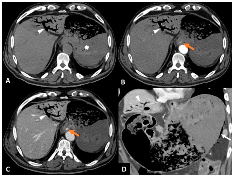 Figure 1
Peer Reviews
No public reviews on file for this paper yet. If you reviewed it on a platform where reviews are public (OpenReview, ICLR, NeurIPS, ICML), you can paste yours below so the community can read it here.
Videos
No videos yet. Explain this paper in a talk, walkthrough, or lecture? Add one.
Taxonomy
TopicsAutoimmune and Inflammatory Disorders · Vascular Malformations and Hemangiomas · Soft tissue tumor case studies