Ultrasonographic and pathological findings of pseudomyogenic hemangioendothelioma
Gui‐Wu Chen, Xiao‐Ling Leng, Shao‐Ming Liu, Xiao‐Min Liao

Abstract
Genes, proteins, chemicals, diseases, species, mutations and cell lines named across the full text — each resolved to its canonical identifier and authoritative record.
Click any figure to enlarge with its caption.
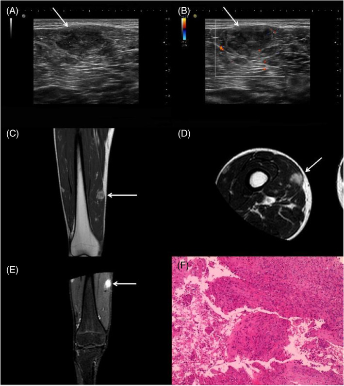 Figure 1
Peer Reviews
No public reviews on file for this paper yet. If you reviewed it on a platform where reviews are public (OpenReview, ICLR, NeurIPS, ICML), you can paste yours below so the community can read it here.
Videos
No videos yet. Explain this paper in a talk, walkthrough, or lecture? Add one.
Taxonomy
TopicsVascular Tumors and Angiosarcomas · Sarcoma Diagnosis and Treatment · Bone Tumor Diagnosis and Treatments
Pseudomyogenic hemangioendothelioma (PHE) is an indolent and low‐grade tumor that often mimics other benign and malignant lesions, making accurate diagnosis crucial for effective patient management.1 The variable clinical presentations and pathological features of PHE pose diagnostic challenges for inexperienced radiologists.2 Here, we present a 17‐year‐old man with PHE, characterized by ultrasonography and magnetic resonance imaging, and confirmed by pathological examination.
A 17‐year‐old male presented to our hospital with a complaint of pain in his leg while walking for the past 10 months. During the physical examination, multiple palpable masses were detected in the right thigh, which were hard, ill‐defined, and had poor mobility. High‐frequency ultrasound revealed that the largest mass in the muscle of the right thigh was hypoechoic, well‐defined, and irregular (Figure 1A), with a few blood flow signals both inside and around the mass (Figure 1B). Magnetic resonance imaging suggested that the largest mass appeared as a high signal on T1‐weighted (Figure 1C) and T2‐weighted imaging (Figure 1D), with significant enhancement (Figure 1E). Finally, the patient underwent a surgical resection of masses, and a pathological examination confirmed the diagnosis of PHE. Hematoxylin and eosin staining revealed a diffuse growth of epithelioid cells with abundant cytoplasm and slightly off‐centered nuclei (Figure 1F). Immunohistochemical staining results were positive for ERG, Fli‐1, INI‐1, Vim, and partially positive for CD31. Weakly positive staining was observed for CK, SMA, Cal, desmon, while CD34, CD56, Desmin, MyoD1, Myogenin, S‐100, WT‐1, EMA, HMB‐45, Melan‐A were negative. The Ki‐67 proliferation index was approximately 5%.
PHE is a rare vascular tumor that mainly affects young patients, with a male predominance, and can present in various locations throughout the body, including the head, esophagus, neck, chest wall, breast, trunk, limbs, pelvis, and external genitalia. It can involve multiple tissue planes, including the dermis, subcutaneous tissue, bone, and skeletal muscle.3 Given its variable clinical presentations and pathological features, inexperienced radiologists may be prone to misdiagnosing PHE as dermatofibroma, epithelioid sarcoma, rhabdomyosarcoma, or other similar lesions.4 In our case, high‐frequency ultrasound showed PHE was hypoechoic, well‐defined, and irregular with a few blood flow signals both inside and around the mass while magnetic resonance imaging showed a high signal with significant enhancement. Accurate diagnosis is crucial for effective patient management and requires expertise in both clinical and pathological domains. Therefore, it is essential to consider PHE in the differential diagnosis of any soft‐tissue mass with variable clinical and radiological presentations.
CONFLICT OF INTEREST STATEMENT
The authors declare no conflict of interest.
CONSENT FOR PUBLICATION
Written informed consent was obtained from the patient to publish this manuscript in accordance with the journal's patient consent policy.
The reference list from the paper itself. Each links out to its DOI / PubMed record.
- 1Wei JQ , Liao ZC , Zhao G , Nahar N , Zhang C , Lu J , et al. Clinicopathological features of pseudomyogenic hemangioendothelioma and precision therapy based on whole exome sequencing. Cancer Commun. 2020;40(4):197–201.10.1002/cac 2.12020 PMC 717065732227592 · doi ↗ · pubmed ↗
- 2Maximen J , Christory A , Bonneau‐Lagacherie J , Guillin R , Ropars M . Spontaneously regressive multifocal bone pseudomyogenic hemangioendothelioma in a 17‐year‐old boy: a case report. Skeletal Radiol. 2023;52(1):119–127.35780259 10.1007/s 00256-022-04109-2 · doi ↗ · pubmed ↗
- 3Otani S , Nakayama R , Sekita T , Hirozane T , Asano N , Nishimoto K , et al. Pseudomyogenic hemangioendothelioma of bone treated with denosumab: a case report. BMC Cancer. 2019;19(1):872.31481040 10.1186/s 12885-019-6072-8PMC 6724307 · doi ↗ · pubmed ↗
- 4Pasricha S , Sharma A , Pruthi M , Durga G , Jajodia A , Gupta G , et al. Multifocal primary pseudomyogenic hemangioendothelioma of bone managed with denosumab: a rare case with diagnostic and therapeutic challenge. J Cancer Res Ther. 2022;18(3):817–819.35900565 10.4103/jcrt.JCRT_1138_20 · doi ↗ · pubmed ↗