Genome sequences of Klebsiella pneumoniae bacteriophages SF_KL2 and SF_KL25
Sophia Lorenz, Gabriel Abreu, Hugo Oliveira

TL;DR
This paper reports the genome sequences of two bacteriophages that infect drug-resistant Klebsiella pneumoniae, isolated from wastewater in Portugal.
Contribution
The study provides new genome sequences of two Webervirus phages with potential applications in combating antibiotic-resistant bacteria.
Findings
SF_KL2 and SF_KL25 are siphovirus phages with genome sizes of 48,737 and 49,170 bp.
The phages were isolated from wastewater in northern Portugal.
They target encapsulated multidrug-resistant Klebsiella pneumoniae.
Abstract
This report describes the genomes of Klebsiella pneumoniae phages SF_KL2 and SF_KL25, which infect the encapsulated multidrug-resistant K. pneumoniae known to cause nosocomial infections. SF_KL2 and SF_KL25 belong to the genus Webervirus (siphovirus morphotype) and have genomes of 48,737 and 49,170 bp, respectively. The phages were isolated from wastewater in northern Portugal.
Genes, proteins, chemicals, diseases, species, mutations and cell lines named across the full text — each resolved to its canonical identifier and authoritative record.
Click any figure to enlarge with its caption.
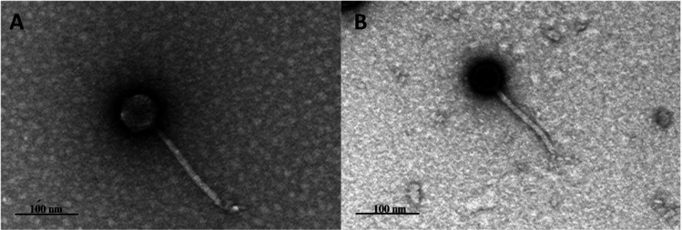 Fig 1
Fig 1Peer Reviews
No public reviews on file for this paper yet. If you reviewed it on a platform where reviews are public (OpenReview, ICLR, NeurIPS, ICML), you can paste yours below so the community can read it here.
Videos
No videos yet. Explain this paper in a talk, walkthrough, or lecture? Add one.
Taxonomy
TopicsBacteriophages and microbial interactions · Microbial infections and disease research · Genomics and Phylogenetic Studies
ANNOUNCEMENT
Klebsiella pneumoniae often causes untreatable nosocomial infections in patients with weakened immune systems (1). The difficulty in treatment arises from resistance genes that reduce the efficacy of antibiotics (2). Therefore, through the specific infection of bacterial pathogens, bacteriophage therapy represents an alternative to combat antimicrobial resistance (3).
Phages SF_KL2 and SF_KL25 were isolated from the same enriched wastewater sample collected from a wastewater treatment plant in Braga, Portugal (4). Wild-type strains (H66, MJH599) of K. pneumoniae isolated from Hospital of Braga (5) were added to a medium containing an equal volume of sewage water and 2× Tryptic Soy Broth (Sigma-Aldrich) and incubated in a shaker (16 h at 37°C and 120 rpm). After centrifugation (10 min at 4°C and 9,000 × g), a volume of the filtered (0.2 µm) supernatant was spotted on bacterial lawns containing the same strains used for enrichment. Phage plaque purification was ensured by amplifying a single phage plaque in new agar plates three times (4). Transmission electron microscopy (TEM) in a Jeol JEM 1400 with uranyl acetate (Sigma-Aldrich) staining evaluated phage morphologies (6). Phage genomic DNA was isolated using the phenol-chloroform (Sigma-Aldrich) method (7), quantified with NanoDrop 1000 Spectrophotometer, and sequenced at Novogene. A VAHTS Universal Plus DNA Library Prep Kit for Illumina (ND617-02) was used to generate a DNA library (350 bp) sequenced on an Illumina Novaseq platform. Briefly, the sheered genomic DNA was ligated with Illumina adapter, amplified by PCR, and size selected. Base calling accuracy was determined by Phred (8). In Geneious Prime version 2019.2.3 (9), raw data were trimmed using BBDuk and sequence reads (4,251,960 and 5,742,711 reads for SF_KL2 and SF_KL25, respectively) were de novo assembled using the Geneious de novo assembler (medium-low sensitivity setting). PhageTerm version 1.0.12 (10) analyzed phage genome termini. Genomes were annotated with Pharokka version 1.3.2 (11), coupled with Phold version 0.2.0 (12), but also manually annotated using BLASTp (13) (based on the non-redundant protein sequences database) and HHpred (14) (based on the Pfam-A_v36 database with a 10^−5^ < E value < 0). PhageAI version 1.0.2 predicted the phages’ life cycle (https://app.phage.ai/), and PhageDPO version 0.1.0 predicted the existence of depolymerases (bit.ly/phagedpo) (15). Default parameters were mainly used in all bioinformatic tools.
SF_KL2 and SF_KL25 have a Siphoviridae morphology, with a non-contractile tail (166 ± 5.1 nm and 161 ± 1.1 nm, respectively) and an icosahedral capsid (55 ± 1.7 nm and 47 ± 2.3 nm, respectively) (Fig. 1). SF_KL2 genome was assembled with 17,450 reads into a 48,737 bp contig (78× coverage), containing 82 coding sequences (CDSs). SF_KL25 genome was assembled with 27,432 reads into a 49,170 bp contig (89× coverage), with 88 CDSs. In both cases, G + C content is 50%, and only 35 proteins have been assigned a function (no tRNA genes). Depolymerases SF_KL2gp65 and SF_KL25gp80 were detected.
Transmission electron micrograph of K. pneumoniae phages. (A) SF_KL2 and (B) SF_KL25. Phages were stained with 2% uranyl acetate and visualized under a Joel JEM 1400 TEM.
According to BLASTn evaluation (13), SF_KL2 and SF_KL25 share high nucleotide identity (>90%) with many other Klebsiella phages, though they are most similar to Weberviruses PSKP16 (GenBank accession number OW251746) and Sin4 (NC_049847), respectively.
The reference list from the paper itself. Each links out to its DOI / PubMed record.
- 1Podschun R, Ullmann U. 1998. Klebsiella spp. as nosocomial pathogens: epidemiology, taxonomy, typing methods, and pathogenicity factors. Clin Microbiol Rev 11:589–603. doi:10.1128/CMR.11.4.5899767057 PMC 88898 · doi ↗ · pubmed ↗
- 2Martin RM, Bachman MA. 2018. Colonization, infection, and the accessory genome of Klebsiella pneumoniae. Front Cell Infect Microbiol 8:4. doi:10.3389/fcimb.2018.0000429404282 PMC 5786545 · doi ↗ · pubmed ↗
- 3Sulakvelidze A, Alavidze Z, Morris JG Jr. 2001. Bacteriophage therapy. Antimicrob Agents Chemother 45:649–659. doi:10.1128/AAC.45.3.649-659.200111181338 PMC 90351 · doi ↗ · pubmed ↗
- 4Oliveira H, Pinto G, Oliveira A, Oliveira C, Faustino MA, Briers Y, Domingues L, Azeredo J. 2016. Characterization and genome sequencing of a Citrobacter freundii phage Cf P 1 harboring a lysin active against multidrug-resistant isolates. Appl Microbiol Biotechnol 100:10543–10553. doi:10.1007/s 00253-016-7858-027683211 · doi ↗ · pubmed ↗
- 5Castro J, Araújo D, Oliveira H, Fernandes L, Oliveira R, Brinks E, Cho G-S, Franz C, Saavedra MJ, Silva S, Almeida C. 2024. Multidrug-resistant Klebsiella pneumoniae and Klebsiella variicola isolated from patients in Portuguese hospitals: genomic and phenotypic characterization. The Microbe 5:100172. doi:10.1016/j.microb.2024.100172 · doi ↗
- 6Melo LDR, Sillankorva S, Ackermann HW, Kropinski AM, Azeredo J, Cerca N. 2014. Isolation and characterization of a new Staphylococcus epidermidis broad-spectrum bacteriophage. J Gen Virol 95:506–515. doi:10.1099/vir.0.060590-024189619 · doi ↗ · pubmed ↗
- 7Jakočiūnė D, Moodley A. 2018. A rapid bacteriophage DNA extraction method. Methods Protoc 1:27. doi:10.3390/mps 103002731164569 PMC 6481073 · doi ↗ · pubmed ↗
- 8Ewing B, Green P. 1998. Base-calling of automated sequencer traces using phred. II. error probabilities. Genome Res 8:186–194. doi:10.1101/gr.8.3.1759521922 · doi ↗ · pubmed ↗