Miliary Mesothelioma
Dujinthan Jayabalan, Michaela Donaghy, Chloe Charlesworth, Edward Fysh, Y. C. Gary Lee

TL;DR
This paper presents a rare case of miliary mesothelioma, highlighting CT imaging features to improve recognition and management.
Contribution
The paper contributes a case emphasizing CT imaging characteristics of miliary mesothelioma for better clinical awareness.
Findings
Miliary mesothelioma can present with characteristic CT imaging findings.
Awareness of this pattern aids in diagnosis and appropriate patient management.
Abstract
Miliary spread of mesothelioma is a rare but important manifestation of late‐stage disease, likely due to hematogenous dissemination. This case highlights characteristic CT imaging findings, emphasizing the need for awareness among clinicians and radiologists. Recognizing this pattern can aid in diagnosis, prognostication, and appropriate referral for palliative management. This case highlights characteristic CT imaging findings of miliary spread of mesothelioma, emphasising the need for awareness among clinicians and radiologists.
Genes, proteins, chemicals, diseases, species, mutations and cell lines named across the full text — each resolved to its canonical identifier and authoritative record.
Click any figure to enlarge with its caption.
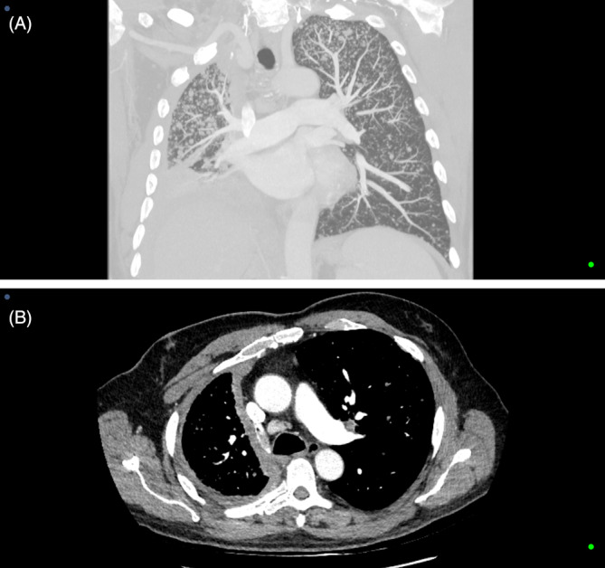 FIGURE 1
- —University of Western Australia 10.13039/501100001801
Peer Reviews
No public reviews on file for this paper yet. If you reviewed it on a platform where reviews are public (OpenReview, ICLR, NeurIPS, ICML), you can paste yours below so the community can read it here.
Videos
No videos yet. Explain this paper in a talk, walkthrough, or lecture? Add one.
Taxonomy
TopicsOccupational and environmental lung diseases · Medical Imaging and Pathology Studies · Pleural and Pulmonary Diseases
Clinical Image
1
A 74‐year‐old man developed right pleural mesothelioma associated with asbestos exposure from working with insulation material as an engineer. Pleural fluid analysis found epithelioid mesothelioma cells, and infection was excluded based on absent clinical signs, normal inflammatory markers, and negative cultures. He received dual immunotherapy (ipilimumab and nivolumab) and radiotherapy for tumour track metastases. Unfortunately, he developed disease progression with increased pleural tumour and lymphadenopathy on computed tomography (CT) 5 months after commencing systemic therapy. Numerous fine bilateral pulmonary deposits were noted, consistent with miliary spread of mesothelioma. He was breathless on exertion but without evidence of infection. The nodules progressed and appeared more confluent on repeat CT after 2 months. The patient was referred to palliative care for symptom‐guided supportive care.
This CT image illustrates the right pleural thickening and contracted hemithorax from mesothelioma and extensive bilateral miliary lung parenchymal involvement (Figure 1A,B). Mesothelioma is generally localised to the pleural cavity [1]. Miliary lung involvement, presumed from hematogenous spread, is rare but should be considered in patients with late‐stage disease, especially with characteristic imaging [2].
(A) Coronal computed tomography scan illustrating miliary mesothelioma. (B) Axial computed tomography scan illustrating miliary mesothelioma.
Author Contributions
The authors confirm their contribution to the paper as follows: Study conception and design: D.J. and Y.C.G.L. Patient data/information collection: D.J., M.D., C.C., and E.F. Manuscript preparation: D.J. and Y.C.G.L. Study supervision: Y.C.G.L. and E.F. All authors reviewed the results and approved the final version of the manuscript, including the authorship list.
Consent
The authors declare that written informed consent was obtained for the publication of this manuscript and accompanying images and attest that the form used to obtain consent from the patient complies with the Journal requirements.
Conflicts of Interest
E.F. and Y.C.G.L. are Editorial Board members of Respirology Case Reports and a co‐authors of this article. They were excluded from all editorial decision‐making related to the acceptance of this article for publication. The other authors declare no conflicts of interest.
The reference list from the paper itself. Each links out to its DOI / PubMed record.
- 1D. O. Hall , C. E. Hooper , J. Searle , et al., “18F‐Fluorodeoxyglucose PET/CT and Dynamic Contrast‐Enhanced MRI as Imaging Biomarkers in Malignant Pleural Mesothelioma,” Nuclear Medicine Communications 39, no. 2 (2018): 161–170.29300270 10.1097/MNM.0000000000000789 · doi ↗ · pubmed ↗
- 2M. Huncharek , “Miliary Mesothelioma,” Chest 106, no. 2 (1994): 605–606.7774346 10.1378/chest.106.2.605 · doi ↗ · pubmed ↗