Author Correction: Identification of novel 7-hydroxycoumarin derivatives as ELOC binders with potential to modulate CRL2 complex formation
Yonghyeok Kim, Seon Jeong Baek, Eun-Kyung Yoon, Minhee Choi, Jung-Hoon Kim, Kyungtae Kim, Chi Hoon Park, Byung Il Lee

Abstract
Genes, proteins, chemicals, diseases, species, mutations and cell lines named across the full text — each resolved to its canonical identifier and authoritative record.
Click any figure to enlarge with its caption.
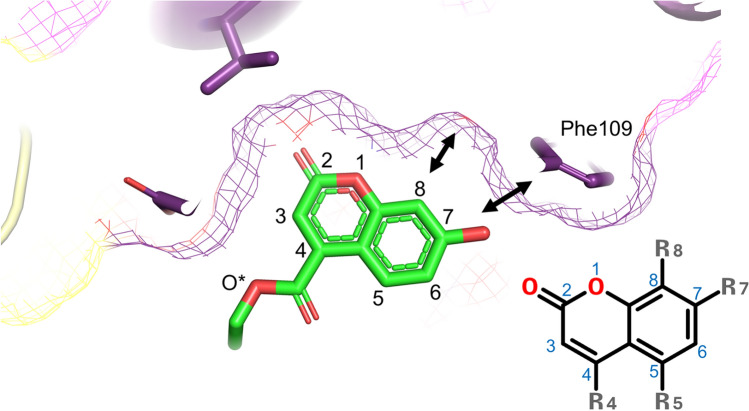 Figure 1
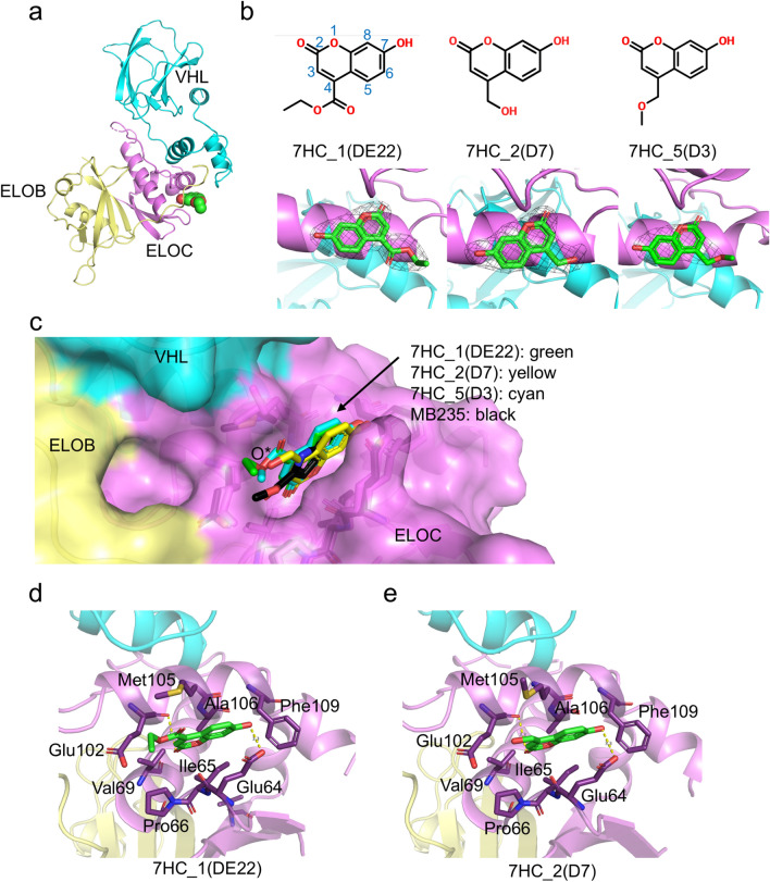 Figure 2
Peer Reviews
No public reviews on file for this paper yet. If you reviewed it on a platform where reviews are public (OpenReview, ICLR, NeurIPS, ICML), you can paste yours below so the community can read it here.
Videos
No videos yet. Explain this paper in a talk, walkthrough, or lecture? Add one.
Taxonomy
TopicsSynthesis of Organic Compounds · Synthesis and biological activity
Correction to: Scientific Reports 10.1038/s41598-025-88166-2, published online 29 January 2025
The original version of this Article contained errors in the Figures. Figure 1 was published as Figure 2 and Figure 2 was published as Figure 1. The Figure legends were correct at the time of publication.
The original Figures 1 and 2 and accompanying legends appear below.Fig. 1. Overall structures of VBC in complex with fragment ligands. (a) Overall structures of the VBC−7HC_1(DE22) complex. Each protein component is colored in cyan (VHL), pale yellow (ELOB), and violet (ELOC). The ligand is shown in the sphere model. (b) Chemical structures and 2mFo-DFc OMIT maps for 7HC_1(DE22), 7HC_2(D7), and 7HC_5(D3). The numbering for the 7HC ring is also shown. The map contour levels were 1.5 σ. (c) Superimposition with ligand-bound VBC structures. 7HC_1(DE22) is drawn in green, 7HC_2(D7) in yellow, 7HC_5(D3) in cyan, and MB235 in black (PDB code: 6GMN)^17^. O*: position of the O3 atom in the 7HC_1(DE22) and 7HC_1(DE22) molecules and O4 atom in the 7HC_5(D3) molecule. (d) Detailed views of interactions between VBC and 7HC_1(DE22). (e) Detailed views of interactions between VBC and 7HC_2(D7).Fig. 2. Sliced view of 7HC derivative binding pocket surface showing the structural basis of no binding activities by C7- and C8-position chemical modifications (R7 and R8). The 7HC_1(DE22) structure is shown as the representative. O*: position of the O3 atom in the 7HC_1(DE22).
The original Article has been corrected.