Ectopic Prostatic Tissue in the Uterine Cervix Following Vaginal Hysterectomy
Abayomi I Alao, Nirav Gandhi, Bhawana Purwar

TL;DR
A rare case of prostatic tissue found in a woman's cervix after a hysterectomy is reported, highlighting unusual tissue development.
Contribution
The paper presents a novel clinical case of ectopic prostatic tissue in the uterine cervix following a vaginal hysterectomy.
Findings
Ectopic prostatic tissue was identified in cervical tissue during histologic examination.
The case suggests possible genetic, hormonal, and developmental factors in tissue misplacement.
Such occurrences are rare and not fully understood embryologically.
Abstract
Ectopic prostatic tissue (EPT) in the female genital tract is a relatively rare histopathological finding. EPT occurs when tissue from one region is found in another region, a phenomenon observed in various medical cases. However, the presence of ectopic tissue specific to one gender in another is uncommon. An 83-year-old woman presented to the gynaecology outpatient clinic with symptoms of uterovaginal prolapse and subsequently underwent a vaginal hysterectomy. Histologic examination of the uterus revealed the presence of mixed glandular and squamous prostatic elements within the cervical tissue. The exact embryological origins of cervical ectopic prostatic tissue remain incompletely understood. It is likely that a combination of genetic, hormonal, and developmental factors contributes to its occurrence.
Genes, proteins, chemicals, diseases, species, mutations and cell lines named across the full text — each resolved to its canonical identifier and authoritative record.
Click any figure to enlarge with its caption.
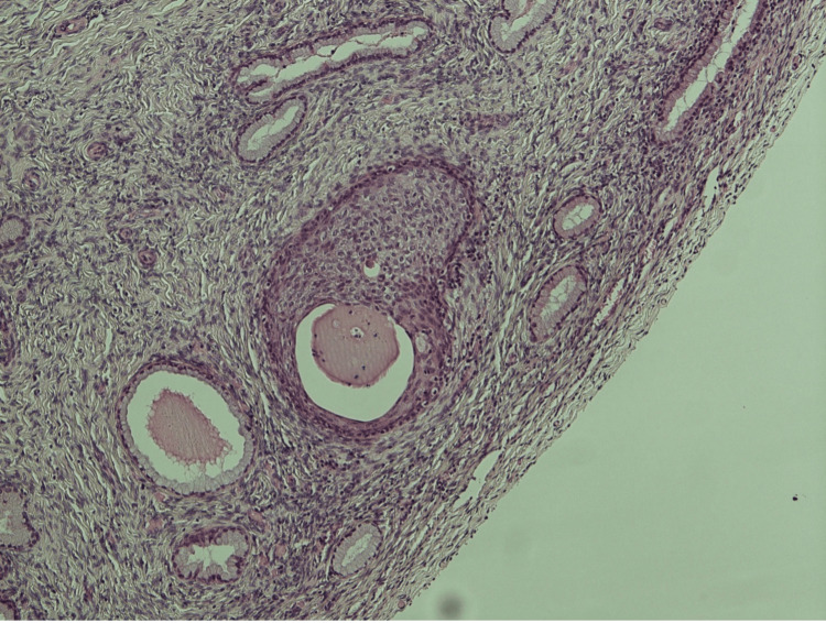 Figure 1
Peer Reviews
No public reviews on file for this paper yet. If you reviewed it on a platform where reviews are public (OpenReview, ICLR, NeurIPS, ICML), you can paste yours below so the community can read it here.
Videos
No videos yet. Explain this paper in a talk, walkthrough, or lecture? Add one.
Taxonomy
TopicsUrologic and reproductive health conditions · Sexual Differentiation and Disorders · Urological Disorders and Treatments
Introduction
Ectopic prostatic tissue (EPT) in the female genital tract is a relatively rare histopathological finding [1]. Ectopic tissue occurs when tissue from one region is found in another region, a phenomenon observed in various medical cases. However, the presence of ectopic tissue specific to one gender in another is uncommon [2].
In male embryonic development, the pelvic urethral endoderm gives rise to the prostate, whereas in females, it develops into the paraurethral Skene glands [2]. Reports have documented instances of EPT in female genital organs such as the ovary, cervix, and uterus. The underlying cause of this occurrence remains incompletely understood and is often an incidental histological finding [3].
Our report details a case in which ectopic prostatic tissue was incidentally discovered in the cervix following a vaginal hysterectomy.
Case presentation
An 83-year-old para 2 woman presented to the gynaecology outpatient clinic with symptoms of uterovaginal prolapse. Despite attempting various sizes of ring pessaries, she experienced no substantial improvement. She did not report any lower urinary symptoms, bowel issues, or unusual vaginal discharge or bleeding. On examination, a stage 2 utero-vaginal prolapse was observed. She was otherwise fit and healthy.
She was counselled on management options and opted for a vaginal hysterectomy with pelvic floor repair. She subsequently underwent a vaginal hysterectomy and anterior repair, and the samples were sent for histology. She recovered well postoperatively and was discharged home on the first postoperative day.
On histology (Figure 1), the uterus and cervix measured 75 × 38 × 23 mm. On slicing the uterus and cervix, a polyp measuring 6 mm was noted at the fundus. Examination revealed the presence of mixed glandular and squamous prostatic elements within the cervical tissue. Histologically, the prostatic component displayed characteristic glandular structures with secretory acini and a distinct epithelial lining of prostatic tissue. Simultaneously, the squamous elements exhibited stratified squamous epithelium with keratinization. There was no evidence of CIN (cervical intraepithelial neoplasia), CGIN (cervical glandular intraepithelial neoplasia), or malignancy.
Photomicrograph (x100) of cervical tissue showing mixed glandular and squamous elements prostatic differentiation (PSA and NKX3.1 positive) with no evidence of malignancy.PSA: prostate-specific antigen stain.
Discussion
Ectopic prostatic tissue (EPT) refers to prostatic tissue that is located outside the prostate gland [4]. This condition was first reported by Jores in 1894 [1]. In men, a common type of non-cancerous EPT is the prostatic-type polyp, which is typically found near the prostate [5]. However, there have been rare cases of EPT found in women, primarily in the cervix, vagina, and ovaries. Unfortunately, these cases are not yet fully recognized by pathologists [1].
The embryological origins of cervical ectopic prostatic tissue remain a subject of ongoing research and speculation. Several theories have been proposed to explain the presence of prostatic tissue in the cervical region [5,6]. Some researchers suggest that abnormalities in the development of the Müllerian ducts, which give rise to the female reproductive tract, could lead to the development of ectopic prostatic tissue in the cervix. It is possible that remnants of male urogenital structures may persist in female embryos, leading to prostatic tissue development in unexpected locations [5]. Genetic factors and mutations may play a role in the development of cervical ectopic prostatic tissue. Certain genetic abnormalities could predispose individuals to the formation of prostatic tissue in unexpected locations [5]. Some experts propose that metaplastic changes in cervical epithelial cells could result in the transformation of these cells into prostatic tissue. This transformation could be driven by factors such as chronic inflammation or hormonal fluctuations [5-7]. The most widely accepted probability is that EPT in the cervix and vagina is probably a developmental anomaly. Cervical EPT may be derivatives of Skene’s gland, possibly homologous with the male prostate gland. Skene’s glands have similar embryogenic origins to the prostate gland, which is the urogenital sinus [8,9].
In 2014, the World Health Organization classified tumours of the female reproductive organs and briefly mentioned EPT. However, there is still a high rate of misdiagnosis due to inadequate knowledge [1]. Cervical EPT has been discovered incidentally in patients who have undergone loop electrosurgical excision or hysterectomy. It has been reported in various women [1]. This was also the case in the present patient, where we discovered cervical EPT following a vaginal hysterectomy.
EPT on histology mimics normal prostatic acini, typically with the epithelial cells on the luminal surface forming inverted papillae or cribriform structures [1]. According to reports, most cases of EPT in the genitourinary tract had lesions below the normal surface of the epithelium. In our specific case, upon examination, mixed glandular and squamous elements were found within the cervical tissue, but there was no sign of invasive growth or malignancy [1].
A possible differential diagnosis for cervical EPT is mesonephric remnants, which usually occur in the cervix and vaginal sidewalls. The ducts are small and relatively round with eosinophilic secretions within the cavity in EPT, while mesonephric remnants have a pathognomonic presence of a single layer of epithelium without peripheral basal cells [1]. Cervical EPT is a rare and often incidental finding, posing diagnostic challenges for pathologists [1]. Misdiagnosis is common due to its resemblance to other cervical lesions, such as mesonephric remnants. Cervical EPT is typically benign; however, its recognition is important to avoid misdiagnosis and unnecessary interventions [1].
Conclusions
In conclusion, cervical ectopic prostatic tissue is a rare finding with unclear embryological origins. This case highlights the importance of histopathological awareness to ensure an accurate diagnosis. Further research is needed to elucidate the precise mechanisms underlying this rare phenomenon.
The reference list from the paper itself. Each links out to its DOI / PubMed record.
- 1Ectopic prostatic tissue in uterine cervix: a case report and literature review Hum Pathol Rep Long W Zhang Q Jiang W 300549262021 https://www.sciencedirect.com/science/article/pii/S 2772736 X 21000086
- 2Ectopic prostatic tissue in vagina - case report Clin Exp Obstet Gynecol Pazin V Dobrosav Ijevic A Stulic J Cimba Ijevic M 22019
- 3Ectopic prostate tissue in the uterine cervix of a female with non-classic congenital adrenal hyperplasia-a case report J Clin Med Tschaidse L Auer MK Dubinski I 11202210.3390/jcm 11154307 PMC 933195235893397 · doi ↗ · pubmed ↗
- 4Prostatic-type tissue in the uterine cervix Diagn Histopathol Sitaraaaman HB Griffin J Warren C 298300262020 https://www.pathsoc.org/_userfiles/pages/files/education/mpdhp_jun_2020_short_case.pdf
- 5Ectopic prostatic tissue in the uterine cervix: a report of four cases and review of ectopic prostatic tissue Am J Surg Pathol Nucci MR Ferry JA Young RH 12241230242000 https://doi.org/10.1097/00000478-200009000-000051097669610.1097/00000478-200009000-00005 · doi ↗ · pubmed ↗
- 6Ectopic prostatic tissue in the uterine cervix. Report of a case and brief overview of basaloid cervical glandular lesions J Clin Pathol Singh M 5735756520122244791610.1136/jclinpath-2011-200455 · doi ↗ · pubmed ↗
- 7Ectopic prostatic tissue in the perineum BMJ Case Rep Hanna B Mc Naught A Chung A Chalasani V 13202010.1136/bcr-2020-239378 PMC 764348333148562 · doi ↗ · pubmed ↗
- 8Ectopic prostatic tissue Gynecologic and Urologic Pathology: Similarities, Differences and Challenges Raspollini MR Montironi R Cheng L Lopez-Beltran A 145149 Cambridge, UK Cambridge University Press 2019