Complete Bilateral Ossification of the Arytenoid Cartilages With Minimal Involvement of the Laryngeal Skeleton: A Unique Anatomical Variation
Nymfodora Malkidou, Aliki Fiska, Katerina Vassiou

TL;DR
An 84-year-old woman had complete bilateral ossification of her arytenoid cartilages, a rare anatomical variation not previously documented.
Contribution
This paper reports a unique case of complete bilateral arytenoid cartilage ossification without laryngeal pathology.
Findings
Bilateral complete ossification of the arytenoid cartilages was observed in a patient with no laryngeal disorders.
The ossification pattern involved the hyaline part and tip of the arytenoid cartilages but spared the vocal processes.
This ossification occurred before that of the thyroid and cricoid cartilages, which is atypical for age-related laryngeal changes.
Abstract
The arytenoid cartilages are distinguished by their mobility and unique mixed composition of hyaline and elastic cartilage. Though laryngeal cartilage ossification is not rare, and its pattern is well-studied, sole complete and bilateral arytenoid cartilage ossification is an aberration. An 84-year-old woman, with no history of voice or breathing issues, was brought to the emergency department for a head-and-neck trauma following a fall. CT scans revealed a subdural hematoma and, incidentally, a bilateral ossification of the arytenoid cartilages, sparing the vocal processes but involving complete ossification of the hyaline part and the tip. The cricoid cartilage was non-ossified, and the thyroid depicted minor ossification. We present a unique, previously undocumented case of both arytenoid cartilages simultaneously ossified in a patient without laryngeal pathology. In previous studies…
Genes, proteins, chemicals, diseases, species, mutations and cell lines named across the full text — each resolved to its canonical identifier and authoritative record.
Click any figure to enlarge with its caption.
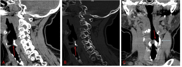 Figure 1
Peer Reviews
No public reviews on file for this paper yet. If you reviewed it on a platform where reviews are public (OpenReview, ICLR, NeurIPS, ICML), you can paste yours below so the community can read it here.
Videos
No videos yet. Explain this paper in a talk, walkthrough, or lecture? Add one.
Taxonomy
TopicsVoice and Speech Disorders · Oropharyngeal Anatomy and Pathologies · Dysphagia Assessment and Management
Introduction
The arytenoid cartilages are pyramidal, three-sided structures situated on the posterior upper margin of the cricoid cartilage. They stand out from other laryngeal cartilages because of their high mobility and their composition of both hyaline and elastic cartilage. The thyroid and cricoid consist only of hyaline cartilage, which typically starts to ossify after the age of 18, whereas epiglottis, made entirely of elastic cartilage, does not undergo ossification [1-2].
The calcification of the laryngeal cartilages is not rare, and it has been thoroughly investigated. Hartley et al conducted the first comprehensive study of laryngeal cartilage ossification patterns, analyzing the largest case series of their era. They established that thyroid cartilage ossification is usually the most prominent and the first to appear, starting at the inferior horns, with the superior horns typically ossifying at a later stage [3]. Cricoid cartilage ossification may occur concurrently or follow the thyroid changes, and the arytenoids usually exhibit none or minor ossification, occurring later. The foci appear on their apices, bodies, and muscular processes, but the vocal processes and the tips are consistently unaffected.
We present the case of an 84-year-old woman without any laryngeal pathology, who was incidentally found to exhibit bilateral ossification of the entire hyaline part of the arytenoid cartilages, combined with minimal ossification of the thyroid cartilage and no ossification of the cricoid. To the best of our knowledge, this is the first documented case of this exceptional event. This anatomical variation was described using the Anatomical Quality Assurance (AQUA) checklist (Appendices) [4].
Case presentation
An 84-year-old woman was brought to the emergency department of a private hospital in Larissa following an accidental fall at home resulting in head and neck trauma. She was on anticoagulant therapy with apixaban for stroke prevention due to atrial fibrillation. Her family reported an episode of acute loss of consciousness after the fall. Given her medical history and symptoms, an urgent computed tomography scan of the brain and cervical spine was performed.
Brain CT revealed a subdural hematoma without midline shift, likely accounting for her loss of consciousness without requiring intubation. Cervical spine CT indicated cervical vertebral osteopenia. Incidentally, the neck visceral space showed bilateral symmetric ossification of the arytenoid cartilages sparing the vocal processes. Limited high-density calcifications were noted in the superior horns of the thyroid cartilage, the thyroid angle, and the thyrohyoid ligament. The remainder of the thyroid cartilage and the entire cricoid cartilage exhibited minimal or no ossification. Notably, the ossified arytenoid cartilage demonstrated on CT the same high density as the mandible (Figure 1).
Arytenoid cartilage ossification with minimal involvement of the laryngeal skeletonConstructed CT of the neck, sagittal plane, shows complete ossification of the arytenoid cartilage, excluding the vocal process. A. with soft tissue density (red arrow) and B. with bone density (red arrow). C. Constructed CT of the neck, coronal plane, with soft tissue density, shows complete ossification of the arytenoid cartilages (red arrow), ossification of the superior thyroid horns (grey arrow), and no sign of cricoid involvement, highlighting the ossification at the tip of the arytenoids.
Upon our inquiry, her family reported she had no history of voice changes, breathing difficulties, or other significant otolaryngological issues. However, due to her impaired level of consciousness, a comprehensive ENT evaluation, including a detailed assessment of laryngeal function and voice, could not be conducted at that time. Following the resolution of the hematoma, the patient resumed her daily activities. As she did not experience any voice or breathing problems, an ENT examination was not considered necessary. The subdural hematoma resolved spontaneously after 14 days, and the patient was discharged after 5 days without any changes to her medication. Regarding the finding on the CT scan of the larynx, an appointment was scheduled with an otolaryngologist, but the patient did not attend. While the finding may be incidental and potentially represent an anatomical variation, the absence of an ENT examination leaves the possibility of an underlying laryngeal pathology, which has not yet manifested, to be carefully considered.
Discussion
Bilaterally ossified arytenoid cartilages, involving complete ossification of the hyaline part, are remarkable, especially with no ossification observed in the cricoid and minimal osseous changes in the thyroid cartilage (Figure 1).
The abnormal ossification pattern of the laryngeal skeleton of our case is unique according to previous studies. Zan et al. reviewed 972 computed tomography (CT) scans to evaluate arytenoid ossification and found no evidence of simultaneous bilateral ossification of the arytenoid cartilages [5]. Yeager et al., who investigated 30 cases of laryngeal cartilage ossification using CT scans, found no evidence of complete arytenoid involvement [6]. Hartley et al., examining 516 neck X-rays, found no signs of ossification in the arytenoids when the thyroid and cricoid cartilages remained unossified [3].
Kahane noted that the ossification process is typically completed by the eighth decade, even if the ossification is partial [7]. Turk et al. in their radiographic study confirmed that the ossification of laryngeal cartilages is age-related; they established specific age-based stages for each cartilage [8]. Given our patient’s old age, we may assume that the laryngeal ossification process is ending, and while the ossification pattern of the arytenoids aligns with the expected final phase, that of the thyroid and cricoid cartilages does not.
However, this finding could either represent an anatomical variation or potentially mask a latent laryngeal pathology that has yet to manifest. Recognizing such cases is crucial, as demonstrated by Morreels et al., where a unilateral ossified arytenoid cartilage was misidentified as a foreign body, leading to the patient undergoing unnecessary surgery [9].
As a concluding remark, from a histologic and physiological perspective, the arytenoid elastic cartilages are located at two sites: the vocal process to facilitate movement and the tip to act as a cushion, protecting the mucosa from mechanical damage [2, 10]. These are typically non-ossifying areas. Interestingly, in our case, while ossification was absent in the vocal process of the arytenoid cartilages, it was observed at the tip of both of them (Figure 1C). This raises the question of whether the vocal process is the only component entirely composed of elastic cartilage.
Conclusions
Our case highlights the complete, bilateral ossification of the arytenoid cartilages, combined with minimal thyroid and the absence of cricoid cartilage ossification. This unique variation of the laryngeal ossification pattern was accompanied by ossification at the tip of the arytenoid cartilages, an area typically spared due to comprising of elastic cartilage. Our findings suggest that arytenoid cartilage ossification may precede that of the thyroid or cricoid cartilage and could be the only notable feature on a CT scan of the neck’s visceral space in a patient without laryngeal pathology. This case could serve as a reference and a basis for future research into laryngeal degeneration processes.
The reference list from the paper itself. Each links out to its DOI / PubMed record.
- 1Gray’s Anatomy: The Anatomical Basis of Clinical Practice Standring S New York Elsevier 2020 https://shop.elsevier.com/books/grays-anatomy/standring/978-0-7020-7705-0
- 2Anatomy, head and neck: larynx arytenoid cartilage Stat Pearls [Internet] Andaloro C Sharma P La Mantia I Treasure Island Stat Pearls Publishing 2025 Jan-https://www.statpearls.com/point-of-care/2828130020624 · pubmed ↗
- 3The pattern of ossification in the laryngeal cartilages: a radiological study Br J Radiol Hartley W Samuel E Evison G 5855913819651432645910.1259/0007-1285-38-452-585 · doi ↗ · pubmed ↗
- 4Development of the Anatomical Quality Assurance (AQUA) checklist: guidelines for reporting original anatomical studies Clin Anat Tomaszewski KA Henry BM Kumar Ramakrishnan P 14203020172780150710.1002/ca.22800 · doi ↗ · pubmed ↗
- 5Asymmetric mineralization of the arytenoid cartilages in patients without laryngeal cancer AJNR Am J Neuroradiol Zan E Yousem DM Aygun N 11131118322011 http://10.3174/ajnr.A 24442149376210.3174/ajnr.A 2444 PMC 8013142 · doi ↗ · pubmed ↗
- 6Ossification of the laryngeal cartilages as it relates to computed tomography Invest Radiol Yeager VL Lawson C Archer CR 1119171982680440910.1097/00004424-198201000-00003 · doi ↗ · pubmed ↗
- 7Connective tissue changes in the larynx and their effects on voice J Voice Kahane J 273011987
- 8Age changes in the human laryngeal cartilages Clin Anat Turk LM Hogg DA 15416261993