Preoperative multiclass classification of thymic mass lesions based on radiomics and machine learning
Yan Zhu, Li Wang, Aichao Ruan, Zhiyu Peng, Zhenzhong Zhang

TL;DR
This study uses CT scans and machine learning to classify thymic mass lesions before surgery, improving early diagnosis and treatment planning.
Contribution
A machine learning model is developed for preoperative classification of thymic mass lesions using radiomic features and clinical parameters.
Findings
The model achieved an accuracy of 0.8547 in classifying thymic mass lesions.
Radiomic features from CT scans and age were used to distinguish between thymic cysts and thymomas.
Abstract
Apart from rare cases such as lymphomas, germ cell tumors, neuroendocrine neoplasms, and thymic hyperplasia, thymic mass lesions (TMLs) are typically categorized into cysts, and thymomas. However, the classification results cannot be determined in advance and can only be confirmed through postoperative pathology. Therefore, the objective of this study is to rely on clinical parameters and radiomic features extracted from chest computed tomography (CT) scans to facilitate the preoperative classification of TMLs. The model development specifically focused on thymic cysts and thymomas, as these are the most commonly encountered anterior mediastinal tumors in clinical practice. This retrospective study included 400 participants from 3 hospitals between September 2017 and September 2024 due to TMLs. The participants were classified into 7 groups based on the ultimately confirmed etiology:…
Genes, proteins, chemicals, diseases, species, mutations and cell lines named across the full text — each resolved to its canonical identifier and authoritative record.
Click any figure to enlarge with its caption.
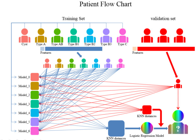 Figure 1
 Figure 2
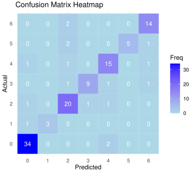 Figure 3
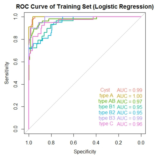 Figure 4
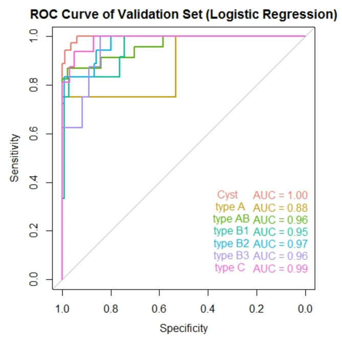 Figure 5
Peer Reviews
No public reviews on file for this paper yet. If you reviewed it on a platform where reviews are public (OpenReview, ICLR, NeurIPS, ICML), you can paste yours below so the community can read it here.
Videos
No videos yet. Explain this paper in a talk, walkthrough, or lecture? Add one.
Taxonomy
TopicsMyasthenia Gravis and Thymoma · Meningioma and schwannoma management · Advanced X-ray and CT Imaging