Cavitary Pulmonary Disease Caused by Mycobacterium kyorinense : A Case Report on a 3‐Year Clinical Course
Masahiro Yanagi

TL;DR
An 80-year-old woman developed cavitary lung disease caused by Mycobacterium kyorinense, highlighting the need for early diagnosis to prevent severe lung damage.
Contribution
Demonstrates the pathogenic potential of Mycobacterium kyorinense in causing cavitary pulmonary disease in a healthy individual.
Findings
Mycobacterium kyorinense was identified as the causative agent of progressive cavitary pulmonary disease.
Conventional diagnostic methods failed to identify the organism, requiring advanced techniques like MALDI-TOF MS.
The case emphasizes the importance of early detection and treatment to prevent irreversible lung damage.
Abstract
An 80‐year‐old female presented with haemoptysis, and chest computed tomography (CT) revealed bronchiectasis in the right middle lobe accompanied by granular and nodular opacities in the right lower lobe. Although acid‐fast bacilli (AFB) smears were consistently positive, polymerase chain reaction (PCR) assays for Mycobacterium tuberculosis yielded negative results, and repeated sputum cultures on conventional solid‐phase media failed to identify the causative organism. Cavitary pulmonary lesions developed progressively over 3 years, accompanied by persistent AFB smear positivity. Ultimately, liquid culture using the Mycobacteria Growth Indicator Tube (MGIT) system isolated nontuberculous mycobacteria, subsequently identified as Mycobacterium kyorinense ( M. kyorinense ) via matrix‐assisted laser desorption/ionisation time‐of‐flight mass spectrometry (MALDI‐TOF MS). This case…
Genes, proteins, chemicals, diseases, species, mutations and cell lines named across the full text — each resolved to its canonical identifier and authoritative record.
Click any figure to enlarge with its caption.
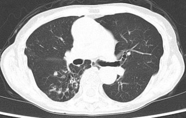 FIGURE 1
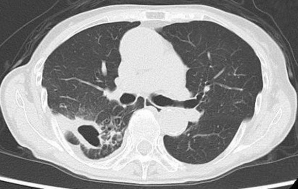 FIGURE 2
| Drug | MIC (μg/mL) |
|---|---|
| CAM | 0.125 |
| AZM | 0.5 |
| EB | 4 |
| KM | ≤ 2 |
| INH | 4 |
| RFP | > 4 |
| RBT | 0.5 |
| MFLX | ≤ 0.25 |
| TH | 1 |
| MINO | 2 |
| DOXY | 8 |
| LZD | 4 |
| STFX | 0.25 |
| AMK | ≤ 8 |
Peer Reviews
No public reviews on file for this paper yet. If you reviewed it on a platform where reviews are public (OpenReview, ICLR, NeurIPS, ICML), you can paste yours below so the community can read it here.
Videos
No videos yet. Explain this paper in a talk, walkthrough, or lecture? Add one.
Taxonomy
TopicsMycobacterium research and diagnosis · Tuberculosis Research and Epidemiology · Infectious Diseases and Mycology
Introduction
1
Nontuberculous mycobacterial infections encompass diverse species, each demonstrating distinct clinical trajectories and necessitating tailored therapeutic approaches. Identifying infections caused by rare species presents a significant challenge, often requiring sophisticated molecular methodologies to achieve precise species differentiation.
Mycobacterium kyorinense ( M. kyorinense ) is a non‐pigmented, slow‐growing, nontuberculous mycobacterium first identified in Japan in 2009 [1]. Approximately 20 cases have been reported, primarily in Japan. However, additional cases identified in Brazil, Australia, Saudi Arabia, and India suggest a broader yet under‐recognised global distribution [2, 3]. Due to its rarity, comprehensive knowledge of its clinical manifestations and disease progression remains limited. In this report, we delineate the clinical trajectory of M. kyorinense infection, which led to cavitary lung disease over 3 years, and discuss the diagnostic challenges associated with its bacterial detection.
Case Report
2
An 80‐year‐old female presented to our hospital with a 5‐day history of haemoptysis. Her medical history was notable for gastro‐oesophageal reflux disease and dyslipidaemia, with no history of smoking or significant pulmonary disease. She had resided in the same region her entire life and had no history of international travel.
Chest computed tomography (CT) revealed bronchiectasis in the right middle lobe and scattered granular and nodular opacities in the right lower lobe (Figure 1). No lymphadenopathy, pleural effusion, or extrapulmonary lesions were noted. Laboratory investigations were unremarkable, with negative results for tuberculosis‐specific interferon‐gamma and anti‐* Mycobacterium avium complex* (MAC) antibodies and no evidence of immunodeficiency. Acid‐fast bacilli (AFB) smears of her sputum remained persistently positive (± or 1 +). However, polymerase chain reaction (PCR) assays for Mycobacterium tuberculosis ( M. tuberculosis ) and MAC were negative, and repeated sputum cultures on conventional solid media failed to identify the pathogen.
Chest computed tomography shows scattered granular and nodular opacities in the right lower lobe.
Although a rare nontuberculous mycobacterial infection was suspected, her haemoptysis resolved with haemostatic agents, and she declined further invasive diagnostic procedures, including bronchoscopy. Consequently, a follow‐up imaging strategy was adopted as the primary management approach.
Over 3 years, a cavitary lesion in the right lower lobe exhibited progressive enlargement (Figure 2). Repeat AFB smears remained persistently positive. However, solid media cultures failed to yield growth, preventing species identification. To overcome this diagnostic limitation, a liquid culture utilising the Mycobacteria Growth Indicator Tube (MGIT) method was performed, resulting in a positive detection of acid‐fast bacilli. Subsequent identification via matrix‐assisted laser desorption/ionisation time‐of‐flight mass spectrometry (MALDI‐TOF MS) confirmed the pathogen as M. kyorinense . The diagnosis was further substantiated by the isolation of M. kyorinense from an additional sputum specimen.
Chest computed tomography demonstrates a cavitary lesion formation in the right lower lobe.
Despite the absence of symptoms such as cough, fever, or fatigue, the progressive enlargement of the cavitary lesion and the persistence of intermittent haemoptysis prompted consideration of pharmacological treatment guided by drug susceptibility testing (Table 1). However, the patient elected to decline treatment, and no further therapeutic interventions were pursued.
TABLE 1: Drug susceptibility testing results of M. kyorinense strains isolated from the patient.
Discussion
3
This case underscores the pathogenic potential of M. kyorinense , a rare, slow‐growing, nontuberculous mycobacterium first identified in Japan in 2009 [1], to independently induce cavitary lung lesions, even without pre‐existing pulmonary abnormalities. Despite its limited characterisation, the phylogenetic similarity of M. kyorinense to Mycobacterium celatum and Mycobacterium branderi presents a diagnostic challenge due to its slow growth [1, 2]. This report highlights its capacity to compromise normal lung architecture over a prolonged disease course, emphasising the critical role of advanced diagnostic modalities such as the MGIT liquid culture system and MALDI‐TOF MS in facilitating timely and accurate identification [2, 3].
A prior study characterising M. kyorinense infections indicates that this pathogen can cause pulmonary disease even in immunocompetent individuals. However, cavitary lesions are typically associated with pre‐existing cysts or structural lung abnormalities [3]. Notably, this case demonstrated that over a three‐year period, M. kyorinense could independently disrupt normal lung architecture and induce cavity formation, highlighting its clinical significance.
At our institution, mycobacterial cultures are routinely performed using solid media, specifically Ogawa medium, due to its cost‐effectiveness and practicality. While sputum smears consistently showed (±) to (1 +), a level typically sufficient for detecting M. tuberculosis or MAC infections, repeated cultures on solid media failed to yield growth, delaying pathogen identification. In contrast, the MGIT liquid culture system offers superior sensitivity and faster detection than solid media. However, its implementation is associated with increased costs and necessitates stringent contamination control. In this case, early utilisation of the MGIT system might have enhanced diagnostic efficiency and facilitated the timely identification of the slow‐growing M. kyorinense .
Due to the limited number of reported cases, no standardised treatment protocol for M. kyorinense has been established. Previous studies consistently indicate that conventional anti‐tuberculosis medications, including isoniazid (INH), rifampicin (RFP), and ethambutol (EB), are ineffective [4]. Resistance to RFP is attributed to a mutation at Ser531 in the rpoB gene, a mechanism analogous to that observed in rifampicin‐resistant M. tuberculosis [4]. Favourable clinical outcomes have been documented with regimens incorporating macrolides (e.g., clarithromycin [CAM]), fluoroquinolones (e.g., moxifloxacin [MFLX]), and aminoglycosides (e.g., amikacin [AMK]), guided by MIC testing when employed as first‐line therapeutic agents [3, 4, 5]. In the present case, MIC testing revealed high resistance to INH and RFP, while low MIC values for CAM, MFLX, and AMK suggested their potential efficacy as first‐line agents.
In conclusion, this case illustrates the ability of M. kyorinense to cause cavitary pulmonary disease over a few years, even in immunocompetent individuals. Early implementation of liquid culture systems such as MGIT and prompt therapeutic interventions guided by susceptibility testing is critical for managing this rare infection.
Author Contributions
The author contributed to the conceptualisation and design of the study, as well as to the acquisition, analysis, and interpretation of the data.
Ethics Statement
The author confirms that written informed consent was appropriately obtained for the publication of this manuscript and its accompanying images.
Conflicts of Interest
The author declares no conflicts of interest.
The reference list from the paper itself. Each links out to its DOI / PubMed record.
- 1M. Okazaki , K. Ohkusu , H. Hata , et al., “ Mycobacterium kyorinense Sp. Nov., a Novel, Slow‐Growing Species, Related to Mycobacterium celatum , Isolated From Human Clinical Specimens,” International Journal of Systematic and Evolutionary Microbiology 59 (2009): 1336–1341.19502312 10.1099/ijs.0.000760-0 · doi ↗ · pubmed ↗
- 2B. Varghese , M. Enani , M. Shoukri , S. Al Thawadi , S. Al Johani , and S. Al‐Hajoj , “Emergence of Rare Species of Nontuberculous Mycobacteria as Potential Pathogens in Saudi Arabian Clinical Setting,” P Lo S Neglected Tropical Diseases 11 (2017): e 0005288.28076350 10.1371/journal.pntd.0005288 PMC 5261817 · doi ↗ · pubmed ↗
- 3G. Nagao , S. Okuzumi , T. Kakimoto , and N. Minematsu , “Pulmonary Mycobacterium Kyorinense Infection Secondary to Cavitary Pulmonary Tuberculosis,” ID Cases 28 (2022): e 01476.35313667 10.1016/j.idcr.2022.e 01476 PMC 8933579 · doi ↗ · pubmed ↗
- 4H. Ohnishi , S. Yonetani , S. Matsushima , et al., “ Mycobacterium Kyorinense Infection,” Emerging Infectious Diseases 19 (2013): 508–510.23750358 10.3201/eid 1903.12-0591 PMC 3647647 · doi ↗ · pubmed ↗
- 5R. Saranathan , C. Padmapriyadarsini , G. N. Sivaramakrishnan , et al., “Pulmonary Mycobacterium kyorinense Disease: A Case Report and Review of Literature,” Indian Journal of Medical Microbiology 37 (2019): 127–131.31424025 10.4103/ijmm.IJMM_19_94 · doi ↗ · pubmed ↗