Effects of royal jelly on ovary cancer cells proliferation and apoptosis
Ender Deniz Asmaz, Sabire Güler, Berrin Zık

TL;DR
This study explores how royal jelly affects the growth and death of ovarian cancer cells, finding that high doses can inhibit cancer cell proliferation and induce apoptosis.
Contribution
The study identifies that 50 mg/ml royal jelly for 72 hours induces apoptosis in ovarian cancer cells, suggesting potential as an alternative treatment.
Findings
1 mg/ml royal jelly for 24 hours had no effect on cell proliferation or apoptosis.
50 mg/ml royal jelly for 72 hours inhibited cancer cell proliferation and induced apoptosis.
Royal jelly's effects were confirmed using multiple methods including TUNEL, Ki-67, and cleaved-Caspase-3/PARP.
Abstract
The aim of the present study is to investigate the proliferative or apoptotic effects of different doses and durations of Royal jelly (RJ) on serous type epithelial ovarian cancer, which is the most common epithelial ovarian cancer. For this purpose, cells of the Skov-3 human ovarian adenocarcinoma cell line were grown in McCoy medium and seeded in 6-well plates. RJ was prepared as a stock solution (1000 mg RJ/10 ml dH2O) and 1, 5, 10, 20, and 50 mg/ml RJ doses from the prepared stock solution were added to the medium for 24, 48, and 72 h incubated. After the treatment of RJ, the cell viability test (Tripan Blue), Ki-67 to determine the proliferative effect, cleaved-Caspase-3 and cleaved PARP expressions to determine its apoptotic effect were examined by immunocytochemical and immunofluorescence methods. In addition, findings were supported by the TUNEL method. As a result of the…
Genes, proteins, chemicals, diseases, species, mutations and cell lines named across the full text — each resolved to its canonical identifier and authoritative record.
Click any figure to enlarge with its caption.
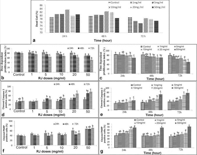 Figure 1
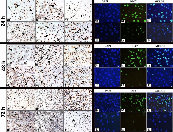 Figure 2
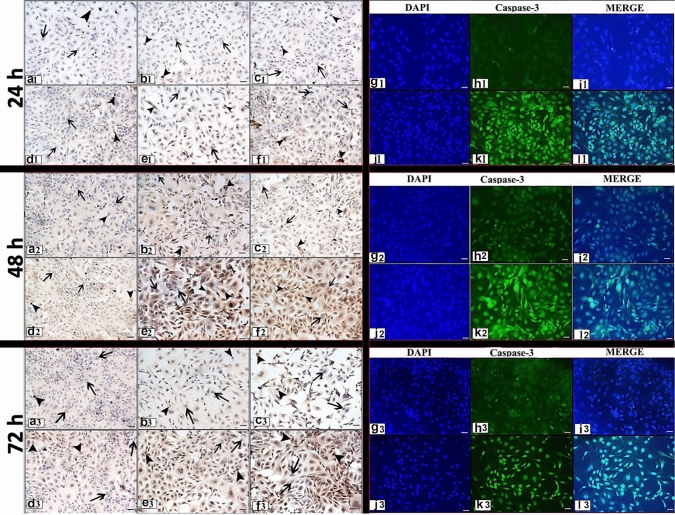 Figure 3
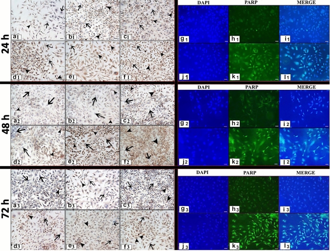 Figure 4
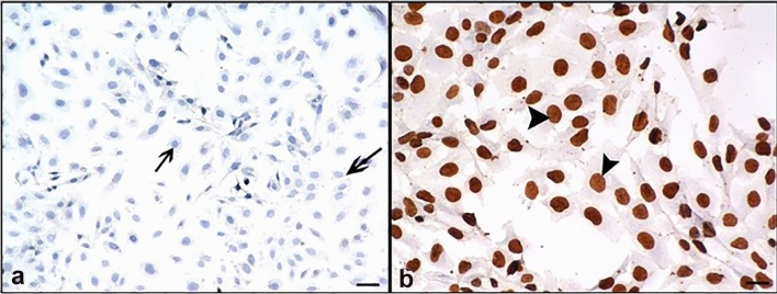 Figure 5
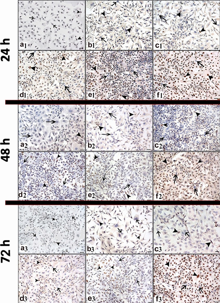 Figure 6
- —Bursa Uludag University Scientific Research Committee (BAP)
- —Bursa Uludag University
Peer Reviews
No public reviews on file for this paper yet. If you reviewed it on a platform where reviews are public (OpenReview, ICLR, NeurIPS, ICML), you can paste yours below so the community can read it here.
Videos
No videos yet. Explain this paper in a talk, walkthrough, or lecture? Add one.
Taxonomy
TopicsBee Products Chemical Analysis · Microbial Inactivation Methods · Saffron Plant Research Studies
Introduction
Cancer ranks second after cardiovascular diseases as the cause of death threatening human health [1]. Ovarian cancer, one of these cancer types, ranks fourth among the most common cancer types in women and is of clinical importance due to its high mortality rates [2, 3]. Ovarian cancers are classified into 3 groups: epithelial ovarian cancers (EOC), gonadal stromal tumors and germ cell tumors. Epithelial ovarian cancer is approximately 90% of all ovarian malignant diseases and is one of the leading causes of death in gynecological cancers [4]. EOCs, according to the cell types in the reproductive system classified into five groups; Serous (incidence: 7/10), mucinous (incidence: 1/10), endometrioid (incidence: 1/20), clear cell (incidence: 3/100), and transitional cell (incidence: 1/10). Treatment is based on a combination of surgery and chemotherapy. Most advanced ovarian cancer patients develop resistance to chemotherapy drugs used after surgery and tumor recurrence occurs over time [3].
Invasion of cancerous cells to surrounding tissues during treatment, development of resistance to chemotherapeutic drugs, and tumor recurrence over time lead researchers to seek the use of a natural compound with strong antioxidant, antitumor, or anticarcinogenic properties that can be protective before or alongside chemotherapeutic agents. Aronia melanocarpa (black currant cherry) fruit, which has antioxidant and anthocyanin properties, causes antiproliferative effects in colon cancer [5, 6], Hypericum olympicum (Uludağ cantoron) and Hypericum adenotrichum (Cranberry grass) strains are reported to show cytotoxic, genotoxic, and apoptotic properties in human lung cancer cell lines (A549 and PC3) [7].
It is reported that Royal jelly (RJ) has many biological activities such as antitumor [8, 9], antiviral, antibacterial, anticarcinogenic [10–13], antiallergic [14], immunomodulatory [15, 16], estrogenic [17, 18] and antidiabetic [19]. RJ is the best food of queen honey bee (Apis mellifera) larvae and is secreted from the hypopharyngeal and mandibular glands of worker honey bees between the sixth and twelfth days of life [20]. RJ is a viscous, bone-colored substance with a distinctive smell and burning taste. Chemically, RJ consists of 50–60% water, 18% proteins (Glutamic acid, Aspartic acid, Serine, Leucine, Phenylalanine, Tyrosine, Threonine, Lysine, Valine, Glycine, Methionine, Isoleucine, Proline, Cystine), 15% carbohydrates (Fructose, Glucose, Sucrose), 3–6% lipids and other fatty acids, 1.5% minerals (Potassium, Calcium, Magnesium, Zinc, Iron, Copper, Sodium, Selenium), and many vitamins (Thiamine, Riboflavin, Pantothenic acid, Pyridoxine, Niacin, Folic acid, Inositol, Biotin) [21]. In addition, RJ contains 10-hydroxy-2-decenoic acid (10-HDA), a major and unique fatty acid, which is not found in other bee products (propolis, pollen). 10-HDA has many potential pharmacological effects such as antitumor and angiogenesis inhibition [22], and immunomodulatory activities [23]. Researchers observed that the combination of RJ and interferon-alpha (HuIFN-αN3) showed an antiproliferative effect in human colorectal adenocarcinoma cells (Caco-2) [24]. There are many studies on MCF-7 cancer cells with RJ in combination with other chemotherapeutic drugs. It has been reported that combinations with RJ and Bisphenol A [9], as well as RJ and Tamoxifen [25] suppress the proliferation of MCF-7 cancer cells. In another study with the glioblastoma multiform (U87MG) cell line, it was shown that the combination of 30 mg/mL RJ with 20 mM Temozolomide (TMZ) inhibited DNA synthesis in cancer cells [26]. In an in vivo study, it was found that RJ administered orally to experimental animals with fibrocarcinoma at doses of 100, 200, and 300 mg/kg reduced tumour sizes in the experimental group compared to the control group [27]. It also has a therapeutic effect of RJ on leukemia cells [28]. Additionally, a presented study reported that RJ administered to mice with Ehrlich-Lettre acid carcinoma at doses of 500, 1000, and 1500 mg/kg for 33 days both had antitumor effects and modulated the body immune system [8].
In addition to the positive effects of RJ on cancer cells, it also positively affects fertility in both males and females by increasing the quality of oocyte and sperm in the reproductive system [29]. In a study by Kridli et al. (2003) in sheep, it was stated that RJ plays an important role in female fertility with its estrogenic effect [30], while in another study, it was stated that RJ induced spermatogenesis by increasing sperm volume, spermatozoon mobility, and concentration [31]. RJ affects ovarian hormone levels in immature female rats, increasing progesterone/estrogen levels and inducing folliculogenesis [18]. However, RJ has positive effects on oocyte maturation [17].
In recent years, RJ, which has been used both in the reproductive system and in the treatment of different types of cancer, there is no study has been found on its effect on ovarian cancer cells. Therefore, the aim of the presented study is to determine for the first time the time- and dose-dependent effects of RJ on Skov-3, one of the ovarian cancer cell lines, by evaluating both in terms of proliferation and apoptosis.
Material and method
Experimental procedure
Cell culture
The RJ used in our study was obtained purely from the same hive from Bursa Uludağ University Beekeeping Development-Application and Research Center. Cells in the Skov-3 human ovarian adenocarcinoma cell line were incubated in McCoy [32] medium containing 10% fetal bovine serum (FBS) and 0.1% penicillin–streptomycin in an incubator at 37 °C and 5% CO2 until they proliferated in sufficient numbers. When the cells proliferate sufficiently, they are seeded on 6-well plates and when it reaches 30% prevalence, 1000 mg of RJ is dissolved in 10 ml of distilled water (stock solution) [24] at appropriate concentrations (1, 5, 10, 20, 50 mg/ml) was added to the medium. Cells were incubated in media containing RJ for 24, 48, and 72 h.
Trypan blue exclusion assay
Following treatment cells were collected. Samples were centrifuged at 1300 rpm for 5 min. Then, pellets were resuspended in medium, and trypan blue (Sigma T6146) was added to the cell suspension in a 1:1 ratio. After, cells were counted using a Thoma slide under the inverted microscope at 10 × magnification. Cells stained blue were counted as dead, and results are expressed as a percentage of total cells. Cell viability was reported as an average of two independent experiments, each condition in duplicates.
Immunostaining
Cells were grown on cover slips in 24-well plates. After 24, 48, and 72 h, cells on the cover slips were washed three times in phosphate-buffered saline (PBS). The cells then were fixed in 10% Neutral buffer formaldehyde at room temperature for 15 min and permeabilize with 0.1% Triton X-100 for 10 min. For immunocytochemistry (ICC), the cells were blocked (Vector Lab.; MP7401) for 20 min and followed by incubation with proliferation markers Ki-67 (Lab Vision SP6 (dilution 1/500)), cleaved caspase-3 (Cell Signaling ASP175-9661 (dilution 1/200) and cleaved Poly (ADP-ribose) polymerase (PARP) (Santa-Cruz Biotechnology, sc-23461-R (dilution 1/300)) antibody at 4 °C overnight, then incubation with secondary antibody (Vector Lab.; MP7401) for 30 min. Cells were then treated with 3,3′- Diaminobenzidine (DAB) (Invitrogen:00-2020) for 5 min and counterstained with Harris Hematoxilen for 2 min, and the images were observed under the Nikon Eclipse 80i microscope.
For immunofluorescence (IF), the cells were blocked with 5% bovine serum albumin (BSA) (in PBS) for 1 h and followed by incubation with Ki-67 (dilution 1:500), cleaved caspase-3 (dilution 1:200) antibody at 4 °C overnight, then incubation with secondary antibody (dilution 1:2000) (Alexa Fluor 488 Conjugate 4412S) for 1 h in a dark room. Then, cover slips mounted and visualized under the fluorescence attached Nikon Eclipse 80i microscope.
TUNEL assay
To confirm the expression of cleaved caspase-3 and cleaved PARP in cells, Terminal Deoxynucleotidyl Transferase Mediated dUTP Nick End Labeling (TUNEL) method was applied according to the protocol in TUNEL Apoptosis Kit (Roche Diagnostics GmbH, Mannheim, Germany). The cells were seeded in cover slides (4 × 10^4^ cells per slide) and then treated with RJ at different concentrations (1, 5, 10, 20, and 50 mg/ml). Following this, the cells were washed in PBS and prepared 10% formaldehyde was added for cell fixation for 20 min. The cells were rewashed in PBS, before being permeabilized in permeabilization solution (0.1% Triton X-100) for 5 min on ice. The permeabilized cells were incubated in the dark at 37 °C for 1 h with the TUNEL reaction mixture containing terminal deoxynucleotidyl transferase (TdT) plus dUTP label. After labeling, cells were washed with PBS and incubated with TUNEL-POD solution for 30 min at 37 °C that to view the cells under light microscopy. Cells washed in PBS were treated with DAB (3.3 diaminobenzidine) for 10 min for peroxidase coloring reaction. Cells were then counterstained with Harris hematoxylin rinsed in distilled water, and examined under microscope (Nikon Eclipse 80i; Tokyo, Japan). Negative control sections were incubated only in TUNEL label solution without adding enzyme (terminal deoxynucleotidyl transferase (TdT)). Positive control sections received the same treatment but were pre-treated with DNase I (Roche, Indianapolis, USA) for 30 min at 37 °C.
Quantitative evaluation of immunostaining and TUNEL
Cells treated with RJ at the end of each dose and time were evaluated by two independent observers after ICC, IF, and TUNEL staining.
-Calculation of the proliferation index; Ki-67 positive and negative cells were counted in 5 randomly selected areas on at least 3 coverslips; The number of Ki-67 positive cells/total cell count was calculated as × 100.
-Calculation of the apoptotic index; TUNEL, cleaved caspase-3 and cleaved PARP positive and negative cells were counted on at least 3 coverslips in 5 randomly selected areas; The number of apoptotic cells/total cell number was calculated as × 100 [33–35].
Statistical analysis
Statistical analyzes of the data obtained from the study were performed using the SPSS 23.0 (Statistical Package for Social Sciences) program. Data were examined for normality distribution and variance homogeneity assumptions (Shapiro–wilk test). Statistical significance between the groups was analyzed by one-way ANOVA test followed by Dunn’s post hoc test. The symbols p ≤ 0.05 were used to indicate the level of confidence.
Results
The SKOV-3 ovarian cancer cell line was treated at different concentrations of RJ (1, 5, 10, 20, and 50 mg/ml) and different time (24, 48, 72 h). At the end of the period, viability tests were performed with trypan blue. No statistical significance was observed time and concentration-dependent in all groups (p ≥ 0.05) (Fig. 1a).Fig. 1. Effect of RJ on Skov 3; a Time and dose-dependent cell viability values of RJ (%); b Dose-dependent Ki-67 immunoreaction at different times; c Time-dependent Ki-67 immunoreaction of different doses; d Dose-dependent cleaved caspase-3 immunoreaction at different times; e Time-dependent cleaved caspase-3 immunoreaction of different doses; f Dose-dependent cleaved PARP immunoreaction at different times; g Time-dependent cleaved PARP immunoreaction of different doses. Different letters across doses are statistically significant (p ≤ 0.05), h hour
ICC test results
Cell proliferation
The dose- and time-dependent proliferation index (Ki-67) of RJ in Skov-3 cancer cell line was evaluated by ICC and IF staining method. Ki-67 expression was observed in the nuclei of the cells (Fig. 2). At the end of the experiment, the proliferative index differences between the groups are presented depending on both doses and times (Fig. 1b, c).Fig. 2. Ki-67 expression following 24, 48, 72 h RJ treatment. a1, a2, a3: Control group, b1, b2, b3: 1 mg/ml dose RJ, c1, c2, c3: 5 mg/ml dose RJ, d1, d2, d3: 10 mg/ml dose RJ, e1, e2, e3: 20 mg/ml dose RJ, f1, f2, f3: 50 mg/ml dose RJ. g1, g2, g3, h1 h2 h3, i1, i2, i3: Control group, j1, j2, j3, k1, k2, k3, l1, l2, l3: 50 mg/ml dose RJ, g–j: Cell nuclei stained with DAPI, h1, h2, h3, k1, k2, k3: Ki-67 protein expression, i1, i2, i3, l1, l2, l3: Merge. arrow: Ki-67 negative reaction, arrowhead: Ki-67 positive reaction. Immunocytochemical staining/ Immunofluorescence staining Bar 50 µm
The treatment of the only 1 mg/ml dose RJ for 72 h caused a decrease in cell proliferation compared to the administration time of 24 h of same dose (Figs. 1b, 2). However, the other doses of RJ suppressed cell proliferation in a time-dependent manner (p ≤ 0.05). Treatment with 50 mg/ml RJ for 72 h showed the lowest proliferative effect in all doses and times (p ≤ 0.05) (Figs. 1b, c, 2).
After 24 h treatment, the proliferation rate in cells treated with higher doses of RJ was markedly reduced (p ≤ 0.05). It was observed that cell proliferation was suppressed with increasing dose (Figs. 1c, 2).
After 48 h, while a statistical significance was determined between the control group and other doses RJ groups except for lowest dose RJ (p ≤ 0.05), no statistical significance was observed between the doses of 10, 20, and 50 mg/ml RJ (p ≥ 0.05). The lowest proliferation level was determined numerically at the high dose of RJ (Figs. 1b, c, 2).
After 72 h, the proliferation rate of the cells treated with 50 mg/ml RJ was significantly lower than the control and other RJ group’s (p ≤ 0.05) (Figs. 1b, c, 2).
Generally, it was observed that cell proliferation was suppressed with increasing dose and duration of RJ.
Cell apoptosis
The apoptotic effects of different concentrations RJ on Skov-3 ovarian cancer line, the expressions of cleaved caspase-3 and cleaved PARP were determined by ICC and IF staining (Figs. 3, 4). Additionally, the TUNEL method was applied to determine the apoptotic effects of RJ (Figs. 5, 6).Fig. 3. Cleaved caspase-3 expression following 24, 48, 72 h RJ treatment**. a1, a2, a3:** Control group, b1, b2, b3: 1 mg/ml dose RJ, c1, c2, c3: 5 mg/ml dose RJ, d1, d2, d3: 10 mg/ml dose RJ, e1, e2, e3: 20 mg/ml dose RJ, f1, f2, f3: 50 mg/ml dose RJ. g1, g2, g3, h1 h2 h3, i1, i2, i3: Control group, j1, j2, j3, k1, k2, k3, l1, l2, l3: 50 mg/ml dose RJ, g–j: Cell nuclei stained with DAPI, h1, h2, h3, k1, k2, k3: Cleaved caspase-3 protein expression, i1, i2, i3, l1, l2, l3: Merge. arrow: Cleaved caspase-3 negative reaction, arrowhead: Cleaved caspase-3 positive reaction. Immunocytochemical staining/ Immunofluorescence staining Bar 50 µmFig. 4Cleaved PARP expression following 24,48,72 h RJ treatment**. a1, a2, a3:** Control group, b1, b2, b3: 1 mg/ml dose RJ, c1, c2, c3: 5 mg/ml dose RJ, d1, d2, d3: 10 mg/ml dose RJ, e1, e2, e3: 20 mg/ml dose RJ, f1, f2, f3: 50 mg/ml dose RJ. g1, g2, g3, h1, h2, h3, i1, i2, i3: Control group, j1, j2, j3, k1, k2, k3, l1, l2, l3: 50 mg/ml dose RJ, g–j: Cell nuclei stained with DAPI, h1, h2, h3, k1, k2, k3: Cleaved PARP protein expression, i1, i2, i3, l1, l2, l3: Merge. arrow: Cleaved PARP negative reaction, arrowhead: Cleaved PARP positive reaction. Immunocytochemical staining/ Immunofluorescence staining Bar 50 µmFig. 5Result of 24-h RJ treatment. a TUNEL negative control group, where only label solution was applied without adding enzyme (TdT); b TUNEL positive control group, where DNase was applied. arrow: TUNEL negative reaction, arrowhead: TUNEL positive reaction. Bar 50 µmFig. 6TUNEL expression following 24, 48,72 h RJ treatment. a1, a2, a3: Control group, b1, b2, b3: 1 mg/ml dose RJ, c1, c2, c3: 5 mg/ml dose RJ, d1, d2, d3: 10 mg/ml dose RJ, e1, e2, e3: 20 mg/ml dose RJ, f1, f2, f3: 50 mg/ml dose RJ. arrow: Tunel negative reaction, arrowhead: Tunel positive reaction. Immunocytochemical staining, Bar 50 µm
Cleaved caspase-3
Cleaved caspase-3 immunoreaction was observed both in the cell nucleus and cytoplasm by immunocytochemical and immunofluorescent staining (Fig. 3). At the end of the experiment, differences between groups and apoptotic index are presented both times and doses dependent (Fig. 1d, e). When the apoptotic effect of dose-dependent RJ treatment on Skov-3 ovarian cancer cells was examined, no statistical difference was observed time-dependent manner in 1 mg/ml RJ dose group (p ≥ 0.05). However, it was observed that cleaved caspase-3 expression was high in other RJ doses (except 1 mg/ml), especially in the 48 and 72 h treatment (Figs. 1d, e, 3).
After 24 h treatment, a higher cleaved caspase-3 expression was determined at doses of 20 and 50 mg/ml RJ than at all other doses (p ≤ 0.05), (Figs. 1e, 3).
After 48 h treatment, while no significant difference was observed between the control group and 1 and 10 mg/ml RJ doses (p ≥ 0.05), statistical significance was determined between the control group and the other RJ groups (p ≤ 0.05). The highest cleaved caspase-3 expression was determined in treatment of 50 mg/ml RJ (p ≤ 0.05) (Figs. 1e, 3).
After 72 h treatment, While no significant difference was observed between 20 and 50 mg/ml RJ doses (p ≥ 0.05), statistical difference was observed between these two doses and the all other groups (control group, 1, 5, 10 mg/ml RJ) (p ≤ 0.05) (Figs. 1e, 3).
Cleaved PARP
Cleaved PARP which is involved in a different step of the apoptosis pathway, immunoreaction was observed both in the cell nucleus and cytoplasm in immunocytochemical and immunofluorescent staining (Fig. 4).
At the end of the experiment, differences between groups and apoptotic index are presented depending on both doses and times (Fig. 1f, g). No statistical significance was observed in a time-dependent manner in the control group and treatment with 1 and 10 mg/ml RJ doses (p ≥ 0.05). For the 5 mg/ml RJ dose, a statistical difference was observed between the 24 h and 48 h treatment periods (p ≤ 0.05) also 20 mg/ml RJ dose showed statistical significance between all treatment periods (p ≤ 0.05).
After 24 h treatment, statistical significance was observed between the control group and 5, 20, and 50 mg/ml RJ doses (p ≤ 0.05). While no significant difference was observed between the 20 and 50 mg/ml RJ doses (p ≥ 0.05), statistical significance was determined between the 50 mg/ml RJ dose and other groups except 20 mg/ml RJ dose (p ≤ 0.05) (Figs. 1g, 4).
After 48 h treatment, a significant difference was determined between 50 mg/ml RJ and all groups (p ≤ 0.05) (Figs. 1g, 4).
Similar to the 48 h RJ treatment, statistical significance was observed between the highest dose and other groups after 72 h treatment (p ≤ 0.05) (Figs. 1g, 4).
TUNEL results
TUNEL negative and positive controls are presented in Fig. 5a, b, respectively. TUNEL positive reaction was observed in cell nuclei (Fig. 5b).
After 24 h and 72 h treatment, TUNEL positive cell density was less dense in treatment the low dose RJ (1and 5 mg/ml). However, as a result of 48 h treatment, TUNEL positive cell density was observed to be higher in the 20 and 50 mg/ml RJ doses compared to the control group and 1, 5, and 10 mg/ml RJ groups (Fig. 6). The most intense tunnel positive cells were observed in the 50 mg/ml RJ dose treatment for all times.
Discussion
Chemotherapy is widely used for cancer treatment, but the side effects of chemotherapy agents, damage to normal cells and resistance of tumor cells to these agents are the biggest limitations of this treatment method. Therefore, there is a need for supporting natural compounds that may be more effective to overcome these limitations.
It is reported that RJ has many biological activities such as antitumor, antiviral, antibacterial, antiallergic, anticancerogenic, immunomodulatory, estrogenic and antidiabetic [9, 11, 12, 15, 18, 19, 36]. Although the doses and treatment periods used in in vivo/in vitro environments are different, many positive results have been revealed in studies with RJ [12, 18, 36]. However, in studies on cancer, no studies on ovarian cancer were found. For this reason, in the presented study, the proliferative and apoptotic effects of RJ on the serous type epithelial ovarian cancer cell line (Skov-3), the most common epithelial ovarian cancer, were investigated.
When the antiproliferative effect of RJ in different types of cancer is examined, it is reported that RJ has a significant antiproliferative activity in slow-growing cancers, but does not show the same effect in fast-growing cancer types [37]. Nakaya et al. (2007) stated that RJ inhibited the growth-promoting effect of Bisphenol A (BPA), an environmental estrogen, on MCF7 cells, and that RJ disrupted estrogen-induced cell proliferation signals [9].
It has been reported that GE132 + natural combination (Reishi mushroom, RJ, Resveratrol, Lycopene and Sulforaphane containing) showed antiproliferative effects on SW480 (colon cancer cells) and EAhy 926 (normal human endothelial cancer cell line) cell lines at a dose of 750 µg/ml [38]. Although the dose of GE132 + natural combination was lower than the doses used in our study, the antiproliferative effect it showed was due to the RJ combination. In our study, while the antiproliferative effect of RJ was not observed at the lowest dose and duration, antiproliferative effects of RJ on Skov-3 were determined with the increase in duration and dose.
It has been reported that the treatment of 1.5 mM and 5 mM doses of 10HDA, a unique protein compound contained in RJ, to B16F10 melanoma cancer cells for 24 h creates a cytotoxic effect on the cells, but at the same time 0.1, 0.5, and 1 mM 10-HDA concentrations are not cytotoxic in cancer cells [39]. However, since the doses used in our study were lower than the doses used by Peng et al. (2017) a similar result was not found in the cell viability test.
It has been reported that the combination of RJ (0.1 g/ml) with human interferon-alpha (HuIFN-aN3) (1000 I.U. mL-1) at a ratio of 2:1 has a stronger antiproliferative effect on human colorectal adenocarcinoma cells compared to the monotherapeutic treatment of RJ [24]. This study, which supports the antiproliferative efficacy of the highest dose (50 mg/ml) used in our study, suggests that testing RJ with combined drug treatments may produce more effective results.
Apoptosis is one of the main forms of cellular death associated with characteristic morphological changes including chromatin condensation, DNA fragmentation and formation of apoptotic bodies [40]. Cysteine proteases called caspase enzymes (especially caspase 3, 8, and 9) and Bcl-2 family, which contribute to the apoptotic mechanism, play an active role in this process. Caspase-3, the most important member of the caspase family, is responsible for many biochemical mechanisms of apoptosis, leading to cleavage of nuclear and cytosolic substrates, chromatin condensation, DNA fragmentation, and apoptotic bodies [41]. Bee products have been reported to induce in vitro cellular apoptosis in numerous cancer cell lines, including prostate, lung and liver cancers [42]. These biologically active natural products are also recognized as part of an innovative treatment for various types of cancer, including breast and colon cancers [36]. It has been proven in many different studies that RJ has a potential antitumor activity in mice [43], induces apoptotic and antiproliferative pathways in tumor cells, and therefore has an anticarcinogenic activity [44, 45]. In the present study, the expressions of cleaved caspase-3 and cleaved PARP proteins, which are involved in different pathways of apoptosis, were investigated by applying different doses and durations of RJ. The apoptotic effects of RJ on Skov-3 cancer line with the increase in dose concentration and treatment period support the studies conducted with other cancer cells [8, 22].
In a study with the Skov-3 cancer cell line, it was reported that the aqueous extract of the leaves of Solanum nigrum (AE-SN), a natural component such as RJ, induced cleaved caspase-3 [46]. In another study, it was reported that the combination of 30 mg/mL RJ, with 20 mM temozolomide (TMZ) inhibited DNA synthesis in cancer cells (glioblastoma multiform (U87MG)), similar to the dose used in our study [26]. In our study, it was determined that similar doses induced apoptosis on the ovarian cancer cell line.
RJ is used in in vivo studies as well as in vitro studies. It is seen that RJ, given orally to experimental animals with fibrocarcinoma at doses of 100, 200, and 300 mg/kg reduces tumor sizes [27]. In addition, RJ administered to mice with Ehrlich-Lettre acid carcinoma at doses of 500, 1000, and 1500 mg/kg for 33 days was seen to have both anti-tumour effects and regulate the body immune system [8]. Although the doses and durations used in in vivo studies differ according to the doses and durations in our study, the results obtained from our study are consistent with both in vivo and in vitro studies above.
Conclusion
In the presented study, the anticarcinogenic effects of different doses and durations of RJ were evaluated in terms of both cell proliferation and apoptosis, and the anticarcinogenic effect of RJ on Skov-3 cancer cell line was revealed for the first time. In our study, the highest dose and duration of RJ treatment on the cancer cell line suppressed cell proliferation, and therefore induced apoptosis in the cells. In the light of the findings, we think that the use of RJ as an alternative treatment on ovarian cancer, both monotherapeutically and in combination, can form the basis for new experimental protocols in future studies.