Ultrasound Identification of an Atypical Course of the Posterior Intercostal Artery During Paravertebral Block
David Y Chee, Oriana Ng

TL;DR
This paper describes the use of ultrasound to identify an unusual artery during a paravertebral block, helping avoid vascular injury.
Contribution
The study highlights an atypical arterial course identified via ultrasound during a paravertebral block procedure.
Findings
An atypical artery was found near the T7-8 paravertebral space during a TPVB.
Similar arteries were observed one intercostal space above and below the identified area.
Ultrasound enabled real-time needle visualization, reducing the risk of arterial puncture.
Abstract
The thoracic paravertebral block (TPVB), while relatively safe, can be associated with significant complications, including inadvertent vascular injury. We describe an ultrasound-guided TPVB where a pulsatile artery was identified between the two transverse processes and in close proximity to the T7-8 paravertebral space, likely the dorsal branch of the posterior intercostal artery. A similar artery was also noted one intercostal space cephalad and caudal of this area. The use of ultrasound allowed for real-time visualization of the needle, minimizing the risk of arterial puncture.
Genes, proteins, chemicals, diseases, species, mutations and cell lines named across the full text — each resolved to its canonical identifier and authoritative record.
Click any figure to enlarge with its caption.
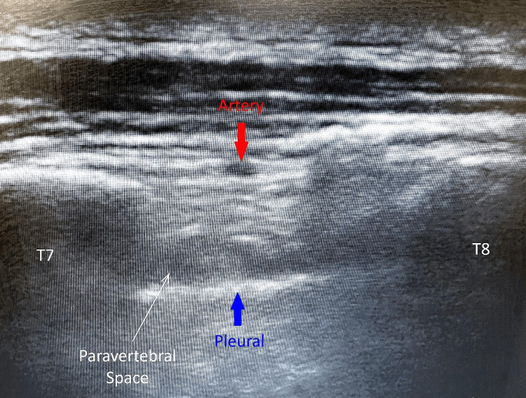 Figure 1
Peer Reviews
No public reviews on file for this paper yet. If you reviewed it on a platform where reviews are public (OpenReview, ICLR, NeurIPS, ICML), you can paste yours below so the community can read it here.
Videos
No videos yet. Explain this paper in a talk, walkthrough, or lecture? Add one.
Taxonomy
TopicsAnesthesia and Pain Management · Shoulder Injury and Treatment · Spine and Intervertebral Disc Pathology