Quantifying morphologic variations as an alternate to standard response criteria for unresectable primary liver tumors after checkpoint inhibition therapy
Laetitia Saccenti, Nicole Varble, Tabea Borde, Andrew S. Mikhail, Michael Kassin, Elliot Levy, Sheng Xu, Lindsey A. Hazen, Ifechi Ukeh, Cyndi Vasco, Austin G. Duffy, Changqing Xie, Cecilia Monge, Donna Mabry, Tim F. Greten, Bradford J. Wood

TL;DR
This study explores using tumor shape changes during immunotherapy as a potential alternative to standard response criteria for liver cancer patients.
Contribution
The study introduces morphologic tumor shape analysis as a novel approach to assess treatment outcomes in liver cancer.
Findings
Low tumor solidity at 6-month follow-up was associated with poorer prognosis.
Decreased tumor solidity over time correlated with worse survival outcomes.
Baseline or 3-month shape features did not correlate with survival.
Abstract
The aim of this study was to assess the feasibility of quantifying morphologic changes in tumors during immunotherapy, as a reflection of response or survival. A retrospective single-center analysis was performed in patients with unresectable liver cancer previously enrolled in clinical trials combining immunotherapy (tremelimumab ± durvalumab) and locoregional treatment (either ablation or transarterial chemoembolization). Conventional response (RECIST 1.1) was assessed at 6-month follow-up. For morphologic assessment, the largest target lesion was manually segmented on axial slices in two dimensions using contrast-enhanced CT. Solidity and circularity of tumors were calculated at baseline, 3-month follow-up, and at 6-months follow-up. Survival analysis was performed. From the 68 patients enrolled in clinical trials, 28 did not have target lesions separate from lesions treated by…
Genes, proteins, chemicals, diseases, species, mutations and cell lines named across the full text — each resolved to its canonical identifier and authoritative record.
Click any figure to enlarge with its caption.
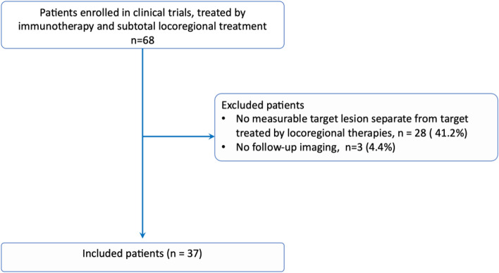 Figure 1
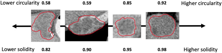 Figure 2
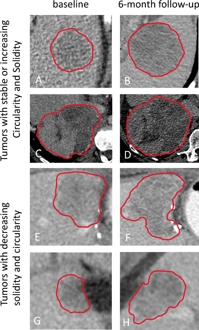 Figure 3
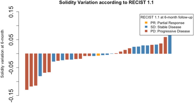 Figure 4
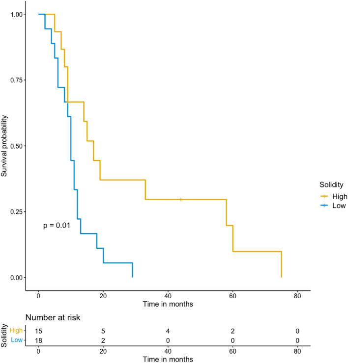 Figure 5
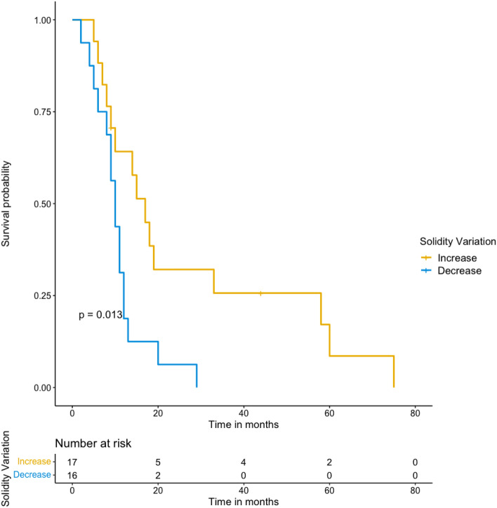 Figure 6
Peer Reviews
No public reviews on file for this paper yet. If you reviewed it on a platform where reviews are public (OpenReview, ICLR, NeurIPS, ICML), you can paste yours below so the community can read it here.
Videos
No videos yet. Explain this paper in a talk, walkthrough, or lecture? Add one.
Taxonomy
TopicsCancer Genomics and Diagnostics · Radiomics and Machine Learning in Medical Imaging · Mathematical Biology Tumor Growth