Analysis of Retinal Thickness in Patients With Chronic Diseases Using Standardized Optical Coherence Tomography Data: Database Study Based on the Radiology Common Data Model
ChulHyoung Park, So Hee Lee, Da Yun Lee, Seoyoon Choi, Seng Chan You, Ja Young Jeon, Sang Jun Park, Rae Woong Park

TL;DR
This study shows how standardizing retinal scan data allows researchers to efficiently compare retinal thickness in patients with chronic diseases across multiple hospitals.
Contribution
The novelty is demonstrating the feasibility of multi-institutional research using harmonized OCT data linked with EMR data via R-CDM and OMOP-CDM.
Findings
Standardized OCT data from two hospitals were successfully used to analyze retinal thickness in chronic disease patients.
Significant reductions in central macular thickness were observed in type 2 diabetes patients at both hospitals during follow-up.
A significant reduction in central macular thickness was also found in the hypertension cohort at one hospital.
Abstract
The Observational Medical Outcome Partners-Common Data Model (OMOP-CDM) is an international standard for harmonizing electronic medical record (EMR) data. However, since it does not standardize unstructured data, such as medical imaging, using this data in multi-institutional collaborative research becomes challenging. To overcome this limitation, extensions such as the Radiology Common Data Model (R-CDM) have emerged to include and standardize these data types. This work aims to demonstrate that by standardizing optical coherence tomography (OCT) data into an R-CDM format, multi-institutional collaborative studies analyzing changes in retinal thickness in patients with long-standing chronic diseases can be performed efficiently. We standardized OCT images collected from two tertiary hospitals for research purposes using the R-CDM. As a proof of concept, we conducted a comparative…
Genes, proteins, chemicals, diseases, species, mutations and cell lines named across the full text — each resolved to its canonical identifier and authoritative record.
Click any figure to enlarge with its caption.
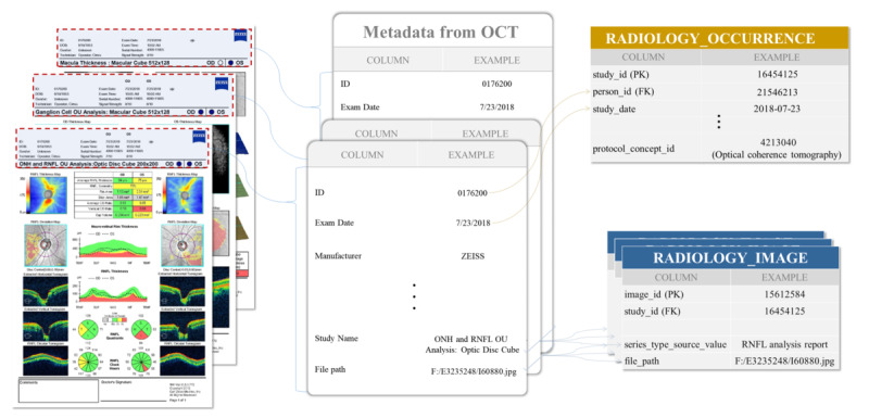 Figure 1
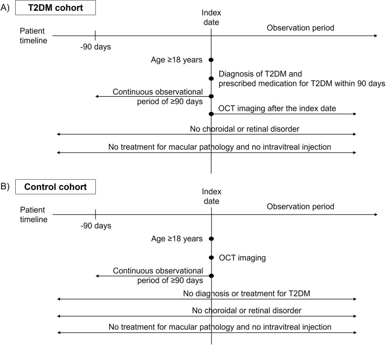 Figure 2
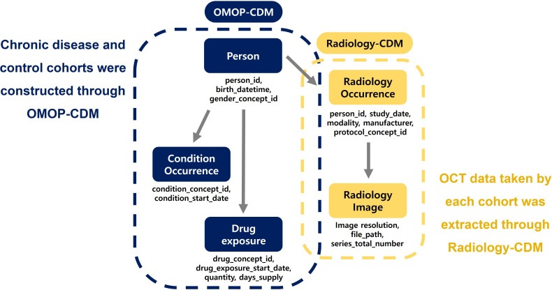 Figure 3
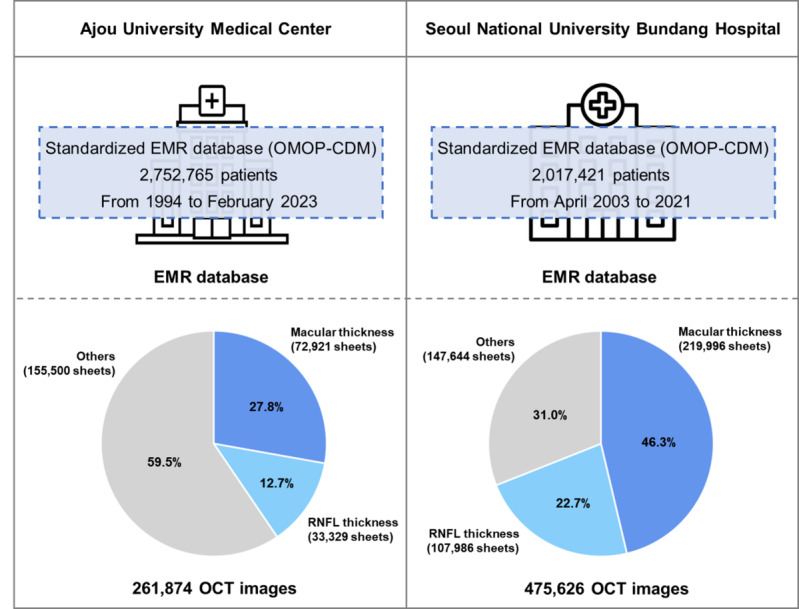 Figure 4
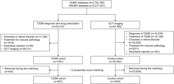 Figure 5
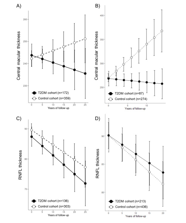 Figure 6
Peer Reviews
No public reviews on file for this paper yet. If you reviewed it on a platform where reviews are public (OpenReview, ICLR, NeurIPS, ICML), you can paste yours below so the community can read it here.
Videos
No videos yet. Explain this paper in a talk, walkthrough, or lecture? Add one.
Taxonomy
TopicsRetinal Imaging and Analysis · Retinal and Optic Conditions · Retinal Diseases and Treatments