Teledermatology viewpoint: Sudden onset of a widespread rash
Alexis R. Bernat, Robert T. Brodell, Lindsey B. Dolohanty

Abstract
Genes, proteins, chemicals, diseases, species, mutations and cell lines named across the full text — each resolved to its canonical identifier and authoritative record.
Click any figure to enlarge with its caption.
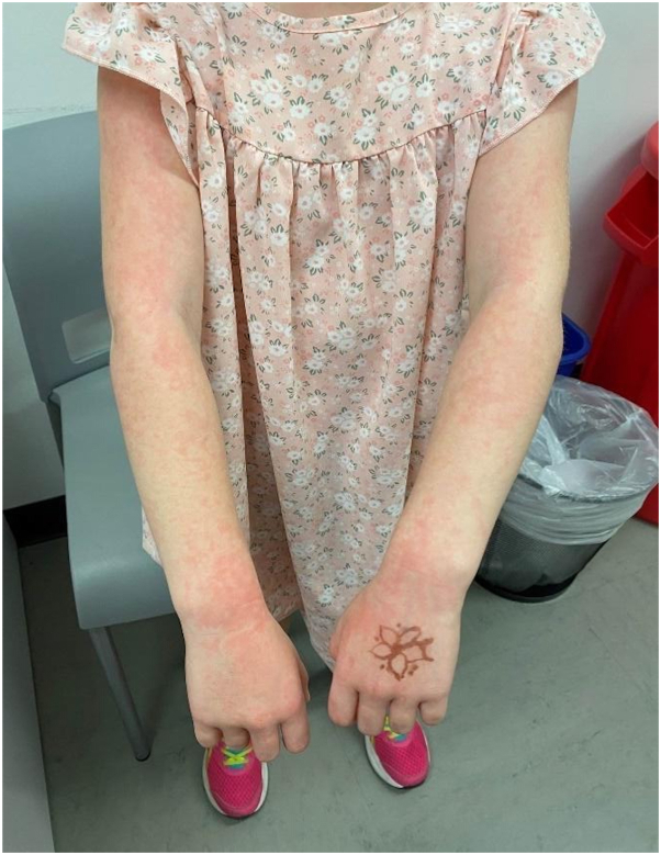 Figure 1
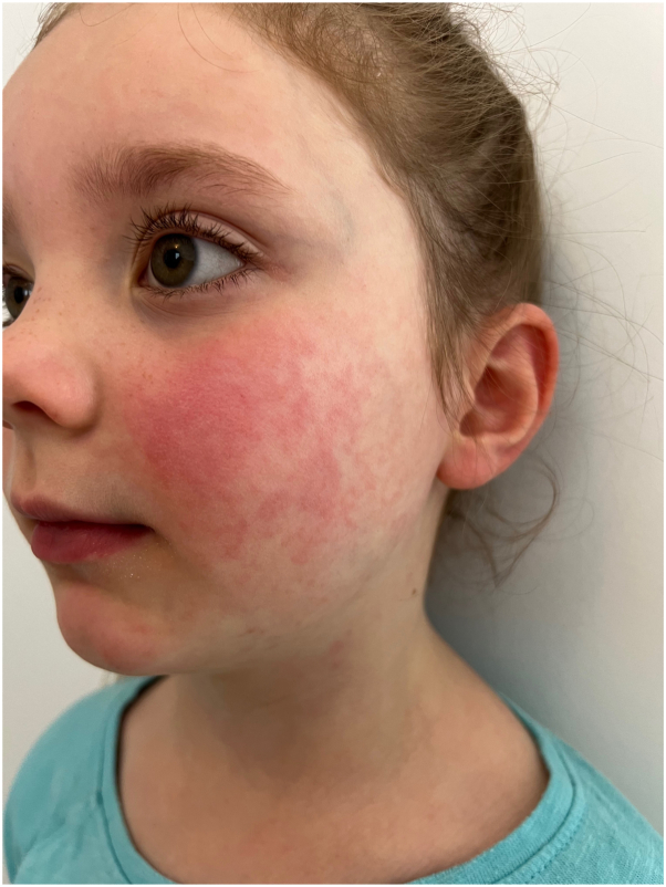 Figure 2
Peer Reviews
No public reviews on file for this paper yet. If you reviewed it on a platform where reviews are public (OpenReview, ICLR, NeurIPS, ICML), you can paste yours below so the community can read it here.
Videos
No videos yet. Explain this paper in a talk, walkthrough, or lecture? Add one.
Taxonomy
TopicsTumors and Oncological Cases · Parvovirus B19 Infection Studies · Cutaneous Melanoma Detection and Management
Introduction
Fifth disease, or erythema infectiosum (EI), is a childhood exanthem that is benign in nature and commonly occurs in school-age children in the early spring and late winter months. The viremia is a result of a parvovirus B19, nonenveloped single-stranded DNA virus. It is highly infectious and spreads in the air through respiratory droplets.1 Interestingly, by the time the rash appears, children are no longer infectious.2^,^3 Patients usually exhibit a nonspecific prodrome or are entirely asymptomatic before the classic skin findings of “slapped-cheeks” and a pruritic, erythematous net-like rash arise.4 As the rash fades, the reticular or lace-like pattern may become confluent. Although the disease is self-limited, exercise leading to overheating can lead to reappearance of the erythema, but usually not pruritus, in the areas where rash occurred for weeks to months.5 Pramoxine-containing moisturizers, low-potency topical corticosteroids, and oral antihistamines may be used to control any associated pruritus.
EI requires consideration of certain special populations. In adolescents with sickle cell disease, it is associated with red cell aplasia—otherwise known as aplastic crisis. Complications such as acute chest syndrome, splenic sequestration crisis, nephrotic syndrome, stroke, and acute severe pain occur after the viremia phase of EI.1 EI can also cause harmful effects to both pregnant mothers and the fetus. Risk of adverse fetal outcomes has been associated with EI, including the development of fetal anemia, fetal hydrops, and fetal death.6 No specific treatment is available; however, counseling of at-risk mothers and monitoring of confirmed fetal infections throughout pregnancy will likely decrease fetal mortality.6 Here, we present a case that uniquely uses teledermatology as a way to identify and diagnose EI.
Case report
An otherwise well child was sent to the school nurse after suddenly developing a net-like, pruritic rash on the arms and thighs associated with a bright red confluent rash on the cheeks. After calling the parents, a teledermatology consultation was sought for this dramatic rash to determine if it was infectious. The teledermatology reader report stated that the history of present illness included a 7-year-old child presented to the school nurse with sudden lace-like erythema on the bilateral arms and profoundly erythematous cheeks that suggested the patient was slapped. Prodrome: none. Prior treatment: none. Primary symptom: pruritus. Two images were provided with this teledermatology consultation. They demonstrated a lace-like erythematous rash along the lateral and dorsal aspects of the child’s arms, which extended onto the dorsum of the hands (Fig 1). A brightly erythematous confluent rash was noted on the cheeks (Fig 2). Based on the report, the consulted dermatologist determined that this viral exanthem is benign and self-limiting. Images showed that findings are pathognomonic for EI (“Fifth disease”). Reassurance and symptomatic treatment with over-the-counter pramoxine-containing moisturizing lotion, mild potency topical corticosteroid (hydrocortisone 2.5% cream), and an oral antihistamine (hydroxyzine 25 mg by mouth every night at bedtime) was recommended as needed for pruritus. Although this exanthem is not infectious at the time of the eruption, the child was sent home so that she could be aggressively treated for her pruritus with frequent topical applications of the prescribed medications.Fig 1. Image provided for teledermatology consultation demonstrating lace-like erythematous rash along the upper extremities of the patient.Fig 2. Image provided for teledermatology consultation demonstrating erythematous confluent rash in “slapped cheek” pattern.
Discussion
Although there are limitations to teledermatology because of lighting, focus, composition, and bias to which lesions have been photographed,7 which can mask important clues, this case showcases how store-and-forward teledermatology was used to allow for important recommendations to be made to the school nurse. Research has indicated that accuracy in diagnosis and effectiveness in management have been comparable to in-person dermatological care, especially in regard to skin cancers.7, 8, 9 Pathognomonic features were visible in this EI case, demonstrating the potential usefulness of store-and-forward teledermatology in diagnosing inflammatory skin conditions as well. In other less clear-cut cases, research shows that teledermatology is beneficial for triage to either provide feedback for management to the referring clinician or recommending further in-person evaluation.7^,^8 Although there are studies that claim the clinical usefulness of store-and-forward teledermatology is limited,10 this case supports the functionality of teledermatology for diagnosis of distinct inflammatory conditions.
Conflicts of interest
Dr Brodell has participated in multicenter clinical trials with CorEvitas (formerly Corrona) Psoriasis Registry, Sanofi, and Novartis and holds stock in Veradermics, Inc. Author Bernat and Dr Dolohanty have no conflicts of interest to declare.
The reference list from the paper itself. Each links out to its DOI / PubMed record.
- 1Saad A.A.Beshlawi I.Al-Rawas A.H.Zachariah M.Nazir H.F.Wali Y.Human parvovirus B 19 in children with sickle cell disease; poking the spleen Oman Med J 325201742542810.5001/omj.2017.7929026475 PMC 5632704 · doi ↗ · pubmed ↗
- 2Servey J.T.Reamy B.V.Hodge J.Clinical presentations of parvovirus B 19 infection Am Fam Phys 753200737337617304869 · pubmed ↗
- 3Kostolansky S.Waymack J.R.Erythema Infectiosum Stat Pearls [Internet]2023 Stat Pearls 30020681 · pubmed ↗
- 4Mc Neely M.Friedman J.Pope E.Generalized petechial eruption induced by parvovirus B 19 infection J Am Acad Dermatol 525 Suppl 12005 S 109S 11310.1016/j.jaad.2004.11.04015858505 · doi ↗ · pubmed ↗
- 5Subiabre D.Martinez L.Ortiz J.M.Esteve A.Vasculitis-like erythema infectiosum in children J Am Acad Dermatol 7662017 AB 28010.1016/j.jaad.2017.04.1087 · doi ↗
- 6Giorgio E.De Oronzo M.A.Iozza I.Parvovirus B 19 during pregnancy: a review J Prenat Med 442010636622439064 PMC 3279187 · pubmed ↗
- 7Wang R.H.Barbieri J.S.Nguyen H.P.Clinical effectiveness and cost-effectiveness of teledermatology: where are we now, and what are the barriers to adoption?J Am Acad Dermatol 831202029930710.1016/j.jaad.2020.01.06532035106 PMC 7302990 · doi ↗ · pubmed ↗
- 8Peracca S.B.Jackson G.L.Weinstock M.A.Oh D.H.Implementation of teledermatology: theory and practice Curr Derm Rep 82019354510.1007/s 13671-019-0252-2 · doi ↗