The genome sequence of a cranefly, Diogma glabrata (Meigen, 1818)
James McCulloch, Liam M. Crowley, Marcus Stensmyr, Saverio Brogna

TL;DR
This paper provides the genome sequence of the cranefly Diogma glabrata, including a detailed assembly of its chromosomes and mitochondrial DNA.
Contribution
The novel contribution is the high-quality genome assembly of Diogma glabrata, including chromosomal pseudomolecules and mitochondrial DNA.
Findings
The genome assembly is 1,328.70 megabases in total length.
90.7% of the assembly is organized into 4 chromosomal pseudomolecules.
The mitochondrial genome is 17.5 kilobases in length.
Abstract
We present a genome assembly from a specimen of Diogma glabrata (cranefly; Arthropoda; Insecta; Diptera; Cylindrotomidae). The genome sequence has a total length of 1,328.70 megabases. Most of the assembly (90.7%) is scaffolded into 4 chromosomal pseudomolecules. The mitochondrial genome has also been assembled and is 17.5 kilobases in length.
Genes, proteins, chemicals, diseases, species, mutations and cell lines named across the full text — each resolved to its canonical identifier and authoritative record.
Click any figure to enlarge with its caption.
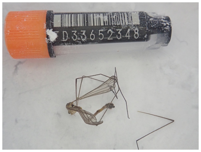 Figure 1
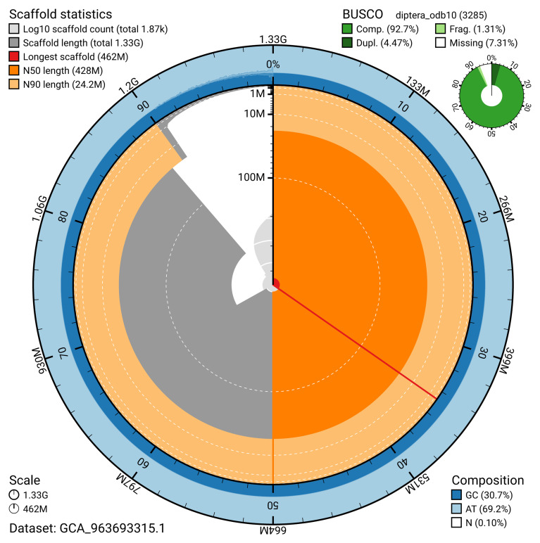 Figure 2
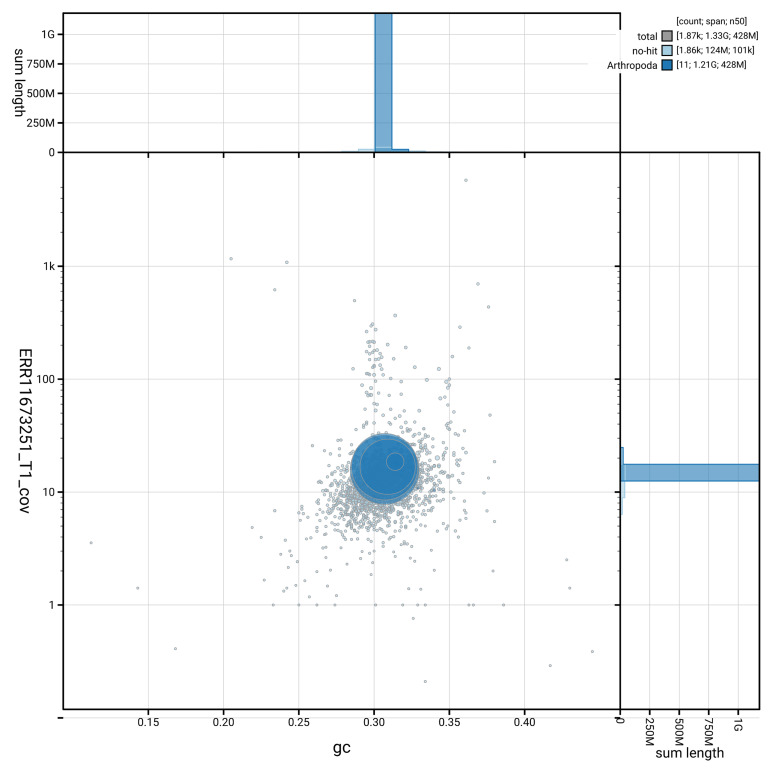 Figure 3
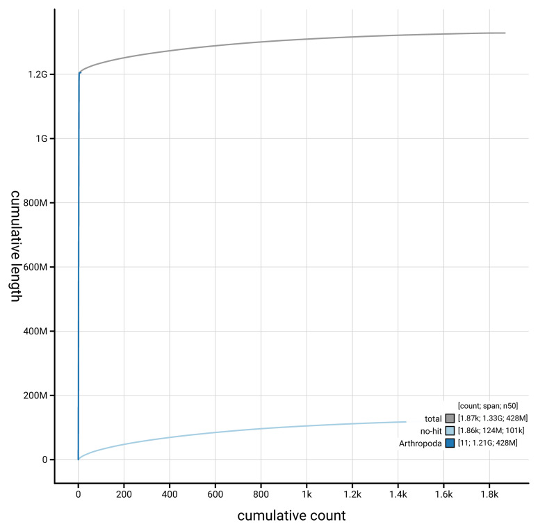 Figure 4
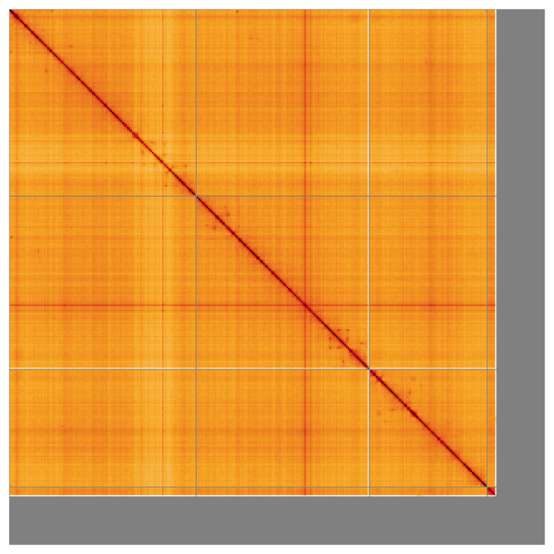 Figure 5
| Project information | |||
|---|---|---|---|
|
| Diogma glabrata | ||
|
| PRJEB64102 | ||
|
|
| ||
|
| SAMEA112232520 | ||
|
| 2715158 | ||
| Specimen information | |||
|
|
|
|
|
|
| idDioGlab1 | SAMEA112232964 | Whole organism |
|
| idDioGlab1 | SAMEA112232964 | Whole organism |
| Sequencing information | |||
|
|
|
|
|
|
| ERR11679415 | 6.48e+08 | 97.89 |
|
| ERR11673251 | 2.35e+06 | 21.07 |
| Genome assembly | ||
|---|---|---|
| Assembly name | idDioGlab1.1 | |
| Assembly accession | GCA_963693315.1 | |
|
|
| |
| Span (Mb) | 1,328.70 | |
| Number of contigs | 8,151 | |
| Number of scaffolds | 1,867 | |
| Longest scaffold (Mb) | 462.97 | |
| Assembly metrics
|
| |
| Contig N50 length (Mb) | 0.3 |
|
| Scaffold N50 length (Mb) | 427.9 |
|
| Consensus quality (QV) | 53.0 |
|
|
| primary: 80.76%; alternate:
|
|
| BUSCO
| C:92.6%[S:88.1%,D:4.5%],
|
|
| Percentage of assembly mapped
| 90.7% |
|
| Sex chromosomes | Not identified |
|
| Organelles | Mitochondrial genome: 17.5 kb |
|
| INSDC
| Name | Length
| GC% |
|---|---|---|---|
| 1 | 461.91 | 30.5 | |
| 2 | 427.87 | 30.5 | |
| 3 | 290.09 | 31.0 | |
| 4 | 24.21 | 31.5 | |
| MT | 0.02 | 20.5 |
| Software tool | Version | Source |
|---|---|---|
| BEDTools | 2.30.0 |
|
| BLAST | 2.14.0 |
|
| BlobToolKit | 4.3.7 |
|
| BUSCO | 5.4.3 and 5.5.0 |
|
| bwa-mem2 | 2.2.1 |
|
| Cooler | 0.8.11 |
|
| DIAMOND | 2.1.8 |
|
| fasta_windows | 0.2.4 |
|
| FastK | 427104ea91c78c3b8b8b49f1a7d6bbeaa869ba1c |
|
| Gfastats | 1.3.6 |
|
| GoaT CLI | 0.2.5 |
|
| Hifiasm | 0.19.8-r587 |
|
| HiGlass | 44086069ee7d4d3f6f3f0012569789ec138f42b84
|
|
| Merqury.FK | d00d98157618f4e8d1a9190026b19b471055b22e |
|
| MitoHiFi | 3 |
|
| MultiQC | 1.14, 1.17, and 1.18 |
|
| NCBI Datasets | 15.12.0 |
|
| Nextflow | 23.04.0-5857 |
|
| PretextView | 0.2.5 |
|
| purge_dups | 1.2.5 |
|
| samtools | 1.16.1, 1.17, and 1.18 |
|
| sanger-tol/
| - |
|
| sanger-tol/
| 1.1.1 |
|
| sanger-tol/readmapping | 1.2.1 |
|
| Seqtk | 1.3 |
|
| Singularity | 3.9.0 |
|
| TreeVal | 1.0.0 |
|
| YaHS | 1.2a.2 |
|
- —Wellcome Trust
Peer Reviews
No public reviews on file for this paper yet. If you reviewed it on a platform where reviews are public (OpenReview, ICLR, NeurIPS, ICML), you can paste yours below so the community can read it here.
Videos
No videos yet. Explain this paper in a talk, walkthrough, or lecture? Add one.
Taxonomy
TopicsGenomics and Phylogenetic Studies · Evolution and Genetic Dynamics · Insect symbiosis and bacterial influences
Species taxonomy
Eukaryota; Opisthokonta; Metazoa; Eumetazoa; Bilateria; Protostomia; Ecdysozoa; Panarthropoda; Arthropoda; Mandibulata; Pancrustacea; Hexapoda; Insecta; Dicondylia; Pterygota; Neoptera; Endopterygota; Diptera; Nematocera; Tipulomorpha; Tipuloidea; Cylindrotomidae; Cylindrotominae; Diogma; Diogma glabrata (Meigen, 1818) (NCBI:txid2715158)
Background
Diogma glabrata is a species of cranefly (Tipuloidea) in the family Cylindrotomidae. This family can be distinguished from the others in the superfamily by the combination of short mouthparts and the wing vein R 1 curving down into to end in R 2+3 as opposed to the costa (the vein running along the wing’s fore margin) ( Stubbs & Kramer, 2016a). The cylindrotomids often also stand out due to their long body relative to the wings, explaining their vernacular name of “long-bodied craneflies”. Diogma glabrata and Cylindrotoma distinctissima are the only two cylindrotomid species with three dark stripes on a yellow thorax; D. glabrata can be further distinguished by the presence of a black spot beneath the eye, alongside characters of the antennae, wing venation, and genitalia ( Stubbs & Kramer, 2016b).
The Cylindrotomidae is the least speciose tipuloid family, with 67 species listed on the Catalogue of the Craneflies of the World ( Oosterbroek, 2024), and not well-studied. However, they are known to be phytophagous as larvae, feeding primarily on bryophytes and herbaceous plants ( Kania-Kłosok et al., 2021). Uniquely among craneflies, cylindrotomid larvae have evolved to mimic their food source, as they live exposed among the mosses and plants rather than hidden within mud. In D. glabrata, this manifests with green colouration of the larva with discrete dark markings, coupled with distinctive cuticular appendages to disrupt the larva’s outline and make it appear more moss-like ( Imada, 2020). It particularly resembles one of its favoured foodplants, the springy turf-moss Rhytidiadelphus squarrosus, a common and widespread species of woodlands and gardens. Accordingly, D. glabrata is similarly widespread in the UK ( NBN Atlas, 2024). Outside of the UK, D. glabrata is distributed across the whole Palearctic, east to Japan ( Imada, 2020).
Here we present a chromosomal-level genome sequence for Diogma glabrata, based on a female specimen from Wytham Woods, Berkshire, United Kingdom ( Figure 1). The genome was sequenced as part of the Darwin Tree of Life Project, a collaborative effort to sequence all named eukaryotic species in the Atlantic Archipelago of Britain and Ireland ( Blaxter et al., 2022).
Photograph of the Diogma glabrata (idDioGlab1) specimen used for genome sequencing.
Genome sequence report
The genome of Diogma glabrata ( Figure 1) was sequenced using Pacific Biosciences single-molecule HiFi long reads, generating a total of 21.07 Gb (gigabases) from 2.35 million reads, providing an estimated 18-fold coverage. Primary assembly contigs were scaffolded with chromosome conformation Hi-C data, which produced 97.89 Gb from 648.25 million reads. Specimen and sequencing details are summarised in Table 1.
Table 1.: Specimen and sequencing data for Diogma glabrata.
Assembly errors, including 77 missing joins or mis-joins and 7 haplotypic duplications, were corrected by manual curation. This reduced the assembly length by 0.54% and the scaffold number by 1.94%, and increased the scaffold N50 by 25.14%. The final assembly has a total length of 1,328.70 Mb in 1,867 sequence scaffolds, with 6,283 gaps, and a scaffold N50 of 427.9 Mb ( Table 2).
The snail plot in Figure 2 provides a summary of the assembly statistics, indicating the distribution of scaffold lengths and other assembly metrics. Figure 3 shows the distribution of scaffolds by GC proportion and coverage. Figure 4 presents a cumulative assembly plot, with separate curves representing different scaffold subsets assigned to various phyla, illustrating the completeness of the assembly.
Table 2.: Genome assembly data for Diogma glabrata, idDioGlab1.1.
Genome assembly of Diogma glabrata, idDioGlab1.1: metrics.The BlobToolKit snail plot provides an overview of assembly metrics and BUSCO gene completeness. The circumference represents the length of the whole genome sequence, and the main plot is divided into 1,000 equal-sized bins around the circumference. The outermost blue tracks display the distribution of GC, AT, and N percentages across the bins. Scaffolds are arranged clockwise from longest to shortest and are depicted in dark grey. The longest scaffold is indicated by the red arc, and the deeper orange and pale orange arcs represent the N50 and N90 lengths. A light grey spiral at the centre shows the cumulative scaffold count on a logarithmic scale. A summary of complete, fragmented, duplicated, and missing BUSCO genes in the diptera_odb10 set is presented at the top right. An interactive version of this figure is available at https://blobtoolkit.genomehubs.org/view/GCA_963693315.1/dataset/GCA_963693315.1/snail.
Genome assembly of Diogma glabrata, idDioGlab1.1: BlobToolKit GC-coverage plot showing sequence coverage (vertical axis) and GC content (horizontal axis).The circles represent scaffolds, with the size proportional to scaffold length and the colour representing phylum membership. The histograms along the axes display the total length of sequences distributed across different levels of coverage and GC content. An interactive version of this figure is available at https://blobtoolkit.genomehubs.org/view/GCA_963693315.1/dataset/GCA_963693315.1/blob.
Genome assembly of Diogma glabrata idDioGlab1.1: BlobToolKit cumulative sequence plot.The grey line shows cumulative length for all scaffolds. Coloured lines show cumulative lengths of scaffolds assigned to each phylum using the buscogenes taxrule. An interactive version of this figure is available at https://blobtoolkit.genomehubs.org/view/GCA_963693315.1/dataset/GCA_963693315.1/cumulative.
Most of the assembly sequence (90.7%) was assigned to 4 chromosomal-level scaffolds. These chromosome-level scaffolds, confirmed by the Hi-C data, are named in order of size ( Figure 5; Table 3).
Genome assembly of Diogma glabrata idDioGlab1.1: Hi-C contact map of the idDioGlab1.1 assembly, visualised using HiGlass.Chromosomes are shown in order of size from left to right and top to bottom. An interactive version of this figure may be viewed at https://genome-note-higlass.tol.sanger.ac.uk/l/?d=KDwtz9dURiiL1pAlCG0A8Q.
Table 3.: Chromosomal pseudomolecules in the genome assembly of Diogma glabrata, idDioGlab1.
While not fully phased, the assembly deposited is of one haplotype. Contigs corresponding to the second haplotype have also been deposited. The mitochondrial genome was also assembled and can be found as a contig within the multifasta file of the genome submission, and as a separate fasta file.
The final assembly has a Quality Value (QV) of 53.0 and k-mer completeness of 97.82% for the combined assembly. BUSCO (v5.4.3) analysis using the diptera_odb10 reference set ( n = 3,285) indicated a completeness score of 92.6% (single = 88.1%, duplicated = 4.5%). The assembly achieves the EBP reference standard of 6.C.53. Other quality metrics are given in Table 2.
Methods
Sample acquisition and DNA barcoding
An adult female specimen of Diogma glabrata (specimen ID Ox002286, ToLID idDioGlab1) was collected from Wytham Woods, Berkshire, United Kingdom (latitude 51.77, longitude –1.34) on 2022-07-08 by netting. The specimen was collected by James McCulloch and Liam Crowley (University of Oxford) and identified by James McCulloch, and then preserved on dry ice.
The initial identification was verified by an additional DNA barcoding process according to the framework developed by Twyford et al. (2024). A small sample was dissected from the specimens and stored in ethanol, while the remaining parts were shipped on dry ice to the Wellcome Sanger Institute (WSI). The tissue was lysed, the COI marker region was amplified by PCR, and amplicons were sequenced and compared to the BOLD database, confirming the species identification ( Crowley et al., 2023). Following whole genome sequence generation, the relevant DNA barcode region was also used alongside the initial barcoding data for sample tracking at the WSI ( Twyford et al., 2024). The standard operating procedures for Darwin Tree of Life barcoding have been deposited on protocols.io ( Beasley et al., 2023).
Nucleic acid extraction
The workflow for high molecular weight (HMW) DNA extraction at the Wellcome Sanger Institute (WSI) Tree of Life Core Laboratory includes a sequence of procedures: sample preparation and homogenisation, DNA extraction, fragmentation and purification. Detailed protocols are available on protocols.io ( Denton et al., 2023b).
The idDioGlab1 sample was prepared for DNA extraction by weighing and dissecting it on dry ice ( Jay et al., 2023). Tissue from the whole organism was homogenised using a PowerMasher II tissue disruptor ( Denton et al., 2023a).
HMW DNA was extracted in the WSI Scientific Operations core using the Automated MagAttract v2 protocol ( Oatley et al., 2023). The DNA was sheared into an average fragment size of 12–20 kb in a Megaruptor 3 system ( Bates et al., 2023). Sheared DNA was purified by solid-phase reversible immobilisation, using AMPure PB beads to eliminate shorter fragments and concentrate the DNA ( Strickland et al., 2023). The concentration of the sheared and purified DNA was assessed using a Nanodrop spectrophotometer and Qubit Fluorometer using the Qubit dsDNA High Sensitivity Assay kit. Fragment size distribution was evaluated by running the sample on the FemtoPulse system.
Hi-C preparation
Whole organism tissue of the idDioGlab1 sample was processed at the WSI Scientific Operations core, using the Arima-HiC v2 kit. Tissue (stored at –80 °C) was fixed, and the DNA crosslinked using a TC buffer with 22% formaldehyde. After crosslinking, the tissue was homogenised using the Diagnocine Power Masher-II and BioMasher-II tubes and pestles. Following the kit manufacturer's instructions, crosslinked DNA was digested using a restriction enzyme master mix. The 5’-overhangs were then filled in and labelled with biotinylated nucleotides and proximally ligated. An overnight incubation was carried out for enzymes to digest remaining proteins and for crosslinks to reverse. A clean up was performed with SPRIselect beads prior to library preparation.
Library preparation and sequencing
Library preparation and sequencing were performed at the WSI Scientific Operations core. Pacific Biosciences HiFi circular consensus DNA sequencing libraries were prepared using the PacBio Express Template Preparation Kit v2.0 (Pacific Biosciences, California, USA) as per the manufacturer's instructions. The kit includes the reagents required for removal of single-strand overhangs, DNA damage repair, end repair/A-tailing, adapter ligation, and nuclease treatment. Library preparation also included a library purification step using AMPure PB beads (Pacific Biosciences, California, USA) and size selection step to remove templates shorter than 3 kb using AMPure PB modified SPRI. DNA concentration was quantified using the Qubit Fluorometer v2.0 and Qubit HS Assay Kit and the final library fragment size analysis was carried out using the Agilent Femto Pulse Automated Pulsed Field CE Instrument and 165kb gDNA and 55kb BAC analysis kit. Samples were sequenced using the Sequel IIe system (Pacific Biosciences, California, USA). The concentration of the library loaded onto the Sequel IIe was in the range 40–135 pM. The SMRT link software, a PacBio web-based end-to-end workflow manager, was used to set-up and monitor the run, as well as perform primary and secondary analysis of the data upon completion.
For Hi-C library preparation, DNA was fragmented to a size of 400 to 600 bp using a Covaris E220 sonicator. The DNA was then enriched, barcoded, and amplified using the NEBNext Ultra II DNA Library Prep Kit following manufacturers’ instructions. The Hi-C sequencing was performed using paired-end sequencing with a read length of 150 bp on an Illumina NovaSeq 6000 instrument.
Genome assembly, curation and evaluation
** Assembly **
The HiFi reads were first assembled using Hifiasm ( Cheng et al., 2021) with the --primary option. Haplotypic duplications were identified and removed using purge_dups ( Guan et al., 2020). The Hi-C reads were mapped to the primary contigs using bwa-mem2 ( Vasimuddin et al., 2019). The contigs were further scaffolded using the provided Hi-C data ( Rao et al., 2014) in YaHS ( Zhou et al., 2023) using the --break option for handling potential misassemblies. The scaffolded assemblies were evaluated using Gfastats ( Formenti et al., 2022), BUSCO ( Manni et al., 2021) and MERQURY.FK ( Rhie et al., 2020).
The mitochondrial genome was assembled using MitoHiFi ( Uliano-Silva et al., 2023), which runs MitoFinder ( Allio et al., 2020) and uses these annotations to select the final mitochondrial contig and to ensure the general quality of the sequence.
** Assembly curation **
The assembly was decontaminated using the Assembly Screen for Cobionts and Contaminants (ASCC) pipeline (article in preparation). Flat files and maps used in curation were generated in TreeVal ( Pointon et al., 2023). Manual curation was primarily conducted using PretextView ( Harry, 2022), with additional insights provided by JBrowse2 ( Diesh et al., 2023) and HiGlass ( Kerpedjiev et al., 2018). Scaffolds were visually inspected and corrected as described by Howe et al. (2021). Any identified contamination, missed joins, and mis-joins were corrected, and duplicate sequences were tagged and removed. The curation process is documented at https://gitlab.com/wtsi-grit/rapid-curation (article in preparation).
** Evaluation of the final assembly **
The final assembly was post-processed and evaluated using the three Nextflow ( Di Tommaso et al., 2017) DSL2 pipelines: sanger-tol/readmapping ( Surana et al., 2023a), sanger-tol/genomenote ( Surana et al., 2023b), and sanger-tol/blobtoolkit ( Muffato et al., 2024). The readmapping pipeline aligns the Hi-C reads using bwa-mem2 ( Vasimuddin et al., 2019) and combines the alignment files with SAMtools ( Danecek et al., 2021). The genomenote pipeline converts the Hi-C alignments into a contact map using BEDTools ( Quinlan & Hall, 2010) and the Cooler tool suite ( Abdennur & Mirny, 2020). The contact map is visualised in HiGlass ( Kerpedjiev et al., 2018). This pipeline also computes k-mer completeness and QV consensus quality values with FastK and MERQURY.FK, and runs BUSCO ( Manni et al., 2021) to assess completeness.
The blobtoolkit pipeline is a Nextflow port of the previous Snakemake Blobtoolkit pipeline ( Challis et al., 2020). It aligns the PacBio reads in SAMtools and minimap2 ( Li, 2018) and generates coverage tracks for regions of fixed size. In parallel, it queries the GoaT database ( Challis et al., 2023) to identify all matching BUSCO lineages to run BUSCO ( Manni et al., 2021). For the three domain-level BUSCO lineages, the pipeline aligns the BUSCO genes to the UniProt Reference Proteomes database ( Bateman et al., 2023) with DIAMOND blastp ( Buchfink et al., 2021). The genome is also divided into chunks according to the density of the BUSCO genes from the closest taxonomic lineage, and each chunk is aligned to the UniProt Reference Proteomes database using DIAMOND blastx. Genome sequences without a hit are chunked using seqtk and aligned to the NT database with blastn ( Altschul et al., 1990). The blobtools suite combines all these outputs into a blobdir for visualisation.
The genome evaluation pipelines were developed using nf-core tooling ( Ewels et al., 2020) and MultiQC ( Ewels et al., 2016), relying on the Conda package manager, the Bioconda initiative ( Grüning et al., 2018), the Biocontainers infrastructure ( da Veiga Leprevost et al., 2017), as well as the Docker ( Merkel, 2014) and Singularity ( Kurtzer et al., 2017) containerisation solutions.
Table 4 contains a list of relevant software tool versions and sources.
Wellcome Sanger Institute – Legal and Governance
The materials that have contributed to this genome note have been supplied by a Darwin Tree of Life Partner. The submission of materials by a Darwin Tree of Life Partner is subject to the ‘Darwin Tree of Life Project Sampling Code of Practice’, which can be found in full on the Darwin Tree of Life website here. By agreeing with and signing up to the Sampling Code of Practice, the Darwin Tree of Life Partner agrees they will meet the legal and ethical requirements and standards set out within this document in respect of all samples acquired for, and supplied to, the Darwin Tree of Life Project.
Further, the Wellcome Sanger Institute employs a process whereby due diligence is carried out proportionate to the nature of the materials themselves, and the circumstances under which they have been/are to be collected and provided for use. The purpose of this is to address and mitigate any potential legal and/or ethical implications of receipt and use of the materials as part of the research project, and to ensure that in doing so we align with best practice wherever possible. The overarching areas of consideration are:
• Ethical review of provenance and sourcing of the material
• Legality of collection, transfer and use (national and international)
Each transfer of samples is further undertaken according to a Research Collaboration Agreement or Material Transfer Agreement entered into by the Darwin Tree of Life Partner, Genome Research Limited (operating as the Wellcome Sanger Institute), and in some circumstances other Darwin Tree of Life collaborators.
The reference list from the paper itself. Each links out to its DOI / PubMed record.
- 1Abdennur N Mirny LA : Cooler: scalable storage for Hi-C data and other genomically labeled arrays. Bioinformatics. 2020;36(1):311–316. 10.1093/bioinformatics/btz 540 31290943 PMC 8205516 · doi ↗ · pubmed ↗
- 2Allio R Schomaker-Bastos A Romiguier J : Mito Finder: efficient automated large-scale extraction of mitogenomic data in target enrichment phylogenomics. Mol Ecol Resour. 2020;20(4):892–905. 10.1111/1755-0998.13160 32243090 PMC 7497042 · doi ↗ · pubmed ↗
- 3Altschul SF Gish W Miller W : Basic local alignment search tool. J Mol Biol. 1990;215(3):403–410. 10.1016/S 0022-2836(05)80360-2 2231712 · doi ↗ · pubmed ↗
- 4Bateman A Martin MJ Orchard S : Uni Prot: the universal protein knowledgebase in 2023. Nucleic Acids Res. 2023;51(D 1):D 523–D 531. 10.1093/nar/gkac 1052 36408920 PMC 9825514 · doi ↗ · pubmed ↗
- 5Bates A Clayton-Lucey I Howard C : Sanger Tree of Life HMW DNA fragmentation: diagenode Megaruptor ®3 for LI Pac Bio. protocols.io. 2023. 10.17504/protocols.io.81wgbxzq 3lpk/v 1 · doi ↗
- 6Beasley J Uhl R Forrest LL : DNA barcoding SO Ps for the Darwin Tree of Life project. protocols.io. 2023; [Accessed 25 June 2024]. 10.17504/protocols.io.261ged 91jv 47/v 1 · doi ↗
- 7Blaxter M Mieszkowska N Di Palma F : Sequence locally, think globally: the Darwin Tree of Life Project. Proc Natl Acad Sci U S A. 2022;119(4): e 2115642118. 10.1073/pnas.2115642118 35042805 PMC 8797607 · doi ↗ · pubmed ↗
- 8Buchfink B Reuter K Drost HG : Sensitive protein alignments at Tree-of-Life scale using DIAMOND. Nat Methods. 2021;18(4):366–368. 10.1038/s 41592-021-01101-x 33828273 PMC 8026399 · doi ↗ · pubmed ↗