Quantitative autofluorescence is increased in clinically unaffected fellow eyes from patients with posterior uveitis
Robert P. Finger, Julie Jungblut, Marie D. Just, Jan H. Terheyden, Frank G. Holz, Raffael Liegl, Thomas Ach, Maximilian W. M. Wintergerst

TL;DR
This study found that eyes of patients with posterior uveitis, even those not showing symptoms, have higher autofluorescence levels compared to healthy eyes, suggesting possible hidden inflammation.
Contribution
The study reveals subclinical changes in unaffected eyes of posterior uveitis patients through increased quantitative autofluorescence.
Findings
Both affected and unaffected eyes of posterior uveitis patients showed significantly higher mean qAF values than healthy controls.
The segment with the highest qAF value also showed increased fluorescence in both affected and unaffected eyes compared to controls.
The findings suggest subclinical inflammation or metabolic changes in unaffected eyes that may precede future disease activity.
Abstract
The purpose of this prospective case-control study is to investigate differences in quantitative autofluorescence (qAF) in clinically affected and unaffected eyes of patients with inactive posterior uveitis compared to healthy, age-matched controls. Patients with posterior uveitis and healthy controls were imaged using fundus autofluorescence (488 nm excitation; Spectralis HRA + OCT; Heidelberg Engineering) to measure qAF values using the proprietary HEYEX software. Mean background qAF (excluding vessels and retinal lesions) across all segments (as previously defined by Delori et al.) and in the segment with the highest mean qAF value were compared between affected and unaffected eyes from patients with posterior uveitis, and healthy age-matched control eyes using the Kruskal-Wallis-test. A total of 83 eyes from 83 patients were included: 33 affected eyes (33 patients with…
Genes, proteins, chemicals, diseases, species, mutations and cell lines named across the full text — each resolved to its canonical identifier and authoritative record.
Click any figure to enlarge with its caption.
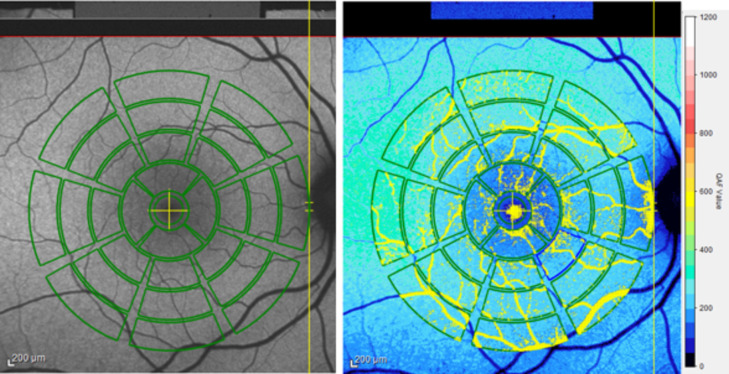 Figure 1
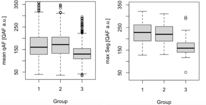 Figure 2
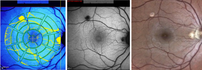 Figure 3
Peer Reviews
No public reviews on file for this paper yet. If you reviewed it on a platform where reviews are public (OpenReview, ICLR, NeurIPS, ICML), you can paste yours below so the community can read it here.
Videos
No videos yet. Explain this paper in a talk, walkthrough, or lecture? Add one.
Taxonomy
TopicsOcular Diseases and Behçet’s Syndrome · Retinal Diseases and Treatments · Retinal and Optic Conditions