Case report: Lobular capillary hemangioma of the right main bronchus: a rare case with calcified and cystic lymph nodes
Xu-ping Chen, Jia Zeng, Wei Zhang, Peng Wu, Yan-ling Zhao, Zhi-shu Li, Ling Shen, Shi-xu He

TL;DR
This case report describes a rare occurrence of a benign blood vessel tumor in the bronchus with unusual calcified and cystic features.
Contribution
The report presents the second documented case of LCH in the bronchus with calcification and cystic changes.
Findings
LCH was found in the right intermediate bronchus with calcification within the lesion.
Cystic changes were observed, which are uncommon in typical LCH cases.
The case highlights the importance of considering LCH in differential diagnoses involving calcified and cystic lesions.
Abstract
Lobular capillary hemangioma (LCH), typically a benign vasoproliferative lesion of the skin or mucosal surfaces, is exceptionally rare in the trachea. Now, we present the second reported case of LCH found in the right intermediate bronchus, characterized by calcification within the lesion and cystic changes. These distinctive features should alert clinicians to consider LCH in the differential diagnosis of other benign vascular tumors and mediastinal lymphadenopathy, particularly when calcified and cystic lesions are observed.
Genes, proteins, chemicals, diseases, species, mutations and cell lines named across the full text — each resolved to its canonical identifier and authoritative record.
Click any figure to enlarge with its caption.
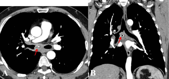 Figure 1
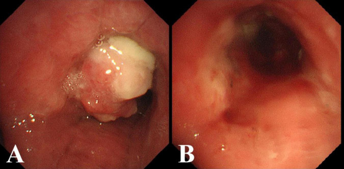 Figure 2
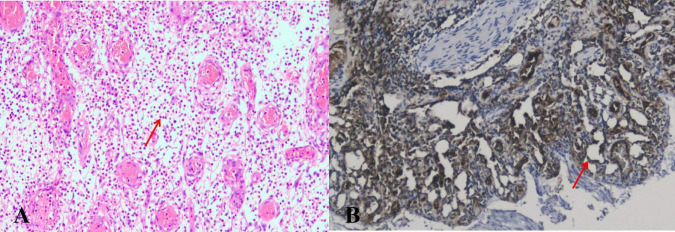 Figure 3
Peer Reviews
No public reviews on file for this paper yet. If you reviewed it on a platform where reviews are public (OpenReview, ICLR, NeurIPS, ICML), you can paste yours below so the community can read it here.
Videos
No videos yet. Explain this paper in a talk, walkthrough, or lecture? Add one.
Taxonomy
TopicsVascular Malformations and Hemangiomas · Vascular Tumors and Angiosarcomas · Medical Imaging and Pathology Studies