Transcriptome and Functional Comparison of Primary and Immortalized Endothelial Cells of the Human Choroid Plexus at the Blood–Cerebrospinal Fluid Barrier
Lea Denzer, Walter Muranyi, Rosanna Herold, Carolin Stump-Guthier, Hiroshi Ishikawa, Carsten Sticht, Horst Schroten, Christian Schwerk, Stefan Weichert

TL;DR
This study compares primary and immortalized human choroid plexus endothelial cells to assess their suitability as models for the blood–cerebrospinal fluid barrier.
Contribution
The study demonstrates that immortalized cells retain barrier function and Wnt signaling features up to high passage numbers.
Findings
Transcriptome analysis showed high concordance between primary and immortalized cells with only minor differences.
Wnt signaling components were preserved in immortalized cells, supporting their barrier function.
Immortalized cells retained barrier function and β-catenin downregulation up to passage 100.
Abstract
The human choroid plexus (CP) is the location of the blood–cerebrospinal fluid (CSF) barrier (BCSFB). Whereas the epithelial cells of the CP mainly contribute to the formation of the BCSFB, the vessels of the CP are built by fenestrated endothelial cells. Still, the CP endothelium can contribute to barrier function. By ectopic expression of human telomerase reverse transcriptase (hTERT) in primary human CP endothelial cells (HCPEnCs), we recently generated and characterized immortalized HCPEnCs (iHCPEnCs). Here, we compared primary cells of the sixth passage (HCPEnCs p6) with a lower (p20) and a higher passage (p50) of iHCPEnCs by transcriptome analysis. A high concordance of HCPEnCs and both passages of iHCPEnCs was observed, as only small proportions of the transcripts examined were significantly altered. Differentially expressed genes (DEGs) were identified and assigned to…
Genes, proteins, chemicals, diseases, species, mutations and cell lines named across the full text — each resolved to its canonical identifier and authoritative record.
Click any figure to enlarge with its caption.
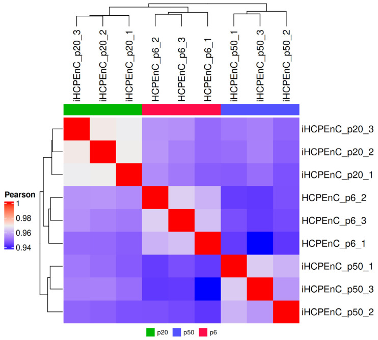 Figure 1
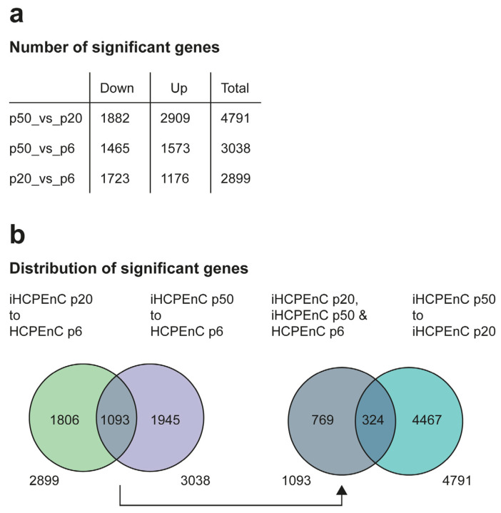 Figure 2
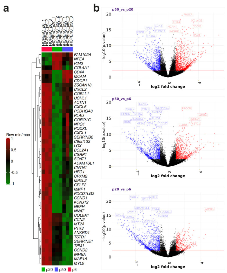 Figure 3
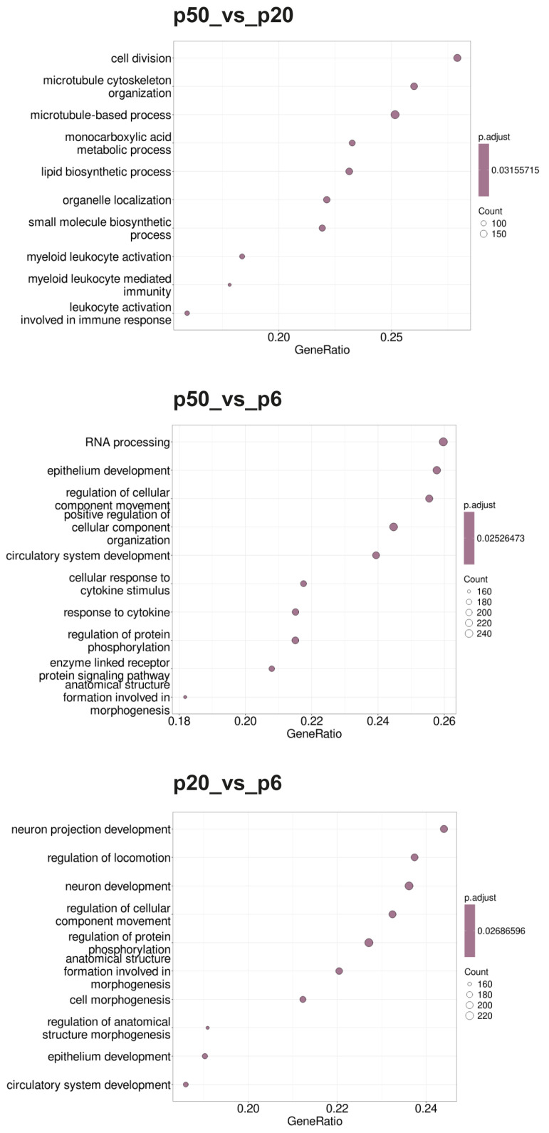 Figure 4
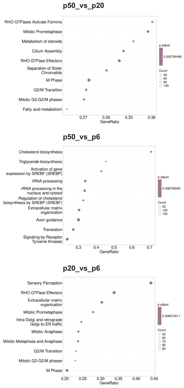 Figure 5
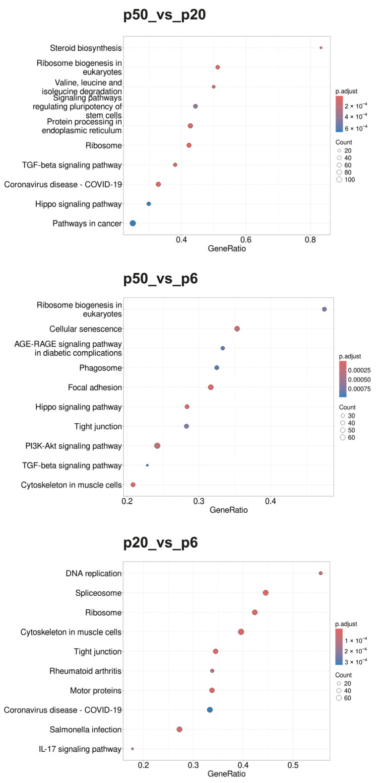 Figure 6
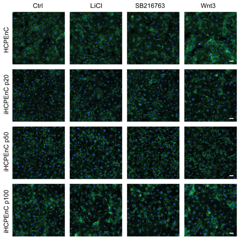 Figure 7
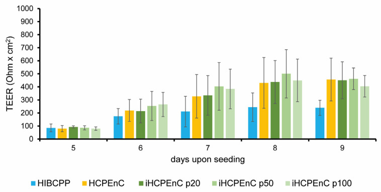 Figure 8
Peer Reviews
No public reviews on file for this paper yet. If you reviewed it on a platform where reviews are public (OpenReview, ICLR, NeurIPS, ICML), you can paste yours below so the community can read it here.
Videos
No videos yet. Explain this paper in a talk, walkthrough, or lecture? Add one.
Taxonomy
TopicsNeurological Disease Mechanisms and Treatments · Barrier Structure and Function Studies