Optical Coherence Tomography in Infectious Keratitis After Femtosecond Keratorefractive Surgery
Antonio Leccisotti, Stefania V. Fields, Giuseppe De Bartolo, Christian Crudale, Matteo Posarelli

TL;DR
This study shows how optical coherence tomography helps diagnose and monitor corneal infections after laser eye surgeries like LASIK and lenticule extraction.
Contribution
The paper presents the first analysis of AS–OCT patterns in infectious keratitis specifically after femtosecond keratorefractive surgeries.
Findings
AS–OCT identified initial infiltrates and interface inflammation in post-surgery infections.
Interface fluid accumulation and stromal reabsorption were observed and documented by OCT.
OCT effectively guided treatment and evaluated healing and scarring after infections.
Abstract
Objectives: Anterior Segment Optical coherence tomography (AS–OCT) can help in the diagnosis and treatment of infectious keratitis, but it has not been studied in cases occurring after corneal refractive surgery procedures such as femtosecond laser in situ keratomileusis (FS–LASIK) and keratorefractive lenticule extraction (KLEx). In these procedures, a surgical interface is created, where infections usually start, thus determining a different AS–OCT pattern compared to non–surgical infections, which begin on the corneal surface. Methods: We retrospectively reviewed 22,756 eyes of 13,564 patients who underwent FS–LASIK and KLEx at our surgical center. Results: Four cases of post–refractive surgery infectious keratitis were included (two after FS–LASIK and two after KLEx), in which the AS–OCT identified an initial infiltrate in the interface, followed by interface inflammation. In one…
Genes, proteins, chemicals, diseases, species, mutations and cell lines named across the full text — each resolved to its canonical identifier and authoritative record.
Click any figure to enlarge with its caption.
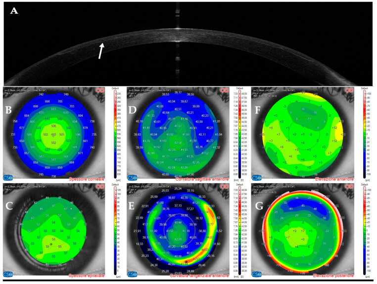 Figure 1
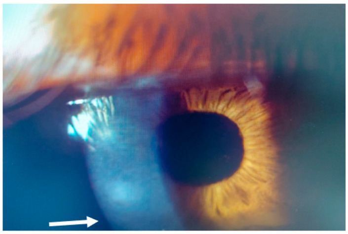 Figure 2
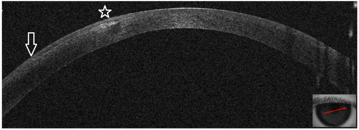 Figure 3
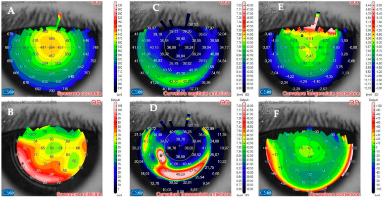 Figure 4
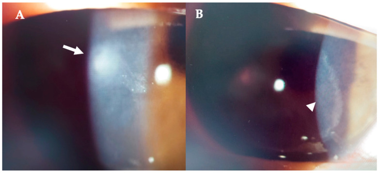 Figure 5
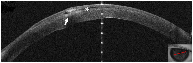 Figure 6
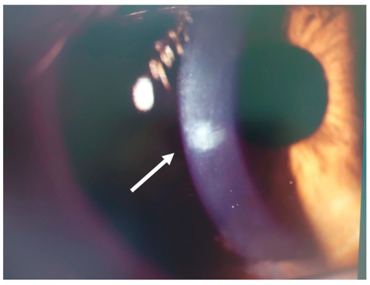 Figure 7
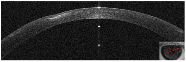 Figure 8
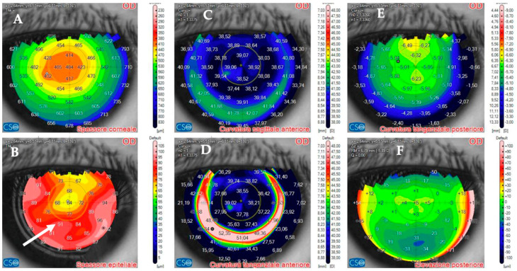 Figure 9
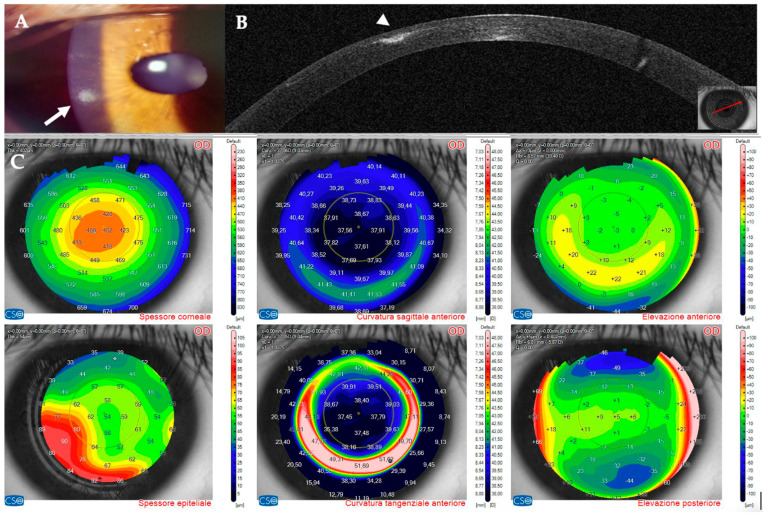 Figure 10
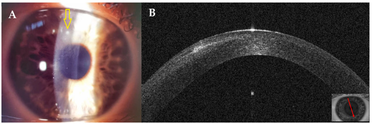 Figure 11
 Figure 12
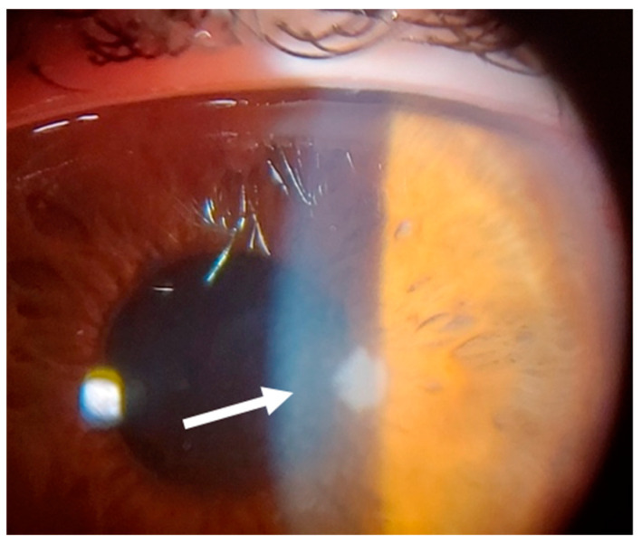 Figure 13
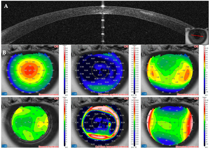 Figure 14
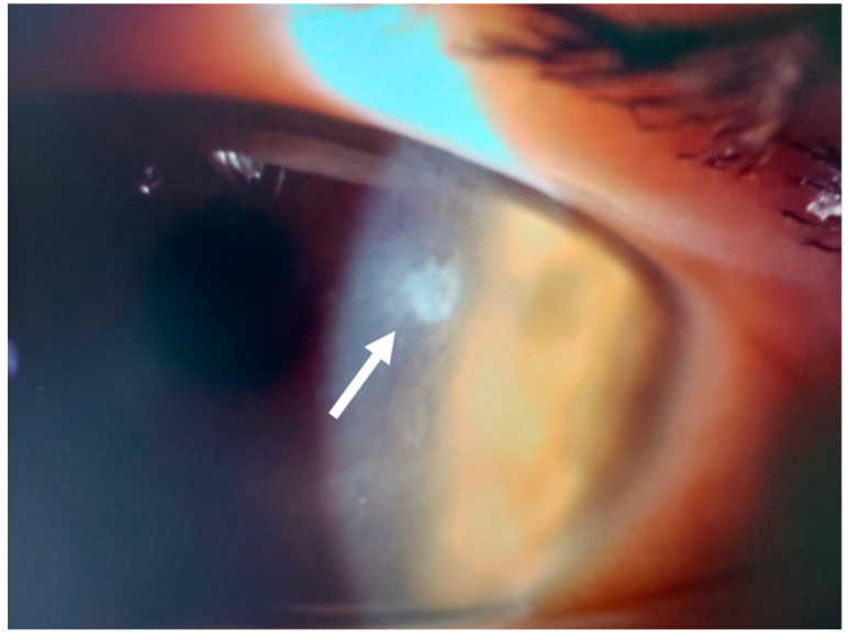 Figure 15
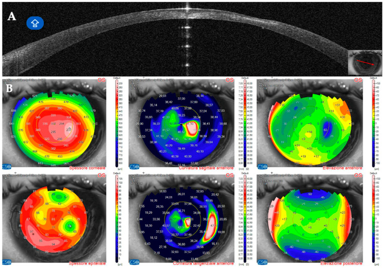 Figure 16
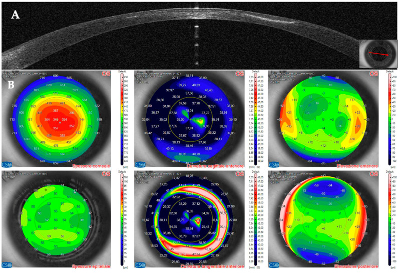 Figure 17
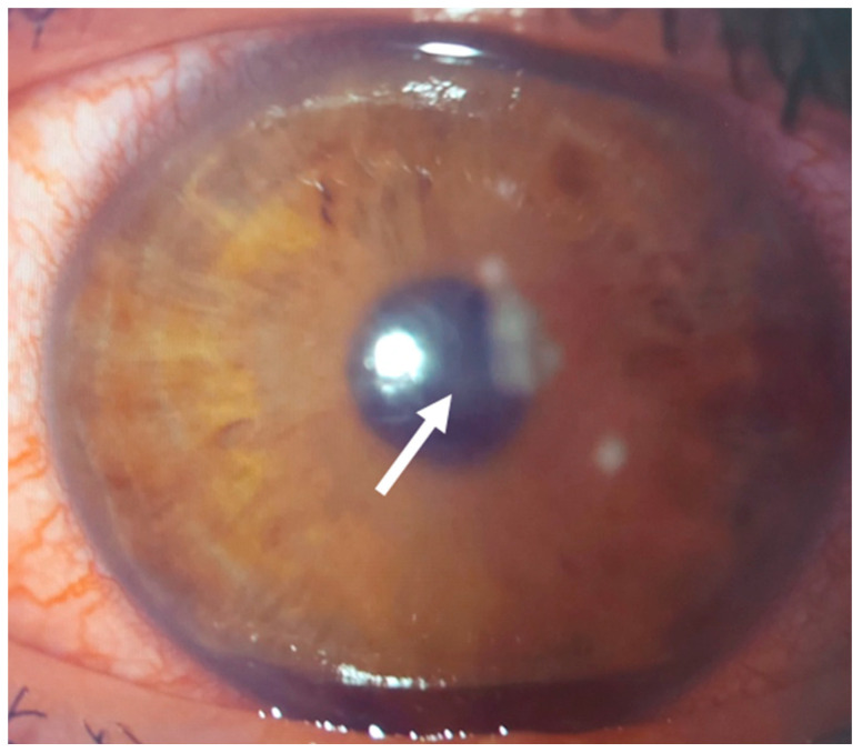 Figure 18
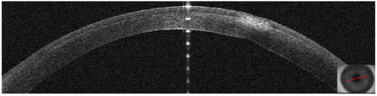 Figure 19
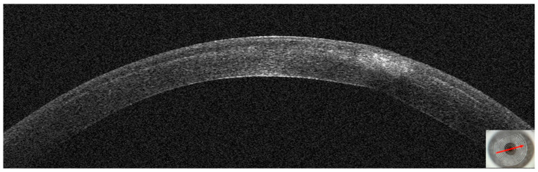 Figure 20
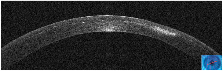 Figure 21
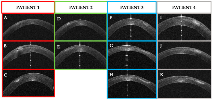 Figure 22
Peer Reviews
No public reviews on file for this paper yet. If you reviewed it on a platform where reviews are public (OpenReview, ICLR, NeurIPS, ICML), you can paste yours below so the community can read it here.
Videos
No videos yet. Explain this paper in a talk, walkthrough, or lecture? Add one.
Taxonomy
TopicsOcular Infections and Treatments · Corneal surgery and disorders · Ocular Diseases and Behçet’s Syndrome