Young Woman With Right Lower Quadrant Abdominal Pain
Gökhan Yılmaz

Abstract
Genes, proteins, chemicals, diseases, species, mutations and cell lines named across the full text — each resolved to its canonical identifier and authoritative record.
Click any figure to enlarge with its caption.
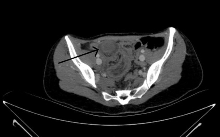 Figure 1
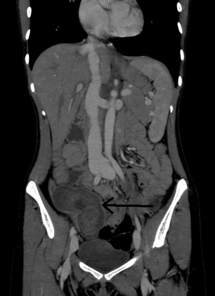 Figure 2
Peer Reviews
No public reviews on file for this paper yet. If you reviewed it on a platform where reviews are public (OpenReview, ICLR, NeurIPS, ICML), you can paste yours below so the community can read it here.
Videos
No videos yet. Explain this paper in a talk, walkthrough, or lecture? Add one.
Taxonomy
TopicsGastrointestinal disorders and treatments · Intestinal Malrotation and Obstruction Disorders · Hernia repair and management
Case Presentation
1
A healthy 26-year-old woman presented with right lower quadrant abdominal pain. The pain was accompanied by bloody diarrhea and vomiting. On examination, there was tenderness in the right lower quadrant. Strawberry jelly-colored stool was detected in her rectal examination. Laboratory tests were completely normal. In our patient's contrast-enhanced computed tomography, the appearance of a triple-layered structure was detected (Figs 1 and 2).Figure 1. Axial view of the 3-layered structure in the ileocecal region suggesting intussusception on the patient's computed tomography (ring structure at the tip of the black arrow).Figure 2. Coronal view of the 3-layered structure in the ileocecal region suggesting intussusception on the patient's computed tomography (ring structure at the tip of the black arrow).
Diagnosis: Intussusception
2
Intussusception is the invagination of one part of the intestine into another more distal.1 It is the most common cause of intestinal obstruction in infants. It usually occurs between 4 and 10 months.1 It can cause intestinal necrosis and death in children.1 Although intussusception is a common disease in infants, it is rare in adults.2 The presenting symptoms and signs are uncharacteristic. Therefore, the diagnosis may be missed in the emergency department.2 Intussusception is present in 1% of patients with intestinal obstruction.3 Although it is idiopathic in 90% of children, it is associated with 3/4 of the malignancies in adults.3 There was no underlying tumor in our patient. This made us think that it might be idiopathic.
Computed tomography with oral and intravenous contrast is the gold standard for intussusception diagnosis. Computed tomography typically shows a 3-layered structure that includes the compacted intestinal wall, its mesentery, and the surrounding intestine.4 This appearance was present in the computed tomography of our patient.
In conclusion, emergency physicians should consider the diagnosis of intussusception when they encounter the 3-layered structure on computed tomography.
Funding and Support
The author received no financial support for the research, authorship, and/or publication of this article.
Conflict of Interest
The author has affirmed he has no conflicts of interest to declare.
The reference list from the paper itself. Each links out to its DOI / PubMed record.
- 1Jiang J.Jiang B.Parashar U.Nguyen T.Bines J.Patel M.M.Childhood intussusception: a literature review P Lo S One 872013 e 6848210.1371/journal.pone.0068482 PMC 371879623894308 · doi ↗ · pubmed ↗
- 2Reiter J.J.Gai H.Ehrlich G.Die [Intestinal invagination in adulthood]Chirurg 55219841111136714006 · pubmed ↗
- 3Honjo H.Mike M.Kusanagi H.Kano N.Adult intussusception: a retrospective review World J Surg 39120151341382519284610.1007/s 00268-014-2759-9PMC 4273082 · doi ↗ · pubmed ↗
- 4Iko B.O.Teal J.S.Siram S.M.Chinwuba C.E.Roux V.J.Scott V.F.Computed tomography of adult colonic intussusception: clinical and experimental studies AJR Am J Roentgenol 14341984769772633248210.2214/ajr.143.4.769 · doi ↗ · pubmed ↗