Self-Resolving Pulsatile Frontal Mass Following Blunt Head Trauma
Yoshihiro Aoki, Koichi Hayakawa, Kazuhiko Suyama

Abstract
Genes, proteins, chemicals, diseases, species, mutations and cell lines named across the full text — each resolved to its canonical identifier and authoritative record.
Click any figure to enlarge with its caption.
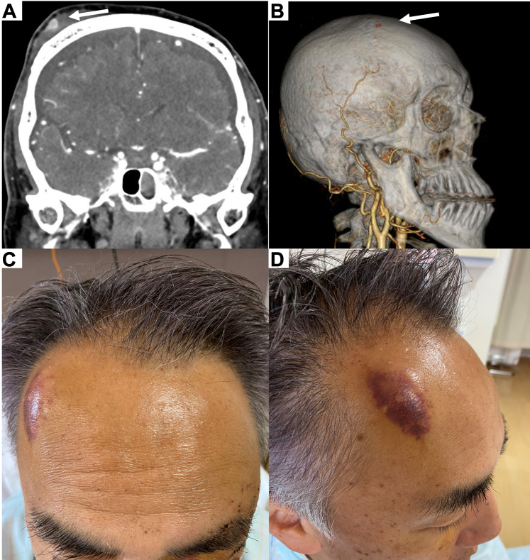 Figure 1
Peer Reviews
No public reviews on file for this paper yet. If you reviewed it on a platform where reviews are public (OpenReview, ICLR, NeurIPS, ICML), you can paste yours below so the community can read it here.
Videos
No videos yet. Explain this paper in a talk, walkthrough, or lecture? Add one.
Taxonomy
TopicsSpinal Fractures and Fixation Techniques · Spinal Hematomas and Complications · Spine and Intervertebral Disc Pathology
Case Presentation
1
A 58-year-old man was hospitalized following a traffic accident involving a light truck. He arrived febrile, SARS-CoV-2 antigen positive, and conscious without neurologic signs. Computed tomography of the head revealed traumatic subarachnoid hemorrhage, basilar skull fracture with pneumocephalus, left periorbital and zygomatic fractures, and multiple left rib fractures. Contrast-enhanced computed tomography showed a right frontal subcutaneous hemorrhage and a 9 mm pseudoaneurysm in the periphery of the frontal branch of the right superficial temporal artery (STA) without an adjacent convexity fracture (Fig A,B). He was admitted for blood pressure regulation, pain management, cerebral monitoring, and rehabilitation.FigureA, Contrast-enhanced computed tomography scan revealing a right frontal subcutaneous hemorrhage with a 9 mm pseudoaneurysm. B, Computed tomography angiography demonstrating a 9 mm pseudoaneurysm at the periphery of the frontal branch of the right superficial temporal artery. C and D, Clinical image of a pulsatile mass that appeared in the right frontal area on the day following admission.
Diagnosis: Traumatic Superficial Temporal Artery Pseudoaneurysm
2
The next day, a pulsatile mass appeared in the right frontal area (Fig C,D), not necessitating surgical intervention during the hospital stay. He was discharged on day 17, and his 2-week follow-up showed that the mass was pulseless and had resolved without surgery. Although the spontaneous resolution of a traumatic STA pseudoaneurysm has been reported following long-term observation, the gold standard is still considered to be surgical or radiologic intervention.1, 2, 3, 4 A rapidly developing closed-traumatic pseudoaneurysm in the periphery of the STA can resolve spontaneously, as demonstrated in our case.
Funding and Support
By JACEP Open policy, all authors are required to disclose any and all commercial, financial, and other relationships in any way related to the subject of this article as per ICMJE conflict of interest guidelines (see www.icmje.org). The authors have stated that no such relationships exist.
Conflict of Interest
All authors have affirmed they have no conflicts of interest to declare.
The reference list from the paper itself. Each links out to its DOI / PubMed record.
- 1Raskin J.Pak K.Lee M.K.Spontaneous resolution of superficial temporal artery pseudoaneurysm BMJ Case Rep 15112022 e 25174610.1136/bcr-2022-251746 PMC 971692536450415 · doi ↗ · pubmed ↗
- 2Ayling O.Martin A.Roche-Nagle G.Primary repair of a traumatic superficial temporal artery pseudoaneurysm: case report and literature review Vasc Endovascular Surg 484201434634810.1177/153857441351971224420058 · doi ↗ · pubmed ↗
- 3Derstine M.Doyle C.Traumatic pseudoaneurysm of the superficial temporal artery J Emerg Med 6142021 e 96e 9710.1016/j.jemermed.2021.07.00734364704 · doi ↗ · pubmed ↗
- 4Park D.H.Lee J.K.Baik B.S.Yang W.S.Kim S.Y.Traumatic hematoma-based pseudoaneurysm of the superficial temporal artery in a 7-year-old boy: a case report Arch Craniofac Surg 2412023323610.7181/acfs.2022.0070336858359 PMC 10009214 · doi ↗ · pubmed ↗