Evidence of Morphological and Morphometric Differences in the Sella Turcica of Pteronotus mesoamericanus and P. mexicanus
M. A. Peralta-Pérez, M. Briones-Salas

TL;DR
This study finds morphological and size differences in the skulls of two bat species from different regions of Mexico, suggesting possible evolutionary divergence.
Contribution
The study provides new morphometric evidence of cranial differences between Pteronotus mesoamericanus and P. mexicanus populations.
Findings
Ten measurements of the sella turcica showed significant differences between Gulf of Mexico and Pacific Coast bat populations.
The dorsum sellae has a truncated pyramidal shape with notable variation in dimensions between the two species.
The study suggests these cranial differences could help distinguish lineages in related bat species across the Americas.
Abstract
Minor modifications in the structure of living beings are signs of the gradual formation of new species. In addition, studying these changes gives us scientific tools, for example, to know whether the species of one region are different from those of another area. In this work, we aim to show these subtle differences in the cranial characteristics in the populations of bats with extensive distributions in Mexico. We now know that there are differences in the populations of Pteronotus mesoamericanus of the Gulf of Mexico and P. mexicanus of the Pacific. Morphological modifications are a potential mechanism for functional species and phylogenetic diversification. The sella turcica in mammals is a structure associated with the basisphenoid bone and serves as the receptacle for the pituitary gland; however, little is known about the morphological variation that may affect functionality in…
Genes, proteins, chemicals, diseases, species, mutations and cell lines named across the full text — each resolved to its canonical identifier and authoritative record.
Click any figure to enlarge with its caption.
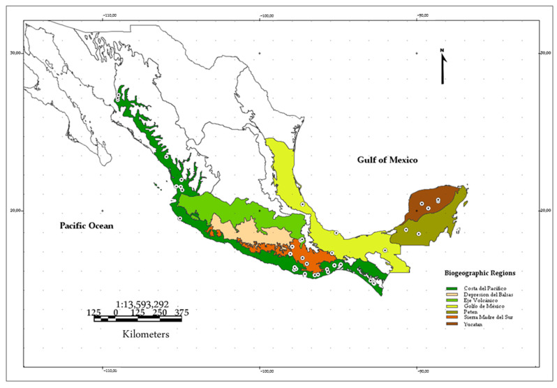 Figure 1
 Figure 2
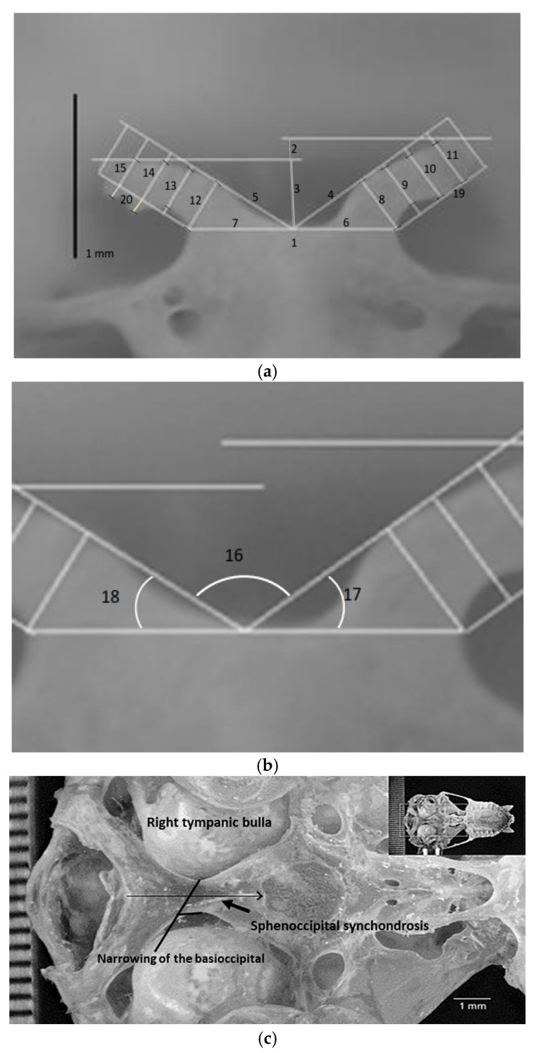 Figure 3
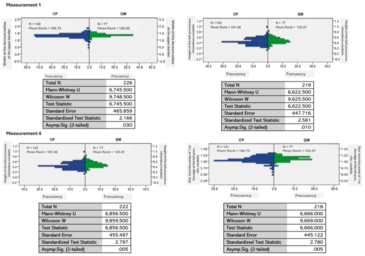 Figure 4
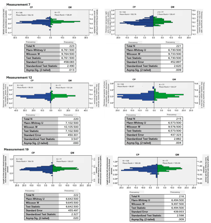 Figure 5
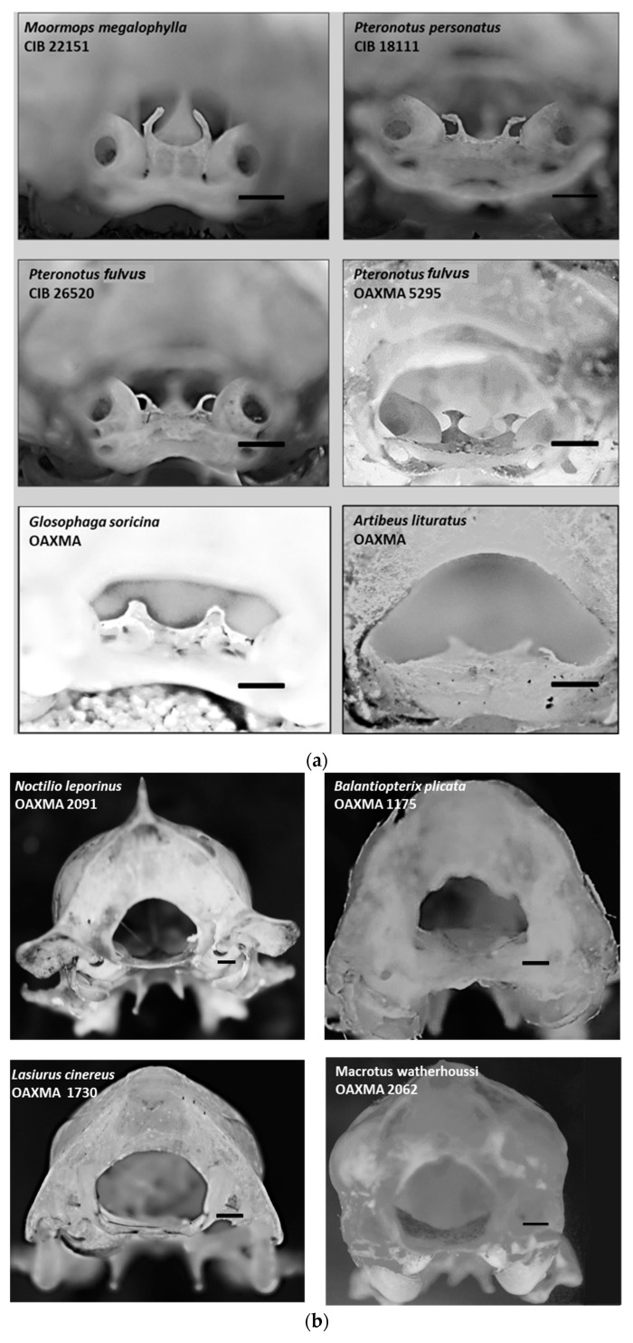 Figure 6
- —Research and Postgraduate Secretariat [SIP: 20220797] of the Instituto Politécnico Nacional
- —Consejo Nacional de Humanidades, Ciencias y Tecnologías [CONAHCYT]
Peer Reviews
No public reviews on file for this paper yet. If you reviewed it on a platform where reviews are public (OpenReview, ICLR, NeurIPS, ICML), you can paste yours below so the community can read it here.
Videos
No videos yet. Explain this paper in a talk, walkthrough, or lecture? Add one.
Taxonomy
TopicsPhysiological and biochemical adaptations · Bat Biology and Ecology Studies · Marine animal studies overview
1. Introduction
The taxonomy of mammals has developed around the morphology and morphometry of their skulls and, consequently, their phylogenetic relationships. Kinship is defined based on the shape and dimensions of the skull. It establishes correlations with the environment and possible preferences in dietary habits and other aspects. Furthermore, the skull is subject to phenotypic modifications due to natural selective interbreeding [1,2,3]. Therefore, modifications in morphological characteristics are a potential mechanism for the functional and phylogenetic diversification of species [4].
The sella turcica in mammals, a structure associated with the basisphenoid bone (os basisphenoidale), plays a significant role. Its variations, particularly in the shape and dimensions of the dorsum sellae, are of great interest. The anterior part presents the tubercle sellae, and continuing towards the middle part is the hypophyseal or pituitary fossa (fossa hypophysialis), which in a non-pathological state houses the pituitary gland. The posterior part contains the dorsum sellae, with the posterior clinoid processes (processus clinoideus caudalis) [5].
While the sella turcica’s role as the pituitary gland’s receptacle is known in humans, its functionality in chiropterans remains a mystery. This structure is known to undergo modifications in its shape according to various pituitary gland pathologies and modifications related to anatomical and dental conditions in humans [6]. Therefore, future studies should focus on unraveling the sella turcica’s functionality in chiropterans, particularly its potential relationship with the pituitary hormone levels in some bat species.
Pteronotus is widely distributed from western and southeastern Mexico, passing through Central America to Brazil. In Mexico, five species are within the genus: Pteronotus davyi, P. personatus, P. gymnonotus, P. mesoamericanus, and P. mexicanus, with the last two species considered subspecies of P. parnellii, P. p. mesoamericanus, and P. p. mexicanus [7]. Genetic studies show that there are different lineages of P. parnelli across its distribution range, and they propose that the subspecies mexicanus y mesoamericanus be recognized at the species level [8,9,10].
The identification of cryptic species, such as those in the genus Pteronotus, has been a significant achievement. These species, with their subtle differences, have been identified using molecular and genetic tools, bioacoustics, behavior, and ecological requirements [11,12,13]. Therefore, further evidence supporting the differentiation of these cryptic species will provide valuable insights into the evolutionary processes across the various populations within the distribution range of Pteronotus.
The variations in the shape and dimensions of the dorsum sellae in microchiropterans, particularly in P. mexicanus and mesoamericanus, and their relationship with the environment are crucial. These variations could potentially be applied to differentiating populations of the species, establishing the significance of these populations in evolutionary, ecological processes, and most importantly, conservation strategies for the species. This underscores the urgency and importance of our work in understanding and preserving these species.
This article aims to provide morphological and morphometric evidence of the differences among the cryptic species P. mexicanus and mesoamericanus. By describing the variations in the dimensions of the dorsum sellae and the processus clinoideus caudals of the sella turcica across different collection sites in Mexico, we hope to contribute significantly to the understanding of these species and their conservation.
2. Materials and Methods
A search for information was conducted without chronological restrictions of the specialized literature on the skulls of Pteronotus mexicanus, mesoamericanus, and other chiropterans to verify whether there are any records of variations in the structures described herein in national or international publications in Web of Science, Google Scholar, Latindex, and SciELO, using the following keywords in both Spanish and English: silla turca, sella turcica, morphology, cranial anatomy, and basicranial anatomy, all in combination with the terms bat, chiropteran, the species name, its synonyms, and the name of the family Mormoopidae.
2.1. Specimen Review
Twenty measurements designed for the sella turcica were taken from 243 skulls of P. mexicanus and mesoamericanus, including 105 male and 138 female specimens. The collection sites for these specimens cover most of the species distribution area in Mexico (Figure 1; Appendix A Table A1). Additionally, to verify whether a developed or at least distinguishable dorsum sellae exists and its shape in other bat species, the skulls of 25 other chiropteran species were examined (although no measurements of the dorsum were made for these species). This review included three species phylogenetically related to P. mexicanus and mesoamericanus: P. fulvus, P. psilotis, and Mormoops magalophylla.
The 243 specimens were organized by region within Mexico, resulting in 150 speci-mens from the Mexican Pacific region and 77 specimens from the Gulf of Mexico region. For this analysis, specimens from the Petén and Yucatán regions were included in the Gulf of Mexico region analysis. Specimens from the Sierra Madre del Sur and Eje Volcáni-co regions were excluded from the analysis due to sample size limitations. They have not yet undergone a genetic analysis that distinguishes them according to one of the two populations proposed by López-Wilchis et al. [8]. However, their measurement values are used to assign them to one of the two species.
The museum specimens analyzed are in two scientific collections that house significant numbers of specimens of the species: the Regional Collection of Mammals of the Interdisciplinary Center for Regional Research and Development, the Oaxaca Unit, of the National Polytechnic Institute (OAX.MA.026.0497), and the Collection of the Center for Biological Research of the Northeast, S.C. (CIBNOR).
2.2. Photographs
Images were taken using a CANON EOS REBEL XS^®^ camera with an RMS^®^ microscope objective adapter, to which an AmScope 4X Planachromatic^®^ objective was attached. The camera was mounted on a Zouminy^®^ micrometer screw stage and placed on a four-way sliding control rail for macrofocusing by Bewinner^®^, with all components fixed to a steel plate separated from the work surface by synthetic rubber to reduce vibration. Each P. mexicanus and mesoamericanus skull was mounted on a stem inside a lightbox for photography and positioned in a front-occipital view towards the objective lens, focusing the interior through the foramen magnum in orthogonal projection (Figure 2). The anatomical terminology used to describe the structure followed the Veterinary Anatomical Nomenclature [14].
2.3. Measurements
Ad hoc measurements were to be reproducible for the dorsum sellae and the processus clinoideus caudalis. The measurements were taken using ImageJ software, version 1.53k [15], with a scale attached to the image. A diagram of the 15 measured lengths and three angles, along with their descriptions, is shown in Figure 3, Table 1.
A Mann–Whitney U test analysis was applied using SPSS V.21 [16] to identify differences between the distributions of the Pacific and Gulf of Mexico populations.
3. Results
3.1. General Description
The morphological variation in the sella turcica in P. mexicanus and mesoamericanus is a comprehensive study, present in all 243 skulls examined and measured. It starts from the floor of the skull, formed by the basioccipital bone, which is generally broad in mammals and is between the tympanic bullae. Uniquely, the basioccipital bone in the species under study narrows forward (Figure 3c) until it is as narrow as less than half of the proximal end to the foramen magnum.
The described structure is in the occipital view through the foramen magnum. The dorsum of the sella and its caudal clinoid processes are in the spheno-occipital joint. From the occipital view, the sella turcica is by the auditory bullae. In the ventral view, the sella turcica is after the basioccipital fossa in an anteroposterior direction (Figure 2).
Regarding the skulls reviewed and photographed from other bat species, in addition to P. mexicanus and mesoamericanus, only members of its family [Mormoopidae] exhibited conspicuous dorsum sellae and processus clinoideus caudalis. In contrast, two species from the Phyllostomidae family, Artibeus lituratus [frugivore] and Glossophaga soricina [nectarivore/pollinivore], have small protrusions that are barely perceptible under a stereoscopic microscope (Table 2). The dorsum sellae is truncated pyramidal, with the processus clinoideus caudalis at the pyramid’s apex.
3.2. Morphometry
In P. mexicanus and mesoamericanus, spheno-occipital synchondrosis is present. Therefore, the dorsum sellae is at an average distance of 3.4 mm [ranging from 3.25 to 3.5 mm, with a standard deviation of 0.12 mm] from the lower border of the foramen magnum (Figure 3c and Table 3).
A Mann–Whitney U test for independent samples was conducted at an asymptotic significance level of 0.05. This test rejected the null hypothesis in ten measurements taken from the sella turcica, indicating significant differences between the samples from the biogeographic zones known as the Pacific Coast and the Gulf of Mexico [Figure 4, Appendix A Table A2].
Regarding the specimens from Sierra Madre del Sur and the Volcanic Axis, a visual comparison of their measurements versus the average, maximum, and minimum values of each of the regions analyzed achieved the definition that one is within the intervals of the Pacific Coast region with 15 measurements that correspond to the species Pteronotus mexicanus [measurements 2, 3, 4, 8, 9, and 11–20]; two specimens with 12 measurements are within the intervals of P. mesoamericanus [measurements 1,2, 3, 4, 6, 7, 8, 11, 12, 17, 19, and 20]; and finally, the remaining specimens were not defined as belonging to either of the two species.
4. Discussion
Regarding the literature review of books and scientific journals, there were no records concerning the structural variation in the sella turcica as described here for the genera of the Mormopidae family. According to Velazco’s [17] review of the sella turcica in other bat species, Carollia subrufa, Sturnira erythromos, and Uroderma magnirostrum do not possess either the dorsum or these processes. In contrast, the genera Platyrrhinus and Vampyrodes present small, almost imperceptible clinoid processes; however, the dorsum sellae is small and undetectable. There are no measurements in the cited work.
Based on the specimens reviewed, only in the Mormoopidae family does the sella turcica possess an elevated dorsum sellae above the floor of the skull with prominent clinoid processes. The posterior shape of the sella turcica is evident to the observer, much like in other mammals [e.g., Carnivora]. In contrast, in the other bat genera reviewed, the dorsum sellae of the sella turcica is completely reduced. In some cases, only discrete protuberances are discernible, with the area of the spheno-occipital synchondrosis where the sella turcica is being practically smooth [Table 2 and Figure 5a,b].
The description of the basisphenoid provided by Simmons and his collaborators [18] indicates that this bone narrows to one-third of the width of the foramen magnum, the dorsum of the sella turcica being at that narrowest site of the basisphenoid. It is likely that the growth of tympanic bullae, mainly located deep within the skull of the family Mormoopidae, causes the narrowing of the width of the basilesphenoid.
From this sample, we can affirm that the presence of the dorsum of the sella turcica in the Mormoopidae family is conspicuous, and its morphology is typical of any mammal. In Pteronotus mexicanus and mesoamericanus, there are various shapes across all populations, and their dimensions vary among populations. Notably, the populations of the Pacific region and those from the Gulf of Mexico region show apparent differences. According to the hypothesis of the family origin, both populations stem from groups that migrated along different routes to the continent from the Antilles. Therefore, this structure should be like the species of this family in the Antilles.
The dorsum of the sella turcica and the clinoid processes in P. mexicanus and mesoamericanus show a wide range of dimensions in the recorded localities in Mexico, where each region has very different environmental conditions. The growth and shape of the sella turcica dorsum may result from pituitary-level responses and, thus, adaptations to the various climates, types of vegetation, and diets of this species within its distribution range in the country. This is supported by the location of the sella turcica in the sellar region. This small area also includes the cavernous sinus and the suprasellar cistern. It houses the pituitary gland, making this region crucial for neurovascular processes and endocrine and optical functions [19].
While our study suggests a potential sympatry zone between P. mexicanus and mesoamericanus, indicated by the specimens from localities in the Sierra Madre del Sur and the Volcanic Axis, it is important to note that this finding is subject to the reservations that the deficiencies of the sample impose. These reservations are crucial to consider for the credibility of our research.
The hormonal activity in the sella turcica area is intense, as it contains the pituitary gland. The posterior lobe of the pituitary is mainly regulated by neurohypophysis, which has axonal projections from hypothalamic neurons [pituicytes], and their endings, known as Herring bodies, serve as hormone reservoirs, such as the antidiuretic hormone [ADH] and oxytocin. This activity is related to changes in the shape and dimensions of the various components of the sella turcica [20,21,22]. Therefore, we consider it important to investigate the hormonal linkage of the shape of the sella turcica in the Mormoopidae family.
In humans, the dimensions of the sella turcica, including its length and diameter, increase with age. This growth rate slows in preschool and then increases considerably during puberty [23]. Afterward, it decreases and eventually ceases at the onset of adulthood. Therefore, the different shapes and dimensions recorded in the collection localities may be related to the ontogenetic development of the mammal species studied here.
This characteristic, consistently present in the skulls of terrestrial mammals but not in flying ones, could be considered a plesiomorphic trait in both species and the Mormoopidae family, potentially signaling their isolation in the past on the Caribbean islands [8].
The measurements for the dorsum of the sella turcica in P. mexicanus and mesoamericanus were explicitly used for this study. For comparative purposes, these measurements should be used in future studies of Mormoopidae species populations from the Antilles, Central America, and South America.
5. Conclusions
This pioneering work unveils, for the first time, the unique characteristics of the dorsum sellae in the skull of a microchiroptera.
The data gathered here not only confirm the existence of differences in the measurements of the dorsum sellae of the populations of the Gulf and Pacific regions in Mexico but also provide support for the division of P. parnelli into two species: P. mesoamericanus [the Gulf region] and P. mexicanus [the Pacific region]. It is crucial to bear in mind the limitations of the sample when interpreting the findings of this study.
The reference list from the paper itself. Each links out to its DOI / PubMed record.
- 1Evin A. Baylac M. Ruedi M. Mucedda M. Pons J.-M. Taxonomy, skull diversity and evolution in a species complex of Myotis Chiroptera:Vespertilionidae: A geometric morphometric appraisal Biol. J. Linn. Soc.20089552953810.1111/j.1095-8312.2008.01076.x · doi ↗
- 2Noftz L.A. Calede J.J.M. Multivariate analyses of skull morphology inform the taxonomy and evolution of geomyoid rodents Curr. Zool.20236945647410.1093/cz/zoac 05537614926 PMC 10443661 · doi ↗ · pubmed ↗
- 3Lala K. Understanding niche construction and phenotypic plasticity as causes of natural selection Paleontology 20246711010.1111/pala.12719 · doi ↗
- 4Stanchack K.E. Arbour J.H. Santana S.E. Anatomical diversification of a skeletal novelty in bat feet Evolution 2019731591160310.1111/evo.1378631206628 · doi ↗ · pubmed ↗
- 5Infante Contreras C. Fundamentos para la Evaluación del Crecimiento, Desarrollo y Función Craneofacial Universidad Nacional de Colombia Bogotá, Colombia 2009 Available online: https://repositorio.unal.edu.co/bitstream/handle/unal/2386/9789584442864.08.pdf(accessed on 9 July 2023)
- 6Magat S.G. Sener O. Morphometric analysis of the sella turcica in Turkish individuals with different dentofacial skeletal patterns Folia Morphol.20187754355010.5603/FM.a 2018.002229500897 · doi ↗ · pubmed ↗
- 7Ramírez-Pulido J. González-Ruiz N. Gardner A.L. Arroyo-Cabrales J. List of Recent Land Mammals of Mexico Biodiversity Heritage Library Washington, DC, USA 2014 Volume 63169
- 8López-Wilchis R. Flores-Romero M. Guevara-Chumacero L.M. Serrato-Díaz A. Díaz-Larrea J. Salgado-Mejia F. Ibañez C. Salles L.O. Juste J. Evolutionary scenarios associated with the Pteronotus parnellii cryptic species-complex (Chiroptera: Mormoopidae)Acta Chiropterologica 2016189111610.3161/15081109 ACC 2016.18.1.004 · doi ↗