Characterization of Normal and Degenerative Discovertebral Complexes Using Qualitative and Quantitative Magnetic Resonance Imaging at 4.7T: Longitudinal Evaluation of Immature and Mature Rats
Benjamin Dallaudière, Emeline J. Ribot, Aurélien J. Trotier, Laurence Dallet, Olivier Thibaudeau, Sylvain Miraux, Olivier Hauger

TL;DR
This study uses advanced MRI techniques to visualize and monitor spinal disc degeneration in rats, showing how these methods can distinguish healthy from damaged discs.
Contribution
The study introduces a comprehensive MRI protocol using 3D-UTE and T1/T2 mapping to evaluate spinal disc degeneration in rats.
Findings
3D-UTE imaging successfully visualized normal and degenerative disc anatomy in rats.
T2 and T1 mapping showed significant changes in disc tissue post-surgery, confirming degeneration.
MRI findings were consistent with histological results, validating the imaging approach.
Abstract
Purpose: We assessed the feasibility of qualitative, semiquantitative, and multiparametric quantitative magnetic resonance imaging (MRI) using a three-dimensional (3D) ultrashort echo time (3D-UTE) sequence together with 2D-T2 and 3D-T1 mapping sequences to evaluate normal and pathological discovertebral complexes (DVCs). We assessed the disc (nucleus pulposus [NP] and annulus fibrosus [AF]), vertebral endplate (cartilage endplate [CEP] and growth plate [GP]), and subchondral bone (SB) using a rat model of degenerative disc disease (DDD). We also assessed whether this complete MRI cartography can improve the monitoring of DDD. Methods: DDD was induced by percutaneous disc trituration and collagenase injection of the tail. Then, the animals were imaged at 4.7T. The adjacent disc served as the control. The MRI protocol was performed at baseline and each week (W) postoperatively for 2…
Genes, proteins, chemicals, diseases, species, mutations and cell lines named across the full text — each resolved to its canonical identifier and authoritative record.
Click any figure to enlarge with its caption.
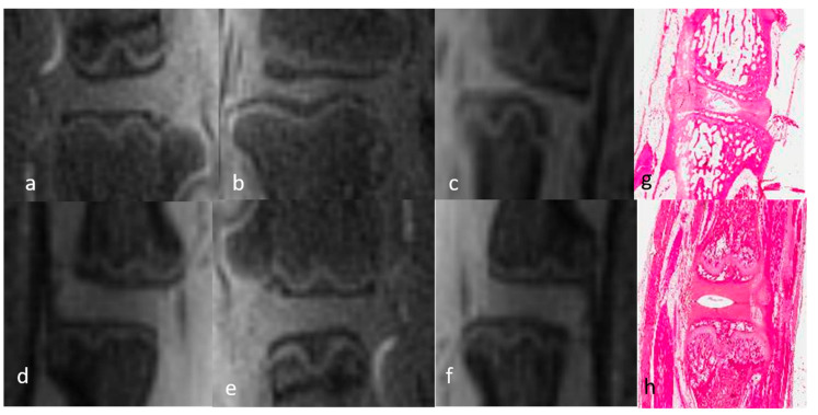 Figure 1
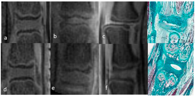 Figure 2
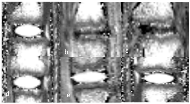 Figure 3
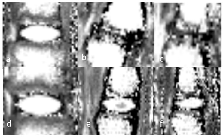 Figure 4
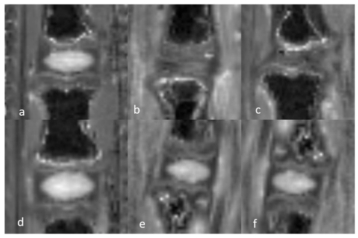 Figure 5
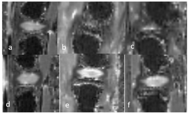 Figure 6
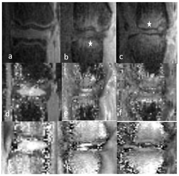 Figure 7
Peer Reviews
No public reviews on file for this paper yet. If you reviewed it on a platform where reviews are public (OpenReview, ICLR, NeurIPS, ICML), you can paste yours below so the community can read it here.
Videos
No videos yet. Explain this paper in a talk, walkthrough, or lecture? Add one.
Taxonomy
TopicsSpine and Intervertebral Disc Pathology · Medical Imaging and Analysis · Spinal Fractures and Fixation Techniques