Synthesis, structure, ionochromic and cytotoxic properties of new 2-(indolin-2-yl)-1,3-tropolones
Yurii A Sayapin, Eugeny A Gusakov, Inna O Tupaeva, Alexander D Dubonosov, Igor V Dorogan, Valery V Tkachev, Anna S Goncharova, Gennady V Shilov, Natalia S Kuznetsova, Svetlana Y Filippova, Tatyana A Krasnikova, Yanis A Boumber, Alexey Y Maksimov, Sergey M Aldoshin

TL;DR
This paper reports the synthesis and properties of new 1,3-tropolone compounds with ionochromic and cytotoxic effects.
Contribution
The study introduces new 2-(indolin-2-yl)-1,3-tropolones with dual ion-sensing and anticancer potential.
Findings
The compounds show emission switching in response to CN− and Hg2+ ions.
One compound shows high cytotoxic activity against A431 and H1299 cancer cell lines.
X-ray analysis confirmed the structure of a specific tropolone derivative.
Abstract
The acid-catalyzed reaction of benzo[e(g)] derivatives of 2,3,3-trimethylindolenines with o-chloranil leads to new 2-(benzo[e(g)]indolin-2-yl)-5,6,7-trichloro-1,3-tropolones and 2-(benzo[e(g)]indolin-2-yl)-4,5,6,7-tetrachloro-1,3-tropolones. Based on the results of PBE0/6-311+G(d,p) calculations, the structural and energetic characteristics of the tautomeric forms of the obtained 1,3-tropolones were determined. The structure of 2-(3,3-dimethyl-3H-benzo[g]indolin-2-yl)-5,6,7-trichloro-1,3-tropolone was determined by X-ray diffraction analysis. The compounds obtained are capable of switching emission at 420–440 nm and 476–530 nm upon successive exposure to CN− and Hg2+ ions in an acetonitrile solution. 2-(1,1-Dimethyl-1H-benzo[e]indolin-2-yl)-5,6,7-trichloro-1,3-tropolone exhibited high in vitro cytotoxic activity against A431 skin cancer and H1299 lung cancer cell lines.
Genes, proteins, chemicals, diseases, species, mutations and cell lines named across the full text — each resolved to its canonical identifier and authoritative record.
Click any figure to enlarge with its caption.
 Scheme 1
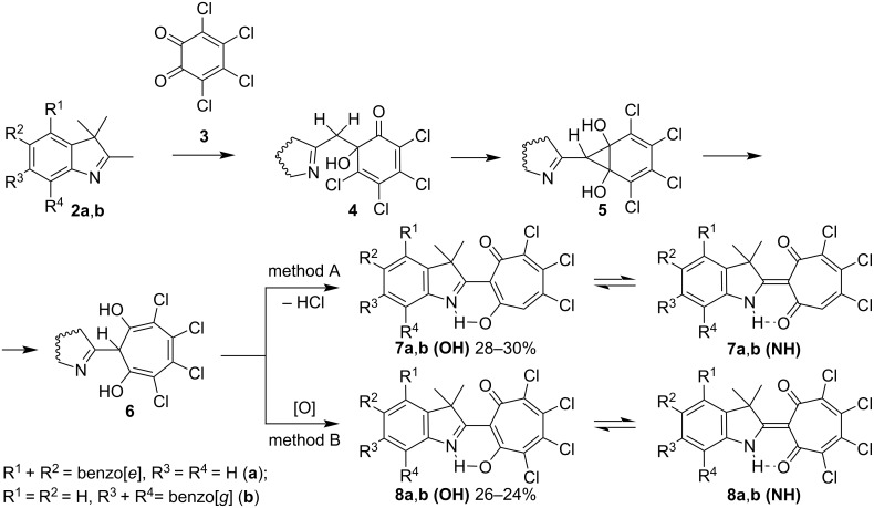 Scheme 2
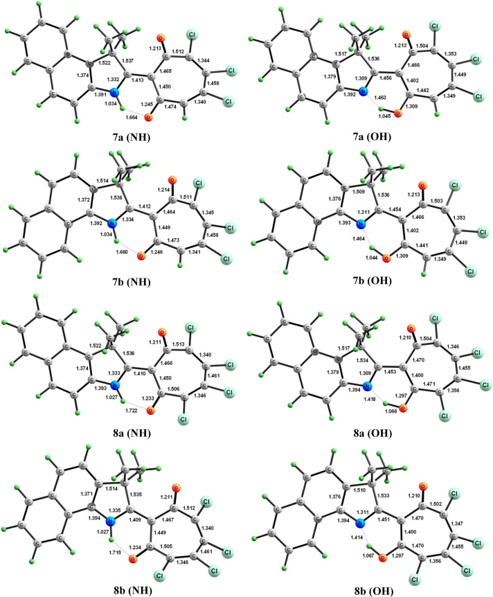 Figure 1
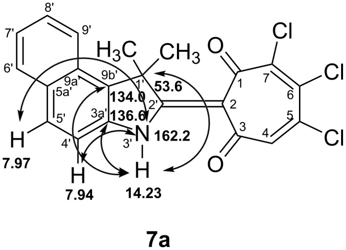 Figure 2
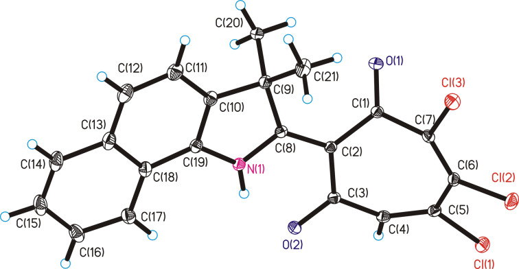 Figure 3
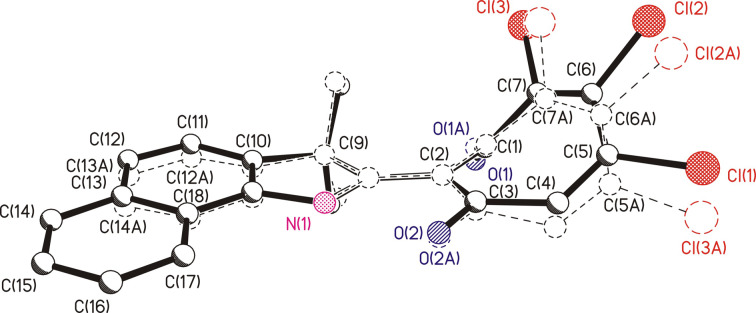 Figure 4
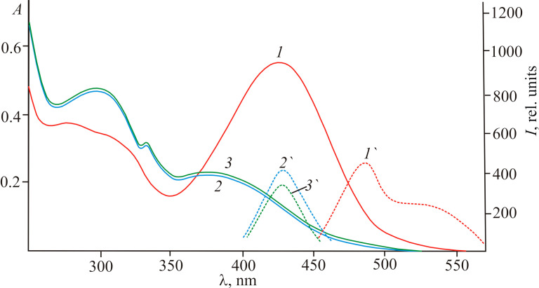 Figure 5
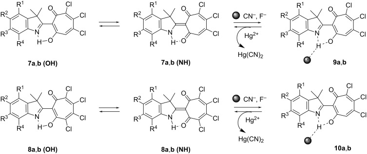 Scheme 3
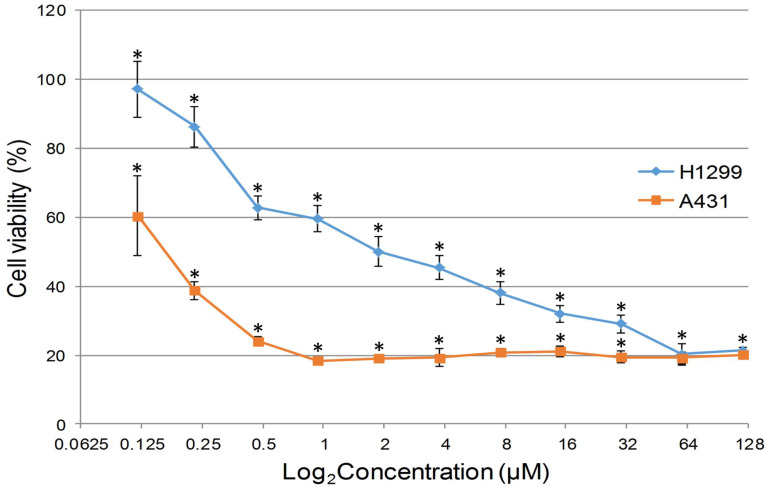 Figure 6
| compound | Δ | Δ | ||
|
| ||||
| −2392.806710 | 0 | −2392.818223 | 0 | |
| −2392.803386 | 2.1 | −2392.812918 | 3.3 | |
| −2392.809986 | 0 | −2392.821048 | 0 | |
| −2392.806819 | 2.0 | −2392.816317 | 3.0 | |
| −2852.269500 | 0 | −2852.281433 | 0 | |
| −2852.263504 | 3.8 | – | – | |
| −2852.272654 | 0 | −2852.284199 | 0 | |
| −2852.266797 | 3.7 | – | – | |
| compound | absorption, λmax, nm | fluorescence, λmax, nm | Stokes shift, |
|
| |||
|
| 276 (17200), 300 (18400), 427 (20400) | 485 (260), 520 (180) | 2800, 4200 |
|
| 292 (14000), 432 (19100) | 495 (220), 523 (190) | 2950, 4050 |
|
| 300 (14800), 310 (15200), 418 (20000) | 476 (420), 520 (190) | 2900, 4700 |
|
| 300 (14400), 425 (21900) | 491(480), 530 (250) | 3150, 4650 |
Peer Reviews
No public reviews on file for this paper yet. If you reviewed it on a platform where reviews are public (OpenReview, ICLR, NeurIPS, ICML), you can paste yours below so the community can read it here.
Videos
No videos yet. Explain this paper in a talk, walkthrough, or lecture? Add one.
Taxonomy
TopicsSynthesis of Organic Compounds · Multicomponent Synthesis of Heterocycles · Synthesis of Indole Derivatives
Introduction
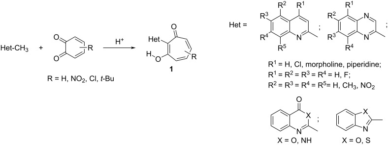
1,2-Benzoquinones represent unique building blocks for various classes of heterocyclic systems. Their structure depends on the reactivity of the initial heterocycles and 1,2-benzoquinones, as well as on the reaction conditions. In the series of 2-methylquinolines [1], 2-methylquinoxalines [2], 2-methylquinazolinones [3], 2-methylbenzoxazinones [4], and 2-methylbenzoxa(thia)zoles [5] the interaction with sterically hindered 1,2-benzoquinones and 3,4,5,6-tetrachloro-1,2-benzoquinone proceeds with the expansion of the o-quinone ring and results in 2-hetaryl-substituted 1,3-tropolones 1 (Scheme 1), which exhibit antibacterial [4] and cytotoxic activity [6–7].
Synthesis of 2-hetaryl-substituted 1,3-tropolones 1.
Existing approaches to the synthesis of 1,3-tropolone derivatives have a number of drawbacks, namely, limited synthesis methodology and low yields of the target compounds [8–15]. Over the past decade, only a few reports have been published [16–17].
Indole derivatives, including those with conjugated aromatic and alicyclic rings, are of considerable interest because of their diverse biological activities (antibacterial, antimicrobial, anticancer, antidiabetic, etc.) [18–22]. For this reason, the synthetic goal of this work is to obtain heterocyclic indole compounds conjugated with a 1,3-tropolone moiety.
The variability of the products of acid-catalyzed reactions in the series of 2,3,3-trimethylindolenine with 1,2-benzoquinone derivatives depends on the nature of the substituents in the 1,2-benzoquinone. Thus, the interaction of 2,3,3-trimethylindolenine with 3,5-di(tert-butyl)-1,2-benzoquinone leads to the formation of indolo[1,2-a]indoline derivatives [23], while the presence of a nitro group in 4,6-di(tert-butyl)-3-nitro-1,2-benzoquinone the reaction with 2,3,3-trimethylindolenine leads to an o-quinone ring contraction and the formation of 2-azabicyclic products and pyridino[1,2-a]benzo[e]indol-10,11-diones [24]. o-Chloranil was found to be the most efficient 1,2-benzoquinone, which engages in o-quinone ring-expansion reactions with 2,3,3-trimethylindolenines to form hard-to-reach polychlorinated derivatives of 2-(indolin-2-yl)-1,3-tropolones [5]. Unlike the cross-aldol reaction of o-chloranil with methyl ketones [13–15], which is always accompanied by the removal of one of the chlorine atoms from the seven-membered ring, the acid-catalyzed reaction between methylene-active heterocyclic compounds and o-chloranil depends on the reaction conditions and can proceed with or without the inclusion of a dehydrochlorination stage and leads to 5,6,7-trichloro- or 4,5,6,7-tetrachlorotropones, respectively [5,25].
The present work reports the synthesis of new 2-(indolin-2-yl)-1,3-tropolones by the reaction of o-chloranil with benzoannelated derivatives of 2,3,3-trimethylindolenine, a comprehensive evaluation of the structure of the compounds obtained using quantum chemical methods, X-ray diffraction analysis, two-dimensional correlation NMR spectroscopy, as well as investigation of their ionochromic properties towards CN^−^ and Hg^2+^ ions and evaluation of in vitro cytotoxic activity against A431 skin cancer and H1299 lung cancer cell lines.
Results and Discussion
We found that boiling of equimolar amounts of benzannelated 2,3,3-trimethylindolenines 2a,b and o-chloranil (3) (method A) in dioxane leads to the formation of trichlorosubstituted 1,3-tropolones 7a,b as the main reaction products (Scheme 2). Heating 2a,b in acetic acid with a two-fold excess of o-chloranil (3) (method B) is accompanied by the formation of tetrachlorosubstituted 1,3-tropolones 8a,b (Scheme 2 and Supporting Information File 1).
Synthesis of 1,3-tropolones 7a,b and 8a,b. Reagents and conditions: method A: dioxane, reflux; method B: AcOH, 40–50 °C.
As shown in Scheme 2, in the initial step, the aldol condensation of 2,3,3-trimethylindolenine 2 with o-chloranil (3) leads to the intermediate compounds, 6-(2-hetarylmethylene)-6-hydroxy-2,4-cyclohexadien-1-ones 4. Such intermediates were isolated preparatively and structurally characterized in the reactions of 2-methylquinolines with 3,5-di-(tert-butyl)-1,2-benzoquinone [1] and benzophenones with o-chloranil [15]. The norcaradiene derivatives 5, formed in the next step by the intramolecular cyclization reaction of 4, undergo thermal isomerization into 2,3-dihydrotropolones 6. The formation of 2-(indolin-2-yl)-5,6,7-trichloro-1,3-tropolones 7a,b is accompanied by dehydrochlorination of 6 upon boiling in dioxane according to method A. One of the conditions for carrying out the reaction according to method B is the use of a two-fold excess of quinone 3. Oxidation of dihydrotropolones 6 with an excess of quinone 3 leads to the formation of 2-(indolin-2-yl)-4,5,6,7-tetrachloro-1,3-tropolones 8a,b as final products. The detailed reaction mechanism in acetic acid solution was studied by the PBE0/6-311+G(d,p) method on the example of the interaction of 2-methylquinolines and 2-methylbenzazoles with 1,2-benzoquinone and o-chloroanil, respectively [1,25].
The structures of compounds 7 and 8 obtained by methods A and B were confirmed by ^1^H NMR, IR spectroscopy and mass spectrometry (Supporting Information File 2). A distinctive feature of the ^1^H NMR spectra of 7 in CDCl_3_ is the signal of the tropolone ring proton, which appears at 6.9 ppm. A characteristic specificity of the ^1^H NMR spectra of compounds 7 and 8 is the presence of signals of hydroxy group protons forming a strong hydrogen bond with the indoline nitrogen atom, which closes the six-membered chelate cycle. These signals are observed in the weak field at 15.2–15.8 ppm for 7a,b and 14.3–14.8 ppm for 8a,b, respectively, as a broadened singlet peak.
A dynamic equilibrium of tautomeric forms 7, 8 (OH)–7, 8 (NH) exists in solution, which can be detected by the broadening of the hydroxy group proton signal in the ^1^H NMR spectrum (Scheme 2). The structures of tautomeric forms of 7a, 7b, 8a, 8b and their energy characteristics were calculated by PBE0/6-311+G(d,p) method in the gas phase and polar solvent (DMSO) (Table 1, Figure 1 and Supporting Information File 3). According to the obtained data, the (NH) isomers of 7 and 8 are thermodynamically more stable than the corresponding (OH) forms. Increasing the polarity of the medium additionally enhances their stabilization. The introduction of an additional acceptor substituent to the tropolone moiety has a similar effect. In addition, compared to compounds 7a,b, compounds 8a,b show a marked increase in the intramolecular hydrogen bond length NH···O of the (NH) isomers and a decrease in the magnitude of hydrogen bond N···HO of the (OH) forms (Figure 1). On the other hand, the position of the benzoannelated fragment does not significantly affect the structural and energetic parameters of the studied compounds.
Structural characteristics of (NH) and (OH) tautomeric forms of compounds 7 and 8 in the gas phase according to PBE0/6-311+G(d,p) calculations. Bond lengths are given in Ångstrom.
The difference in the energy characteristics of the (NH) and (OH) tautomeric forms of compounds 7a, 7b, 8a, and 8b in the polar solvent and the gas phase is small (2.0–3.8 kcal/mol), and probably the nature of the solvent can influence the shift of the equilibrium towards one of the tautomeric forms.
To reveal the predominance of either the (NH) or (OH) tautomeric form of compounds 7a, 7b, 8a, and 8b in solution depending on the nature of the solvent, we carried out a detailed study using compound 7a. A complete signal assignment of ^1^H and ^13^C NMR spectra was carried out, based on characteristic values of chemical shifts and cross-peak analysis in two-dimensional spectra of ^1^H,^1^H COSY correlations, as well as ^1^H,^13^C correlations HSQC, HMBC, and ^1^H,^15^N HMBC spectra in DMSO-d6 and CDCl_3_ (Supporting Information File 2).
In the ^1^H NMR spectrum of 7a in DMSO-d6, the signal of the weak field proton is shifted to a stronger field of 14.23 ppm and appears as a narrow singlet (Supporting Information File 2, Figure S17). Analysis of two-dimensional correlation spectra of heteronuclear NMR spectroscopy shows that compound 7a in DMSO-d6 solution exists in the (NH) tautomeric form. Thus, in the ^1^H,^15^N HMBC spectrum of 7a there are cross peaks of the indolenine nitrogen atom at 162.2 ppm with a weak field proton at δ_H_ 14.23 ppm, as well as aromatic protons H(4') and H(5') with δ_H_ 7.94 ppm and 7.97 ppm, respectively (Supporting Information File 2, Figure S19). In the two-dimensional ^1^H,^13^C HMBC spectrum of 7a, a correlation of the NH proton with the bridging carbon atoms C(3a'), C(9b'), and the quaternary carbon atom C(1') of the indolenine fragment is observed with δ_H_ 136.6 ppm, 134.0 ppm, and 53.6 ppm, respectively (Supporting Information File 2, Figure S20). The most important HMBC correlations for the analysis of structure 7a are schematically depicted in Figure 2.
Scheme of HMBC correlations of compound 7a in DMSO-d6.
At the same time, in the ^1^H,^15^N HMBC spectra of 7a in CDCl_3_, there are no cross-peaks of the proton in the region of δ_H_ 15.15 ppm with the nitrogen atom of the indolenine fragment at 162.7 ppm, and in the ^1^H,^13^C HMBC spectrum, no correlations of the down-field proton with the carbon atoms of the indolenine fragment are observed (Supporting Information File 2, Figures S21 and S22). Thus, in CDCl_3_, compound 7a is predominantly in the (OH) tautomeric form.
The structure of 2-(3,3-dimethyl-3H-benzo[g]indolin-2-yl)-5,6,7-trichloro-1,3-tropolone (7b) was established by X-ray diffraction analysis (Figure 3). The main distances and angles are summarized in Supporting Information File 3.
Molecular structure of 2-(3,3-dimethyl-3H-benzo[g]indolin-2-yl)-5,6,7-trichloro-1,3-tropolone (7b).
To compare the structural characteristics, we combined the molecule 7b (solid lines) and the previously obtained 2-(3,3-dimethylindolin-2-yl)-5,6,7-trichloro-1,3-tropolone [5] (dashed lines) at the positions of N(1), C(2) and C(9) atoms (hydrogen atoms removed) (Figure 4).
Result of matching structures of 7b (solid lines) and 2-(3,3-dimethylindolin-2-yl)-5,6,7-trichloro-1,3-tropolone (dashed lines) at the positions of N(1), C(2), and C(9) atoms.
While maintaining the general configuration of the molecular framework, there are minor differences in the details of their structures, manifested in the observed distances between the atoms C12)···C(13A) = 0.31 Å, Cl(2)···Cl(2A) = 0.76 Å and in the geometric parameters of the tropolone rings. The angle between the planes C(2)C(3)C(6)C(7) and C(1)C(2)C(7) is equal to 42.9° (42.1°) (here and below, when comparing geometric values, similar values are given in parentheses for the previously obtained 2-(indolin-2-yl)-1,3-tropolone)). The angle between the planes C(2)C(3)C(6)C(7) and (3)C(4)C(5)C(6) is equal to 27.6° (34°). In compound 7b, as in 2-(indolin-2-yl)-1,3-tropolone, an intramolecular hydrogen bond N(1)–H(1)···O(2) was realized with parameters: distances N–H = 0.86 (0.84) Å, H···O = 1.79 (1.83) Å, and N···O = 2.513 (2.518) Å, angle N–H–O 140.4° (138.0°).
The electronic absorption spectra of compounds 7a,b and 8a,b in acetonitrile have long wavelength bands in the region of 425–432 nm (Table 2). They exhibit dual-maxima fluorescence at 476–530 nm with normal and large Stokes shift values (Table 2, Figure 5).
Absorption and emission spectra of compound 8b in acetonitrile before (1,1’) (c 2.5 × 10⁻⁵ mol L–1) and after the addition of CN− (2,2’) and F− (3,3’) ions (c 5.0 × 10⁻⁵ mol L−1).
This is consistent with the above conclusion about the existence of tautomeric equilibrium 7,8 (OH)–7,8 (NH) in solutions based on NMR data and DFT quantum chemical calculations (Scheme 2). The emission with a larger Stokes shift appears to correspond to the 7,8 (OH) form and is caused by the excited-state intramolecular proton transfer (ESIPT) process due to intramolecular O→N proton migration in the singlet excited state [26–27].
The ionochromic sensitivity of compounds 7a,b and 8a,b to anions was investigated in acetonitrile upon addition of tetra-n-butylammonium salts (TBAX: F, Cl, Br, I, CN). Exclusively cyanide and fluoride anions lead to a naked-eye effect due to a change of the solution’s colour from yellow-orange to pale yellow (Figure 5). At the same time, a new fluorescence band appears at 420–440 nm. The Stokes shifts of fluorescence decrease to normal values of 1800–2000 cm^−1^, indicating a complete inhibition of the ESIPT process. These spectral transformations are usually caused by the formation of a strong N–H(O–H)···CN^−^ (F^−^) hydrogen bond up to deprotonation [28]. Using the principle of “relay recognition” [29], we investigated the sensitivity of in situ obtained complexes 9 and 10 with CN^−^ to different cations. It appeared that the addition of an equivalent amount of Hg(ClO_4_)2 to an acetonitrile solution selectively and completely restores the initial absorption and fluorescence spectra (Scheme 3).
Possible binding mode of 7 and 8 with CN− and F−.
Thus, the obtained compounds represent molecular switches of optical and fluorescent properties under sequential addition of CN^−^ and Hg^2+^ ions and the transformation cycle presented in Scheme 3 can be repeated at least 5–6 times.
The resulting compounds 7b and 8a,b have extremely low water solubility, which makes it impossible to prepare concentrations sufficient for studying cellular cytotoxicity. In this regard, 1,3-tropolone 7a, which has an acceptable level of solubility in water, was used for biological testing. The in vitro cytotoxic activity was investigated with A431 skin cancer and H1299 lung cancer cell lines. A standard MTT assay was used to determine the anticancer activity of compound 7a against these cultures (Figure 6). The viability test is an important method to determine which new compounds are able to target cancer cells. The results of this test (IC_50_ value ± 95% confidence interval) allow us to evaluate the possibility of carrying out subsequent stages of research and selecting promising compounds for the further development of anticancer drugs [30].
*Dose–response curves for H1299 and A431 cells treated with compound 7a for 24 h. Significant difference compared to control samples, p < 0.05.
The study showed that the IC_50_ inhibitory concentration of 2-(1,1-dimethyl-1H-benzo[e]indolin-2-yl)-5,6,7-trichloro-1,3-tropolone (7a) against the A431 skin cancer cell culture, at which the number of live cells was reduced by 50%, was 0.172 ± 0.029 μM. For the H1299 culture, this value was 2.18 ± 0.7 μM. In comparison, the IC_50_ value of the standard anticancer drug cisplatin at 24 hour incubation is 137 ± 12 μM for the A431 culture [31] and 34.9 μM for the H1299 culture [32], which is significantly higher than the values obtained for compound 7a.
Conclusion
In summary, new 2-(benzo[e(g)]indolin-2-yl)-5,6,7-trichloro-1,3-tropolones 7a,b and 2-(benzo[e(g)]indolin-2-yl)-4,5,6,7-tetrachloro-1,3-tropolones 8a,b were obtained by acid-catalyzed reactions of benzo[e(g)] derivatives of 2,3,3-trimethylindolenines with o-chloranil. The energetic characteristics of the tautomeric forms of 1,3-tropolones 7 and 8 were determined by PBE0/6-3-311+G(d,p) calculations, and the structure of 2-(3,3-dimethyl-3H-benzo[g]indolin-2-yl)-5,6,7-trichloro-1,3-tropolone (7b) was established by X-ray diffraction analysis. Using two-dimensional correlation NMR spectroscopy methods, it was shown that the nature of the solvent significantly affects the equilibrium of the tautomeric forms of the 1,3-tropolones. The obtained compounds represent molecular switches of optical and fluorescent properties under sequential addition of CN^−^ and Hg^2+^ ions. 2-(1,1-Dimethyl-1H-benzo[e]indolin-2-yl)-5,6,7-trichloro-1,3-tropolone (7a) was found to exhibit high in vitro cytotoxic activity against A431 skin cancer and H1299 lung cancer cell lines. In addition, the IC_50_ values of compound 7a are significantly lower than the IC_50_ values of cisplastin, which is widely used in the therapy of tumors, including lung cancer.
Experimental
General
^1^Н, ^13^С and ^15^N NMR spectra were obtained on the integrated analytical LC–SPE–NMR–MS system AVANCE-600 (Bruker) (600 MHz, ^1^Н; 150 MHz, ^13^С; 60 MHz, ^15^N) at 20 °C in CDCl_3_ and DMSO-d6. The signals were referenced to the signals of residual proton signals of corresponding deutero-solvents. IR spectra were recorded on a Varian Excalibur 3100 FTIR instrument using the attenuated total internal reflection technique. Mass spectra were obtained on a Finnigan Mat Incos 50 spectrometer. Electronic absorption spectra were obtained on a Varian Cary 100 spectrophotometer. Emission spectra were recorded on a Varian Cary Eclipse spectrofluorimeter. Acetonitrile of spectroscopic grade (Aldrich), previously purified by distillation, and tetra-n-butylammonium salts (TBAX: F, Cl, Br, I, CN) (Aldrich) were used to prepare the solutions. Spectral fluorescent experiments were performed using quartz cells (lcuvette 1.0 cm, volume V 2 mL). Stock solutions of compounds 7 or 8 (c 5.0 × 10^−5^ mol L^−1^) and tetrabutylammonium salts (c 1.0 × 10^−4^ mol L^−1^) in acetonitrile were used. The corresponding solution (1 mL) and the tetra-n-butylammonium salt (1 mL) solution were mixed directly in the cell and thoroughly stirred. Hence, the working concentrations of compounds 7 or 8 and anions was 2.5 × 10^−5^ mol L^−1^ and 5.0 × 10^−5^ mol L^−1^, respectively. All spectral experiments were performed at room temperature (23 °C). Chromatography was carried out on columns filled with Al_2_O_3_ of II–III degree of activity according to Brockmann. Melting points were determined on a Fisher-Johns melting point apparatus. 1,1,2-Trimethyl-1H-benzo[e]indolenine (2a, 98%, Alfa Aesar), 2,3,3-trimethyl-3H-benzo[g]indolenine (2b, 98%, Alfa Aesar) and tetrachloro-o-benzoquinone (o-chloranil, 3) (97%, ACROS organics) were used as the starting reagents. NMR and IR spectra were recorded on the equipment of the Center for Collective Usage “Molecular Spectroscopy” of Southern Federal University.
X-ray diffraction study
The unit cell parameters and reflection intensities for compound 7b (a three-dimensional set) were measured on an Xcalibur EOS autodiffractometer (Mo Kα irradiation, graphite monochromator, 150 K). Orange monoclinic crystals, chemical formula C_21_H_14_NO_2_Cl_3_, М = 418.68, a = 11.4480(3), b = 7.4883(2), с = 21.7720(6) Å, β = 102.590(3)°, V = 1821.55(9) Å^3^, Z = 4, ρ_calc_ = 1.527 g cm^−3^, μ(Мо Кα) = 0.520 mm^−1^, P2_1_/c space group. Intensities of 15441 reflections were measured in the reciprocal space (2θ ≤ 64.14°) using the ω/2θ-scanning method from a single crystal with dimensions of 0.31 × 0.25 × 0.20 mm. An empirical accounting of absorption was carried out using the Multiscan procedure. After exclusion of systematically cancelled reflections and averaging intensities of equivalent reflections, the working array of measured F^2^(hkl) and σ*(F*^2^) reflections contained 6352 independent reflections, 5089 of which with F^2^ > 4σ*(F*^2^). The structure was solved with the direct method and was refined by the full-matrix least-squares procedure (LSP) with respect to F^2^ in anisotropic approximation for non-hydrogen atoms (hydrogen atoms isotropic) using the SHELXTL program. In the crystal structure, most of the H atoms are localized in the Fourier synthesis of the difference electron density, then the coordinates and isotropic thermal parameters of all H atoms were calculated by the LSP using the “rider” model [33]. The absolute shifts of all 247 varied structure parameters were less than 0.001 σ, the final value of the factor R1 = 0.0385. The CIF file containing atomic coordinates, full tables of bond lengths, bond angles, and thermal parameters of compound 7b have been deposited at the Cambridge Crystallographic Data Centre (CCDC 2040907) and can be obtained upon request on the website: https://www.ccdc.cam.ac.uk/data_request/cif.
Computational methods
Quantum chemical calculations were performed using the Gaussian 09 software package [34] with hybrid functional PBE0 [35–36] and 6-311+G (d,p) basis set. Solvent effects were modeled by using polarizable continuum model (PCM) [37] within the integral equation formalism (IEFPCM) [38].
Biological experiments
The MTT colorimetric test for cell viability assessment is based on the reduction by NADPH-H-dependent cellular oxidoreductase enzymes of the tetrazolium dye 3-(4,5-dimethylthiazol-2-yl)-2,5-diphenyltetrazolium bromide, which has yellow color, into violet-blue formazan, with absorption maximum in the range of 540–560 nm. The optical density of the solution in this wavelength range is an indirect indicator of the number of live cells in the culture. The cytotoxic activity of the test substance was determined by the decrease in optical density of experimental samples compared to control samples [39].
A431 and H1299 cells were seeded into 96-well plates at 15000/well in DMEM medium supplemented with 10% EFV and cultured under standard conditions at 5.0% CO_2_ and 37 °C. The next day, the medium was replaced and test compound 7a was added in a series of two-fold dilutions from 0.12 μM to 120 μM. In the control wells, the medium was replaced without adding the compound 7a. The cells were then incubated under the same standard conditions for 24 h, after which the medium was replaced and 10% MTT solution in DMEM medium supplemented with 10% EFV was added and incubated for another 2 h under CO_2_ incubator conditions. At the end of cultivation, the medium with MTT was completely removed from the wells, and the formed formazan crystals were dissolved in DMSO and the optical density of the resulting solution was measured at 540 nm. The level of cytotoxic activity was determined by the change in optical density at 540 nm in wells treated with the compound 7a or cisplatin compared to control wells. The experiment was carried out in three biological replicates with 6 technical replicates in each. Results were analyzed using one-way ANOVA followed by Tukey’s post hoc test of significance. IC_50_ values were determined in the RStudio development environment using the DRC software package [40].
Supporting Information
File 1Experimental procedures and characterization data for all novel compounds 7a, 7b, 8a and 8b.
File 2^1^H, ^13^C NMR, IR and HRMS spectra of all novel compounds.
File 3X-ray analysis data of 7b and DFT quantum chemical calculations for 7a, 7b, 8a and 8b.
File 4Crystallographic information file for compound 7b.
The reference list from the paper itself. Each links out to its DOI / PubMed record.
- 1Minkin V I Aldoshin S M Komissarov V N Dorogan I V Sayapin Y A Tkachev V V Starikov A G Russ Chem Bull 2006552032205510.1007/s 11172-006-0547-x · doi ↗
- 2Sayapin Y A Komissarov V N Bang D N Dorogan I V Minkin V I Tkachev V V Shilov G V Aldoshin S M Charushin V N Mendeleev Commun 200818418018210.1016/j.mencom.2008.07.002 · doi ↗
- 3Sayapin Y A Gusakov E A Kolodina A A Komissarov V N Dorogan I V Tkachev V V Shilov G V Nosova E V Aldoshin S M Charushin V N Russ Chem Bull 2014631364137210.1007/s 11172-014-0604-9 · doi ↗
- 4Sayapin Y A Gusakov E A Dorogan I V Tupaeva I O Teimurazov M G Fursova N K Ovchinnikov K V Minkin V I Russ J Bioorg Chem 20164222422810.1134/s 1068162016020114 · doi ↗
- 5Sayapin Y A Tupaeva I O Kolodina A A Gusakov E A Komissarov V N Dorogan I V Makarova N I Metelitsa A V Tkachev V V Aldoshin S M Beilstein J Org Chem 2015112179218810.3762/bjoc.11.23626664640 PMC 4661002 · doi ↗ · pubmed ↗
- 6Gusakov E A Topchu I A Mazitova A M Dorogan I V Bulatov E R Serebriiskii I G Abramova Z I Tupaeva I O Demidov O P Toan D N RSC Adv 20211184555457110.1039/d 0ra 10610 k 33996031 PMC 8121267 · doi ↗ · pubmed ↗
- 7Kit O I Minkin V I Lukbanova E A Sayapin Y A Gusakov E A Sitkovskaya A O Filippova S Y Komarova E F Volkova A V Khodakova D V Bull Sib Med 202221606610.20538/1682-0363-2022-2-60-66 · doi ↗
- 8Johns R B Johnson A W Chem Ind 1954192193