Correction: Loss of LKB1 disrupts breast epithelial cell polarity and promotes breast cancer metastasis and invasion
Juan Li, Jie Liu, Pingping Li, Xiaona Mao, Wenjie Li, Jin Yang, Peijun Liu

Abstract
Genes, proteins, chemicals, diseases, species, mutations and cell lines named across the full text — each resolved to its canonical identifier and authoritative record.
Click any figure to enlarge with its caption.
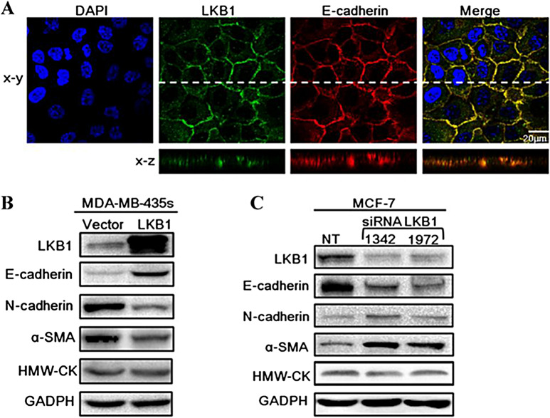 Figure 1
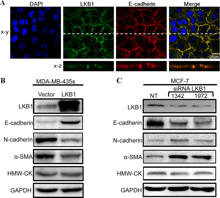 Figure 2
Peer Reviews
No public reviews on file for this paper yet. If you reviewed it on a platform where reviews are public (OpenReview, ICLR, NeurIPS, ICML), you can paste yours below so the community can read it here.
Videos
No videos yet. Explain this paper in a talk, walkthrough, or lecture? Add one.
Taxonomy
TopicsMetabolism, Diabetes, and Cancer · Cancer-related Molecular Pathways · Cancer Cells and Metastasis
Correction: J Exp Clin Cancer Res 33, 70 (2014)
10.1186/s13046-014-0070-0
Following the publication of the original article [1], the authors identified errors in Figure 5c. The GAPDH protein band in Figure 4A and Figure 5C were obtained from two separate replicate experiments. However, the LKB1 protein band in both figures originated from the same experimental set. Therefore, the LKB1 band in Figure 5C requires replacement.
- Fig. 5c: LKB1 band needs replacement.
The corrected figures are provided below:
The corrections do not affect the overall results, discussion, or conclusion of the article.
Incorrect Figure 5
Fig. 5LKB1 was localized at adheren junctions and regulated the expressions of EMT markers. (A) MCF-10 A was stained for LKB1 (green), E-cadherin (red) and DAPI (blue). (B) Expressions of EMT markers in control and LKB1 overexpressing MDA-MB-435 s. (C) MCF-7 cells were transfected with non-targeting siRNA (NT) or siLKB1(1342/1972). The protein levels of E-cadherin, N-cadherin and α-SMA were determined by western blot
Correct Figure 5
Fig. 5LKB1 was localized at adheren junctions and regulated the expressions of EMT markers. (A) MCF-10 A was stained for LKB1 (green), E-cadherin (red) and DAPI (blue). (B) Expressions of EMT markers in control and LKB1 overexpressing MDA-MB-435 s. (C) MCF-7 cells were transfected with non-targeting siRNA (NT) or siLKB1(1342/1972). The protein levels of E-cadherin, N-cadherin and α-SMA were determined by western blot
The reference list from the paper itself. Each links out to its DOI / PubMed record.