Research on Correlations of lncRNA ST7-AS1 with Progression and Therapeutic Targets of Esophageal Cancer
Xiao Lin, Sijia Sun, JiWen Zhang, Yan Cai, Quan Cheng

TL;DR
This study explores how the lncRNA ST7-AS1 contributes to esophageal cancer progression and identifies its potential as a therapeutic target.
Contribution
The study reveals that ST7-AS1 acts as a miR-4262 sponge, offering a new therapeutic target for esophageal cancer.
Findings
ST7-AS1 is overexpressed in esophageal cancer tissues and correlates with shorter patient survival.
Silencing ST7-AS1 reduces cancer cell proliferation, migration, and invasion.
ST7-AS1 functions as a miR-4262 sponge, and miR-4262 inhibition reverses the effects of ST7-AS1 knockdown.
Abstract
Esophageal cancer is a highly prevalent gastrointestinal tumor in China, resulting in a significant number of deaths annually. In this paper, we investigated the regulatory role and therapeutic potential of aberrant ST7-AS1 expression in esophageal cancer. The presence of ST7-AS1 in 125 esophageal cancer tissues was identified through RT-qPCR assays. The application of Kaplan-Meier to evaluate survival rates in patients with esophageal cancer. Cell activity was assessed by both CCK-8 and Transwell assays. The luciferase activity assay verified the association of ST7-AS1 with miR-4262. ST7-AS1 expression in esophageal cancer was noticeably overexpressed compared to the control group. Patients with upregulated ST7-AS1 had shorter survival rates. Silencing ST7-AS1 reduced the proliferation level of esophageal cancer cells, as did the migration and invasion levels. Mechanistically,…
Genes, proteins, chemicals, diseases, species, mutations and cell lines named across the full text — each resolved to its canonical identifier and authoritative record.
Click any figure to enlarge with its caption.
 Figure 1
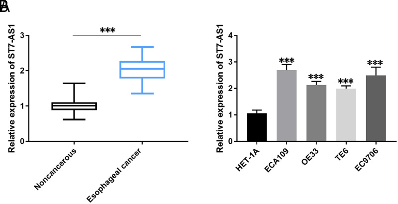 Figure 2
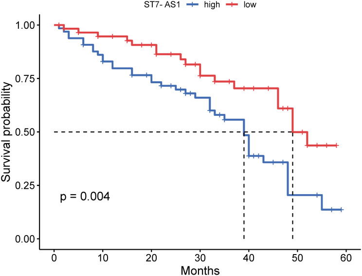 Figure 3
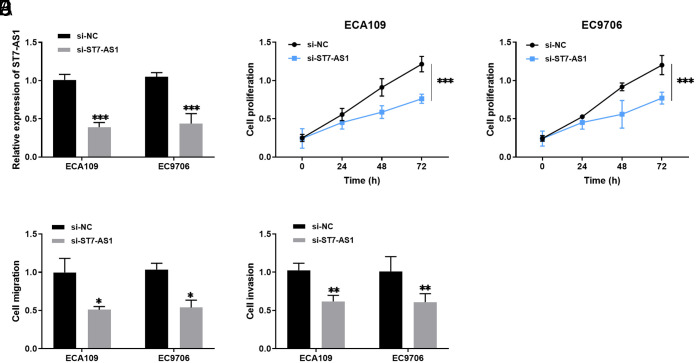 Figure 4
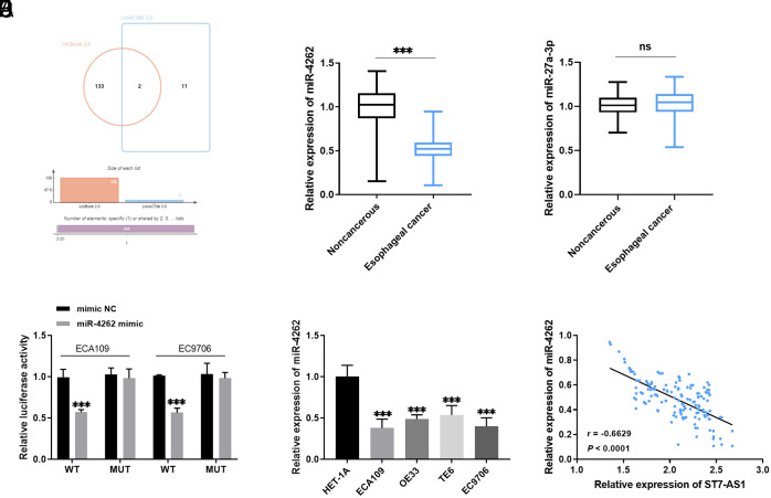 Figure 5
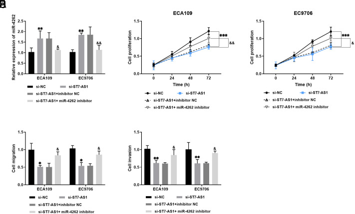 Figure 6
Peer Reviews
No public reviews on file for this paper yet. If you reviewed it on a platform where reviews are public (OpenReview, ICLR, NeurIPS, ICML), you can paste yours below so the community can read it here.
Videos
No videos yet. Explain this paper in a talk, walkthrough, or lecture? Add one.
Taxonomy
TopicsCancer-related molecular mechanisms research · Mycobacterium research and diagnosis · RNA modifications and cancer
Introduction
Esophageal cancer is a highly lethal disease with a high incidence in the digestive system. It ranks sixth among global malignant tumors,^1^ with over 90% of the cases being squamous cell carcinoma and adenocarcinoma.^2^ Despite significant attention and funding dedicated to cancer prevention and treatment in China, the incidence of esophageal cancer remains high. Patients with esophageal cancer often experience difficulty swallowing, a distinct foreign body sensation, sternal pain, or progressive dysphagia.^3^ The standard treatment is surgical resection with preoperative radiotherapy. However, many patients present nonspecific early symptoms, leading to diagnoses in intermediate or advanced stages of the disease. This results in less effective treatment outcomes, poor prognosis, and low quality of life. The advent of molecular targeted therapy over recent decades offers new hope for cancer treatment. In-depth analysis of esophageal cancer pathogenesis and studies on molecular targeted therapy may present a potential treatment method. Hence, identifying specific biomarkers for therapeutic intervention in esophageal cancer patients is a promising avenue to explore.
Long non-coding RNAs (lncRNAs) have intricate secondary or tertiary spatial structures, enabling complex gene regulation.^4^ LncRNAs are linked to the development of various tumors, including esophageal cancer, and are useful for its early detection and prognostic assessment.^5^ LncRNA LUESCC, DGCR5, and LINC00680 have all been shown to mediate the development of esophageal cancer.^6-8^ The dysregulated ST7-AS1, a novel lncRNA, has been associated with tumor progression. For instance, Hu et al pointed out that ST7-AS1 accelerates lung adenocarcinoma metastasis by targeting the miR-181b-5p/KPNA4 axis.^9^ Its oncogenic role has also been emphasized in cervical and gastric cancers.^10^ However, no studies have yet examined ST7-AS1’s role in esophageal cancer. Given the ST7 gene’s widespread expression in human tissues, we hypothesize that a thorough understanding of ST7-AS1’s mechanisms in esophageal cancer could offer promising therapeutic prospects.
This study primarily investigated the levels of ST7-AS1 and its target gene miR-4262 in esophageal cancer tissues and cell samples. Additionally, the potential effect of the abnormal expression of ST7-AS1 on the prognosis and survival of patients was evaluated. The potential mechanisms underlying their regulatory network were further examined, with the goal of providing novel insights for tumor treatment.
Materials and Methods
General Information and Inclusion Criteria
This study involved 125 esophageal cancer patients admitted to Shanghai Baoshan Luodian Hospital from August 2015 to February 2017. None of the participants had undergone antitumor therapies like radiation or chemotherapy prior to diagnosis. Patients with a history of coexisting immune diseases or other malignancies were excluded. Esophageal cancer tissue samples, along with some adjacent normal tissue samples (≥ 5 cm), were collected by surgical resection. These were promptly stored in a -80°C refrigerator after being treated with liquid nitrogen. The study was conducted with the consent of the patients and their families and under the accreditation of the Shanghai Baoshan Luodian Hospital ethics committee (approval number: KY2015-02, date: 19 March 2015). All the participants signed the written informed consent.The collected tissue samples were confirmed by the pathologist, and the relevant clinical data were recorded in Table 1.
Patients were monitored for 60 months through telephone communication and outpatient follow-ups. This included an assessment of clinical symptoms, recurrence, and overall prognosis.
Culture of Experimental Cells
In vitro cell experiments, esophageal epithelial cells HET-1A, and esophageal cancer cells ECA109, OE33, TE6, and EC9706 were purchased from the Cell Bank of Shanghai Academy of Biological Sciences. All cells were maintained in RPMI-1640 medium supplemented with FBS (Gibco, Carlsbad, CA, USA). The incubator conditions were set at 37°C with 5% CO_2_, and the culture medium was changed every 24 hours.
Transfection
The negative control siRNA (si-NC), silencing ST7-AS1 (si-ST7-AS1), mimic negative control (mimic NC), inhibitor negative control (inhibitor NC), miR-4262 mimic, and miR-4262 inhibitor were produced by GenePharma (Shanghai, China). The primer sequences were (5’-3’): si-NC UCUCCGAACGUGUCACG; si-ST7-AS1 GGGTAACTCAAAAAGCCTG; mimic NC UCACAACCUCCUAGAAAGAG; inhibitor NC CAGUACUUUUGUGUAGUACA; miR-4262 mimic GACAUUCAGACUACCUG; miR-4262 inhibitor AGATTGCTGGGTCACACC.
RNA Extraction and RT-qPCR
TRIzol reagent (Invitrogen, Carlsbad, CA, USA) was added to tissue samples and esophageal cancer cells for RNA extraction. The RNA was then reverse-transcribed into cDNA. Subsequently, the SYBR Premix EX Taq Kit (TAKARA, Shiga, Otsu, Japan) was applied to real-time quantitative PCR detection. Following amplification, GAPDH and U6 were used as internal parameters for ST7-AS1 and miR-4262. The relative expression was calculated using the 2^-ΔΔCt^ method. The primer sequences were: ST7-AS1 F-5’-ACCCTACTCTGCCTCCCTTA-3’, R-5’-TAGCATCTGCCACCCAA-3’; miR-4262 F-5’-TGCGGGACATTCAGA-3’, R-5’-CCAGTGCAGGGTCCGAGGT-3’; GAPDH F-5’-AGGTCGGTGTGAACGGATT-3’, R-5’-GGGGTCGTTGATGGCAA-3’; U6 F-5’-TGCGGGTGCTCGCTTCGG-3’, R-5’-CCAGTGCAGGGTCCGAGGT-3’.
In Vitro Cell Assay
ECA109 and EC9706 cells were chosen and cultured in 96-well cell culture plates after being washed with PBS. Following incubation periods of 0, 24, 48, and 72 hours, CCK-8 reagent (Dojindo, Tokyo, Japan) was added to each well. After an additional incubation of 2 hours, the absorbance value was measured at 450 nm using a microplate reader.
The upper chamber of the Transwell was filled with RPMI-1640 medium without serum and inoculated with an esophageal cancer cell suspension, while the lower layer was filled with medium only. After a 24-hour incubation period, the migrated cells were fixed in formaldehyde for 10 minutes and then stained with crystal violet for 20 minutes. The cells were then photographed under a microscope and counted. For the cell invasion assay, Matrigel (BD Biosciences, USA) was spread in the upper chamber of the Transwell. The remaining steps followed the same procedure as the migration assay.
Luciferase Activity Assay
LncBook 2.0 and LncACTdb 3.0 online databases were used to predict the possible downstream target of ST7-AS1 and draw a Venn diagram. The ST7-AS1 fragment carrying the miR-4262 binding sites was amplified and inserted into the pmirGLO luciferase reporter plasmid (Promega, Madison, WI, USA) to construct ST7-AS1-WT/-MUT. The recombinant plasmids were then combined with either mimic NC or miR-4262 mimic and co-transfected into ECA109 and EC9706 cells. The luciferase activity was measured using the Lipofectamine 2000 reagent.
Statistical Analysis
The statistical analysis was performed using SPSS 26.0 (SPSS, Chicago, Ill, USA) and GraphPad Prism 9.0 (La Jolla, Calif., USA). Student’s t-test was used for comparing 2 data groups, and ANOVA was used for comparisons among multiple groups. The log-rank method was applied for the statistical test of survival analysis, and Kaplan-Meier was used for survival curve plotting. The correlation between ST7-AS1 and miR-4262 expression was evaluated using the Pearson correlation coefficient. All results were statistically significant with *P *< .05.
Results
ST7-AS1 Was Relatively Highly Expressed in Esophageal Cancer
ST7-AS1 was markedly enhanced in esophageal cancer tissues, as observed using RT-qPCR (Figure 1A). Additionally, ECA109, OE33, TE6, and EC9706 cells exhibited significantly higher expression of ST7-AS1 compared to HET-1A cells (Figure 1B).
In Table 1, the outstanding level of ST7-AS1 was not noticeably correlated with gender, age, tumor size, histological classification, or the smoking and drinking habits of esophageal cancer patients. However, it was influenced by lymph node metastasis and TNM stage.
Patients with High ST7-AS1 Expression Have Poor Prognosis
The Kaplan-Meier method and log-rank test were utilized to analyze the relationship between the relative expression of ST7-AS1 and the prognosis of patients with esophageal cancer. This analysis was conducted through follow-up and summary of esophageal cancer patients. Results are depicted in Figure 2.
The follow-up and summary of esophageal cancer patients revealed a decreased survival rate for those with high-expression ST7-AS1 esophageal cancer. This suggests that high expression of ST7-AS1 predicts a poor prognosis for esophageal cancer patients (P = .004, Figure 2).
Regulation of Cell Viability by Knockdown of ST7-AS1
In vitro cell experiments resulted in the transfection of si-NC and si-ST7-AS1 into ECA109 and EC9706 cells, respectively. The transfection efficiency is shown in Figure 3A. The proliferation levels of ECA109 and EC9706 cells were down-regulated after transfection with si-ST7-AS1(Figures 3Band3C). Moreover, the migration number of ECA109 and EC9706 cells decreased after silencing ST7-AS1, as seen in Figure 3D. As illustrated in Figure 3E, the knockdown of ST7-AS1 slowed down the cells’ invasion ability.
ST7-AS1 Sponge miR-4262 in Esophageal Cancer
Two downstream targets of ST7-AS1 were found by Venn diagram verification (Figure 4A). miR-4262 level was downregulated in esophageal cancer tissues (Figure 4B), while the miR-27a-3p level was not significantly changed (Figure 4C). The luciferase activity in the WT group decreased, while there was no significant change in the MUT group, which suggested that ST7-AS1 directly adsorbs miR-4262 and regulates its expression (Figure 4D). miR-4262 expression was also decreased in esophageal cancer cells (Figure 4E). Additionally, there was an inverse correlation between the levels of ST7-AS1 and miR-4262 (Figure 4F; r = −0.6629, *P *< .0001).
Effect of miR-4262 Inhibitor on Cell Viability
In Figure 5A, the relative reduction of miR-4262 levels is shown after the transfection of si-ST7-AS1 + miR-4262 inhibitor in ECA109 and EC9706 cells, compared to the transfection of si-ST7-AS1. The miR-4262 inhibitor counteracts the suppressive effect of si-ST7-AS1 on ECA109 and EC9706 cells (Figure 5B-C). Furthermore, the miR-4262 inhibitor compensates for the inhibitory function of the ST7-AS1 knockdown on cell function (Figure 5D-E).
Discussion
In China, esophageal cancer incidence and mortality rates are high, with most patients diagnosed at advanced stages. Despite advancements in treatments such as surgery and radiotherapy, their clinical effectiveness often falls short due to the cancer’s high malignancy and patients’ poor prognosis.^11^ The research elaborated that molecular targeted therapy targeting the regulation of related genes to inhibit cancer cell metastasis and proliferation can be effective treatments.^12^ In recent times, the utility of lncRNAs in the context of diseases has become an increasingly focal point of interest. LncRNAs have been proven to have great potential in regulating cell activity, participating in tumor treatment, and affecting the prognosis of patients.^13,14^ There is also evidence that lncRNAs bring new possibilities in the treatment and prognosis of digestive tumors.^15^ For instance, Huang et al proposed that lncRNA PDIA3P1 promotes the progression of esophageal squamous cell carcinoma by modulating OCT4 in a recent report.^16^
ST7-AS1 is an lncRNA located on 7q31.2, and its copy number variation may cause abnormal expression of downstream targeted genes and regulate changes in tumor cell activity.^17^ Existing evidence shows that ST7-AS1 is upregulated in gastric cancer, promoting cell proliferation and inhibiting apoptosis. Conversely, knocking down ST7-AS1 inhibits cell cycle progression.^18^ Furthermore, ST7-AS1 is overexpressed in laryngeal squamous cell carcinoma, propelling carcinogenesis through the CARM1-Sox-2 axis signaling pathway.^19^ In this study, we found that ST7-AS1 is elevated in esophageal cancer, its significantly high expression influenced by lymph node metastasis and TNM staging. Moreover, the Kaplan-Meier curve indicated that increased ST7-AS1 in esophageal cancer could adversely affect patient survival.
In vitro cell assays showed that the growth and viability of esophageal cancer cells decreased after silencing ST7-AS1. This is consistent with previous studies on esophageal cancer, where silencing TUG1 impeded the proliferation and invasion of cancer cells by affecting miR-1294.^20^ Additionally, knockdown of FAM83A-AS1,^21^ ELFN1-AS1,^22^ and SNHG7^23^ reportedly has the potential to inhibit esophageal cancer cell activity. Therefore, it is reasonable to conclude that ST7-AS1 in esophageal cancer can target downstream genes, influencing the tumor process.
LncRNAs interact with miRNAs to function as competing endogenous RNAs, which can abnormally regulate miRNA expression.^24^ Online predictions illustrated a potential interaction between miR-4262 and ST7-AS1, suggesting that ST7-AS1 might directly target miR-4262. This was later confirmed through a luciferase reporter gene assay. As a novel miRNA, miR-4262 was first shown to be associated with cisplatin resistance in adenocarcinoma cells in 2013.^25^ Current evidence indicates that miR-4262 is under-expressed in cervical cancer,^26^ colon cancer,^27^ and osteosarcoma,^28^ which aligns with our experimental results. miR-4262 expression was reduced in esophageal cancer, and there was a negative correlation between ST7-AS1 and miR-4262. Moreover, research by Liu and colleagues demonstrated that miR-4262 is expressed at lower levels in esophageal cancer and slows tumor progression by negatively controlling the downstream gene KLF6.^29^ The inhibitory effect of silencing ST7-AS1 on cells was counteracted by a miR-4262 inhibitor.
Based on these findings, the ST7-AS1 sponge of miR-4262 appears to play a negative regulatory role, thereby participating in the biological functions of esophageal cancer cells and tumorigenesis. Unfortunately, the number of patients included in this study was limited, and there was a lack of discussion regarding the correlation between ST7-AS1 expression and factors such as the clinical setting. This requires further attention in subsequent work.
Generally, ST7-AS1 expression was elevated in esophageal cancer. Silencing ST7-AS1 suppressed cell viability by acting as a sponge for miR-4262, which subsequently influenced the development of esophageal cancer. ST7-AS1 may serve as a potential therapeutic target for future clinical treatments.
Availability of Data and Materials:
The data that support the findings of this study are available on request from the corresponding author.
The reference list from the paper itself. Each links out to its DOI / PubMed record.
- 1Xu X Xu Y Wang J , et al. The controversy of esophageal carcinosarcoma: a case report and brief review of literature. Medicine. 2019;98(10):e 14787. (10.1097/MD.0000000000014787)30855491 PMC 6417540 · doi ↗ · pubmed ↗
- 2Wang B Li M Su A , et al. Prognostic value of GPNMB, EGFR, p-PI 3K, and Ki-67 in patients with esophageal squamous cell carcinoma. Anal Cell Pathol (Amst). 2022;2022:9303081. (10.1155/2022/9303081)36090016 PMC 9452951 · doi ↗ · pubmed ↗
- 3Wang J Zhao Q Cai L Li J Chen S . Efficacy of bevacizumab and gemcitabine in combination with cisplatin in the treatment of esophageal cancer and the effect on the incidence of adverse reactions. Bio Med Res Int. 2022;2022:2317181. (10.1155/2022/2317181)35480138 PMC 9038406 · doi ↗ · pubmed ↗
- 4Lai IL Chang YS Chan WL , et al. Male-specific long noncoding RNA TTTY 15 inhibits non-small cell lung cancer proliferation and metastasis via TBX 4. Int J Mol Sci. 2019;20(14). (10.3390/ijms 20143473)PMC 667859031311130 · doi ↗ · pubmed ↗
- 5Yang C Chen K . Long non-coding RNA in esophageal cancer: a review of research progress. Pathol Oncol Res. 2022;28:1610140. (10.3389/pore.2022.1610140)35241975 PMC 8885534 · doi ↗ · pubmed ↗
- 6Xue ST Cao SQ Ding JC , et al. Lnc RNA LUESCC promotes esophageal squamous cell carcinoma by targeting the mi R-6785-5p/NRSN 2 axis. Cell Mol Life Sci. 2024;81(1):121. (10.1007/s 00018-024-05172-9)38457049 PMC 10924007 · doi ↗ · pubmed ↗
- 7Li Y Chen B Jiang X , et al. A Wnt-induced lnc RNA-DGCR 5 splicing switch drives tumor-promoting inflammation in esophageal squamous cell carcinoma. Cell Rep. 2023;42(6):112542. (10.1016/j.celrep.2023.112542)37210725 · doi ↗ · pubmed ↗
- 8Xue ST Zheng B Cao SQ , et al. Long non-coding RNA LINC 00680 functions as a ce RNA to promote esophageal squamous cell carcinoma progression through the mi R-423-5p/PAK 6 axis. Mol Cancer. 2022;21(1):69. (10.1186/s 12943-022-01539-3)35255921 PMC 8900330 · doi ↗ · pubmed ↗