Effects of 100 IU versus 150 IU hCG supplementation on oocyte and embryo quality in patients aged ≥ 35 years: a propensity score-matched analysis
Nguyen Thi Lien Huong, Tran Thu Thuy, Phi Thi Tu Anh, Hoang Bao Long, Le Duc Thang, Vu Thi Ngoc, Le Quang Do, Nguyen Xuan Anh Duy, Bui Thi Hanh, Do Van Loi, Le Hoang

TL;DR
This study compares two hCG doses in women over 35 undergoing IVF, finding that a higher dose increases total oocytes but not embryo quality.
Contribution
A propensity score-matched analysis reveals that 150 IU hCG increases total oocyte yield in older IVF patients without compromising safety.
Findings
150 IU hCG resulted in significantly more total oocytes compared to 100 IU.
No significant difference in mature oocyte numbers or embryo development between the two doses.
Only one case of moderate ovarian hyperstimulation occurred in the 150 IU group.
Abstract
Background: The optimal dose of human chorionic gonadotropin (hCG) supplementation during controlled ovarian stimulation (COS) remains controversial, particularly in women of advanced reproductive age. Objective: To compare the efficacy and safety of 150 IU versus 100 IU daily hCG supplementation from stimulation day 6 in women aged ≥ 35 years undergoing in vitro fertilization/intracytoplasmic sperm injection (IVF/ICSI). Methods: In this retrospective cohort study, we analyzed data from 438 patients aged ≥ 35 years undergoing IVF at a single center. Propensity score matching was performed at a 1:1 ratio, yielding 192 patients per group who received either 100 IU or 150 IU daily hCG supplementation from stimulation day 6 during GnRH antagonist cycles. Results: After propensity score matching, baseline characteristics were comparable between groups. The 150 IU group yielded…
Genes, proteins, chemicals, diseases, species, mutations and cell lines named across the full text — each resolved to its canonical identifier and authoritative record.
Click any figure to enlarge with its caption.
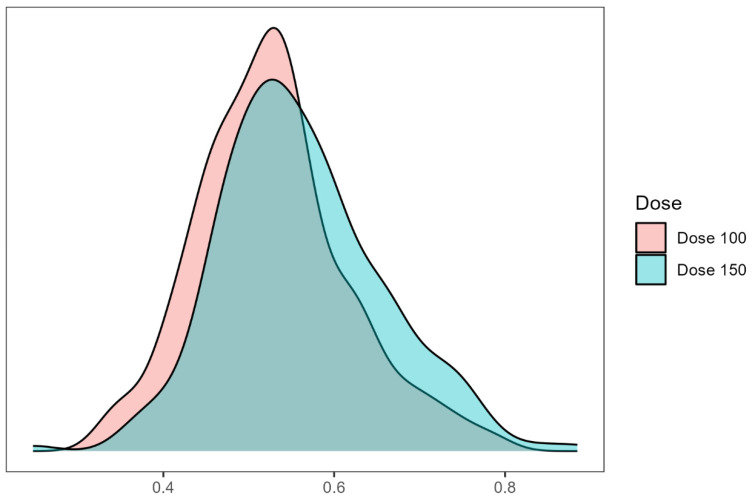 Figure 1
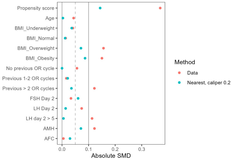 Figure 2
Peer Reviews
No public reviews on file for this paper yet. If you reviewed it on a platform where reviews are public (OpenReview, ICLR, NeurIPS, ICML), you can paste yours below so the community can read it here.
Videos
No videos yet. Explain this paper in a talk, walkthrough, or lecture? Add one.
Taxonomy
TopicsReproductive Biology and Fertility · Ovarian function and disorders · Assisted Reproductive Technology and Twin Pregnancy
Introduction
In vitro fertilization (IVF) success rates decline substantially with advancing maternal age, particularly in women aged 35 years and older, primarily due to diminished ovarian reserve and reduced oocyte quality1,2. During controlled ovarian stimulation (COS) for IVF, adequate luteinizing hormone (LH) activity plays a crucial role in follicular development and oocyte maturation, with its importance potentially magnified in older women who often exhibit altered gonadotropin receptor expression and decreased ovarian sensitivity3,4.
The "two-cell, two-gonadotropin" theory emphasizes that both follicle-stimulating hormone (FSH) and LH are essential for optimal folliculogenesis. FSH promotes granulosa cell proliferation and aromatase activity, while LH stimulates theca cells to produce androgens for subsequent conversion to estradiol5. Recent evidence suggests that LH/human chorionic gonadotropin (hCG) signaling also directly influences oocyte quality through mitochondrial function and oxidative stress regulation pathways6. hCG shares structural homology with LH but exhibits distinct pharmacological advantages, including a longer half-life (24-36 hours versus 60 minutes) and stronger receptor binding affinity7. Recent molecular studies have shown that hCG induces more robust activation of ERK1/2 and AKT pathways crucial for follicular survival and steroidogenesis compared to LH8.
Several studies have investigated low-dose hCG supplementation during COS with promising results. Filicori et al. demonstrated that adding 200 IU/day of hCG from the mid-follicular phase could effectively complete follicular maturation while reducing FSH requirements9. The MERIT trial found that preparations containing hCG-derived LH activity resulted in higher top-quality embryo rates compared to recombinant FSH alone10. More recently, the MEGASET study showed improved ongoing pregnancy rates with hCG-supplemented protocols in high responders11.
Women aged ≥35 years, a subgroup known to benefit from LH activity supplementation during ovarian stimulation, may require different hCG doses due to age-related changes in ovarian function. These changes, including diminished androgen production, altered steroidogenesis, and reduced LH/hCG receptor expression, can lead to a suboptimal response to standard stimulation protocols12. The higher biological potency of hCG compared to LH may be particularly beneficial for women of advanced reproductive age due to its anti-apoptotic effects on follicles and ability to sustain androgen production. hCG has been shown to upregulate anti-apoptotic pathways in granulosa cells, including ERK1/2 and AKT signaling, which may rescue smaller follicles that would otherwise undergo atresia8. This follicular rescue effect could be especially important for older women with diminished ovarian reserve. Additionally, hCG's stronger LH receptor binding affinity compared to LH itself allows it to more potently stimulate theca cell androgen production13, which may help counteract the age-related decline in ovarian androgen levels that can impair folliculogenesis14.
The optimal dose of supplemental hCG remains debatable, particularly in women of advanced reproductive age. Previous studies have utilized doses ranging from 50 to 200 IU/day, with most protocols using either 100 or 150 IU/day15-23. A randomized controlled trial by Thuesen et al. suggested that 100 IU/day may represent the minimum effective dose, while doses up to 150 IU/day appeared safe and potentially more effective16. However, these studies included women across all age groups, and the specific requirements for older patients remain unclear.
Given the unique endocrine challenges faced by older women undergoing IVF, such as altered gonadotropin receptor expression and reduced ovarian sensitivity12, age-specific investigations are needed to determine the most effective and safe hCG dosing strategies for this subgroup. Therefore, we conducted this propensity score-matched retrospective cohort study to compare the effects of 100 IU versus 150 IU daily hCG supplementation from stimulation day 6 on oocyte and embryo outcomes in women aged ≥35 years undergoing IVF. Our findings may help establish evidence-based recommendations for hCG supplementation protocols in this challenging patient population.
Materials and Methods
Study Design and Population
This retrospective cohort study was conducted at our academic fertility center between January 2020 and September 2023. The study protocol was approved by the Institutional Review Board (IRB.TAHN.024/03). Patient data was collected from two distinct time periods corresponding to different hCG supplementation protocols: Period 1 (January 2020-July 2022) when 100 IU hCG was used and Period 2 (July 2022-September 2023) when 150 IU hCG was administered.
Eligible patients were women aged ≥35 years undergoing IVF/ICSI, using GnRH antagonist protocol and receiving hCG supplementation from stimulation day 6. Exclusion criteria were endometrioma, prior ovarian surgery, severe male factor requiring surgical sperm retrieval or severe oligoasthenoteratozoospermia, and parental chromosomal abnormalities.
Ovarian Stimulation Protocol
Individualized starting doses of recombinant FSH (Follitrope, LG Chem, Korea) ranging from 225-300 IU were administered based on patient characteristics including age, body mass index (BMI), anti-Müllerian hormone (AMH) level and previous response. GnRH antagonist cetrorelix (Cetrotide 0.25mg, Merck Serono, Germany) was started on stimulation day 6 and continued until trigger. In addition to the GnRH antagonist, patients received daily supplementation with either 100 IU (Period 1) or 150 IU (Period 2) of highly purified hCG (Pregnyl, MSD, Netherlands or IVF-C, LG Chem, Korea). When at least two follicles reached 17 mm in mean diameter, final oocyte maturation was triggered with 5,000 IU hCG (IVF-C, LG Chem, Korea) and 0.2 mg triptorelin acetate (Fertipeptil, Ferring, Germany). Transvaginal ultrasound-guided oocyte retrieval was performed 35-36 hours later under intravenous sedation.
Laboratory Procedures
Retrieved oocytes were assessed for nuclear maturity after denudation. Intracytoplasmic sperm injection (ICSI) was performed on mature oocytes (MII) that exhibited the first polar body. Fertilization was confirmed 16-18 hours post-ICSI by the presence of two pronuclei. Embryos were cultured in sequential media (Vitrolife, Sweden) or single-step medium (Irvine Scientific, United States), under standard conditions (37°C, 6% CO_2_, 5% O_2_). Cleavage embryo morphology was assessed daily according to consensus scoring criteria based on cell number, symmetry, fragmentation and multinucleation24. Good-quality day 3 embryos were defined as those with 6-8 cells, <20% fragmentation, and stage-appropriate cell size. The classification of obtained blastocysts was based on Gardner and Schoolcraft's criteria, which consider blastocoel expansion, as well as the morphology of the trophoblast and inner cell mass. Blastocysts with grades of 3BB or higher were identified as being of good quality25.
Outcome Measures and Definitions
The primary outcomes were number of MII oocytes retrieved and number of day 3 embryos obtained. Secondary outcomes included total FSH dose, estradiol level on trigger day, number of follicles 14-24 mm on trigger day, number of good-quality day 3 embryos and blastocysts, and incidence of ovarian hyperstimulation syndrome (OHSS). OHSS was classified according to the American Society for Reproductive Medicine guidelines26.
Statistical Analysis
The sample size calculation was based on previously published data from Thuesen et al., which indicated a mean number of oocytes retrieved of 9.2 ± 4.2 in the 100 IU group and 11.3 ± 5.7 in the 150 IU group16. We assumed a difference of 2.1 in the mean number of oocytes retrieved between the two groups. Setting the two-tailed α level at 0.05 and the power (1-β) at 80%, a minimum of 90 cycles were required for each group. Considering a potential dropout rate of 10%, we aimed to include at least 100 cycles in each group.
Continuous variables were tested for normality using the Shapiro-Wilk test and presented as mean ± standard deviation or median (interquartile range). Categorical variables were presented as number (percentage). Between-group comparisons were conducted using Student's t-test or Mann-Whitney U test for continuous variables and chi-square or Fisher's exact test for categorical variables, as appropriate.
To minimize confounding bias, propensity score matching was performed using a 1:1 nearest neighbor matching algorithm with a caliper of 0.2 and no replacement. Variables included in the propensity score model were age, BMI, previous IVF attempts, infertility factors, baseline FSH, LH, AMH, and antral follicle count (AFC). The kernel density plot (Figure 1) and Love plot (Figure 2) are presented to visually illustrate the propensity score distribution and covariate balance achieved after matching.
The effect of hCG dose on primary and secondary outcomes was evaluated using negative binomial regression models, both unadjusted and adjusted using the propensity score method. Results were expressed as mean ratios with 95% confidence intervals (CIs). For outcomes only available in a subset of patients (blastocyst parameters), the number of patients with data in each group was reported.
All statistical tests were two-tailed, with p-values <0.05 considered statistically significant. Statistical analysis was performed using R version 4.2.1.
Results
After propensity score matching, 384 patients were included in the final analysis (192 per group). Baseline characteristics were comparable between the 100 IU and 150 IU hCG groups (Table 1). The mean age (38.88 ± 3.14 vs 38.89 ± 3.28 years, p=0.975), age distribution (64.6% vs 62.5% aged 35-39 years), BMI (21.69 ± 1.97 vs 21.81 ± 2.19 kg/m^2^), baseline FSH (9.04 ± 3.87 vs 8.80 ± 3.80 mIU/mL), AMH (1.94 ± 1.43 vs 1.84 ± 1.36 ng/mL), and AFC (10.89 ± 7.13 vs 10.68 ± 6.25) did not differ significantly between groups. Compared to the population before matching, the characteristics in the population after matching were very similar.
The duration of ovarian stimulation was similar between groups (10.18 ± 0.63 days), with comparable total FSH dose (2664.84 ± 141.91 vs 2666.02 ± 142.51 IU, p=0.936). End-of-stimulation hormone profiles showed higher levels in the 150 IU group for both estradiol (3281.35 ± 2504.58 vs 3000.74 ± 2336.16 pg/mL, p=0.257) and progesterone (0.95 ± 0.90 vs 0.73 ± 0.51 ng/mL, p=0.004). The number of preovulatory follicles (14-24mm) on trigger day was numerically higher in the 150 IU group (8.01 ± 5.42 vs 6.74 ± 5.42, p=0.141).
The total number of oocytes retrieved was significantly higher in the 150 IU group compared with the 100 IU group (9.23 ± 6.60 vs 7.79 ± 5.52, p<0.001), a difference that persisted after adjusting for potential confounders (adjusted mean ratio - aMR 0.87, 95% CI 0.79-0.96) (Table 3). However, the number of mature MII oocytes did not differ significantly between groups in adjusted analyses (6.25 ± 5.12 vs 5.66 ± 4.58, aMR 0.93, 95% CI 0.82-1.06). The number of day 3 cleavage stage embryos was higher in the 150 IU group (5.22 ± 4.62 vs 4.80 ± 3.96), but the difference did not reach statistical significance (aMR 0.97, 95% CI 0.84-1.11). The proportion of good-quality embryos on day 3 was also similar (3.65 ± 3.60 vs 3.35 ± 3.23, aMR 0.96, 95% CI 0.80-1.14). For the subgroup of patients having blastocyst culture, the higher hCG dose did not significantly impact total blastocyst number (3.89 ± 3.59 vs 3.72 ± 2.92, aMR 0.93, 95% CI 0.77-1.13) or good quality blastocyst formation (2.23 ± 2.62 vs 2.21 ± 2.10, aMR 0.95, 95% CI 0.72-1.24).
Regarding safety, only one case of moderate OHSS (0.5%) occurred in the 150 IU group, with none in the 100 IU group.
Discussion
In this propensity score-matched retrospective cohort study, we found that increasing daily hCG supplementation from 100 IU to 150 IU starting from stimulation day 6 in GnRH antagonist cycles significantly increased the total number of oocytes retrieved in women ≥35 years old undergoing IVF/ICSI. While mature oocyte and day 3 embryo yields also favored the 150 IU group, these differences did not reach statistical significance after adjusting for confounders. Embryo quality parameters and blastocyst development were comparable between the two doses. Only one case of moderate OHSS occurred with 150 IU hCG, suggesting a favorable safety profile in this population.
Our observation that 150 IU hCG produced more oocytes than 100 IU (9.23 vs 7.79, p<0.001) contrasts with some previous findings. Madani et al. compared 100 IU versus 200 IU hCG supplementation in poor responders and found comparable outcomes between doses17. However, their study population differed from ours in including only poor responders. More relevant to our age-focused analysis, Gomaa et al. demonstrated that low-dose hCG supplementation particularly benefited women over 40 years, though they did not directly compare different hCG doses19. The discrepancy between the increased oocyte yield and lack of improvement in mature oocyte and embryo numbers with 150 IU hCG may be explained by a potential ceiling effect, where higher hCG doses can support the growth and maturation of a larger cohort of follicles up to a certain stage, but may not necessarily enhance their final acquisition of developmental competence. The increased oocyte yield in the 150 IU group may have been primarily driven by the improved survival and growth of smaller antral follicles, which are more sensitive to the anti-apoptotic effects of hCG27. These rescued follicles, while contributing to the total oocyte count, might not have had sufficient time or optimal conditions for complete oocyte maturation. Further research is needed to elucidate the molecular mechanisms governing the acquisition of oocyte developmental potential in the context of hCG supplementation.
The estradiol levels observed in the 150 IU group (3281 vs 3001 pg/mL, p=0.257) did not differ significantly from the 100 IU group. This observation aligns with Thuesen et al.'s dose-response study which demonstrated that serum estradiol reached a plateau with hCG doses above 100 IU/day, despite continuing increases in androgen production at higher doses16. The lack of significantly higher estradiol with 150 IU hCG supports the concept of an estradiol ceiling effect, likely due to saturation of aromatase activity in converting androgens to estrogens, as proposed by Smitz and Platteau8.
The significantly higher progesterone levels observed in the 150 IU hCG group (0.95 vs 0.73 ng/mL, p=0.004) warrant further discussion. This finding is consistent with the known dose-dependent effect of hCG on ovarian steroidogenesis, particularly its ability to stimulate progesterone production by luteinized granulosa cells16. The higher progesterone levels in the 150 IU group likely reflect the increased steroidogenic activity of a larger cohort of hCG-responsive follicles, as evidenced by the significantly higher oocyte yield compared to the 100 IU group. While elevated progesterone levels have been associated with impaired endometrial receptivity in fresh embryo transfer cycles, it is important to note that the mean progesterone level in our 150 IU group (0.95 ng/mL) remained below the threshold of 1.5 ng/mL, above which negative effects on clinical outcomes have been consistently reported28. Furthermore, our universal application of a freeze-all strategy mitigates any potential detrimental impact of elevated progesterone on endometrial receptivity, as the endometrium is not exposed to the supraphysiologic hormonal milieu of ovarian stimulation during the subsequent frozen embryo transfer cycle29.
While we did not directly measure androgen levels, the higher progesterone levels may indirectly reflect increased androgen substrate availability30. Elevated intraovarian androgens have been proposed to enhance follicular FSH sensitivity and promote follicular survival14. However, excessive androgens may also impair oocyte quality and embryo development31. The lack of improvement in embryo quality parameters in our study despite higher oocyte yields with 150 IU hCG suggests that any potential benefits of increased androgens were outweighed by other factors governing oocyte competence.
Regarding safety, our finding of only one moderate OHSS case with 150 IU hCG compares favorably with previous studies. Kosmas et al.'s meta-analysis found that low-dose hCG supplementation (50-200 IU) actually reduced OHSS risk compared to conventional protocols32. Although the mechanism remains unclear, it is possible that hCG supports the survival and growth of smaller follicles, allowing them to develop into larger follicles, thereby reducing the number of smaller follicles associated with OHSS risk.
Our findings underscore the importance of prioritizing oocyte quality over quantity in the pursuit of optimal IVF outcomes. While maximizing oocyte yield may be an attractive goal, the lack of a corresponding improvement in embryo quality suggests that this approach may not be universally beneficial. This discrepancy raises questions about the cost-effectiveness of using higher hCG doses to maximize oocyte yield, particularly in patients planning for oocytes cryopreservation. In clinical practice, the decision to use a higher hCG dose should be weighed against the potential risks and benefits for each patient. For those with diminished ovarian reserve or a history of suboptimal response, a modest increase in oocyte yield may prove advantageous, providing more opportunities for embryo selection. However, this potential benefit must be balanced against the lack of improvement in embryo quality and the increased cost associated with higher gonadotropin consumption.
The key strengths of our study include the use of propensity score matching to minimize confounding bias, the large sample size, and standardized protocols. The single-center setting also reduces heterogeneity in clinical and laboratory practices. However, this study has several limitations. The retrospective design and nonrandomized allocation of patients to hCG dosing groups may have introduced bias, although this was mitigated by propensity score matching. The lack of significant differences in mature oocyte and embryo numbers between the two groups may have been influenced by the sample size, which was primarily powered to detect changes in total oocyte yield. Larger studies are needed to further elucidate the impact of hCG dosing on these key outcomes and to assess the potential effects on clinical pregnancy and live birth rates.
In conclusion, this propensity-matched retrospective study suggests that while increasing daily hCG supplementation from 100 IU to 150 IU from stimulation day 6 safely improves total oocyte yield in IVF/ICSI patients ≥35 years old, it may not significantly enhance the more clinically relevant endpoints of mature oocyte yield and embryo outcomes. These findings highlight the need to carefully consider the cost-effectiveness and individualized risk-benefit balance when selecting hCG doses for ovarian stimulation in older women. Further research is needed to evaluate the impact of late follicular hCG dosing on live birth rates and elucidate the molecular mechanisms governing oocyte maturation.
The reference list from the paper itself. Each links out to its DOI / PubMed record.
- 1Wang X Tian P zhe Zhao Y jun Lu J Dong C yue Zhang C lian The association between female age and pregnancy outcomes in patients receiving first elective single embryo transfer cycle: a retrospective cohort study Sci Rep 2024 Aug 19141192163916020310.1038/s 41598-024-70249-1PMC 11333704 · doi ↗ · pubmed ↗
- 2Bao S Yin T Liu S Ovarian aging: energy metabolism of oocytes Journal of Ovarian Research 2024 May 311711183882240810.1186/s 13048-024-01427-y PMC 11141068 · doi ↗ · pubmed ↗
- 3Salehpour S Aleyasin A Moini A Mousavifar N Mohammadhossein N Abdollahi Fard S Luteinizing hormone supplementation in controlled ovarian stimulation: the Iran Delphi consensus Front Reprod Health 2024 May 9613974463878412410.3389/frph.2024.1397446 PMC 11111922 · doi ↗ · pubmed ↗
- 4Bielfeld AP Schwarze JE Verpillat P Lispi M Fischer R Hayward B Effectiveness of recombinant human FSH: recombinant human LH combination treatment versus recombinant human FSH alone for assisted reproductive technology in women aged 35-40 years Reproductive Bio Medicine Online 2024 Jun 14861037253859374510.1016/j.rbmo.2023.103725 · doi ↗ · pubmed ↗
- 5Narayan P Ulloa-Aguirre A Dias JA Chapter 2 - Gonadotropin Hormones and Their Receptors. In: Strauss JF, Barbieri RL, editors. Yen and Jaffe's Reproductive Endocrinology (Eighth Edition)Philadelphia: Elsevier 2019 p. 25-57.e 15
- 6Arroyo A Kim B Yeh J Luteinizing Hormone Action in Human Oocyte Maturation and Quality: Signaling Pathways, Regulation, and Clinical Impact Reprod Sci 2020 Jun 12761223523204645110.1007/s 43032-019-00137-x PMC 7190682 · doi ↗ · pubmed ↗
- 7Choi J Smitz J Luteinizing hormone and human chorionic gonadotropin: Origins of difference Molecular and Cellular Endocrinology 2014 Mar 53831203132436533010.1016/j.mce.2013.12.009 · doi ↗ · pubmed ↗
- 8Smitz J Platteau P Influence of human chorionic gonadotrophin during ovarian stimulation: an overview Reproductive Biology and Endocrinology 2020 Aug 6181803276269810.1186/s 12958-020-00639-3PMC 7409634 · doi ↗ · pubmed ↗