Contrast-enhanced endoscopic ultrasonography for diagnosis of leiomyosarcoma of the inferior vena cava
Wen Zhang, Ming-Yan Cai

Abstract
Genes, proteins, chemicals, diseases, species, mutations and cell lines named across the full text — each resolved to its canonical identifier and authoritative record.
Click any figure to enlarge with its caption.
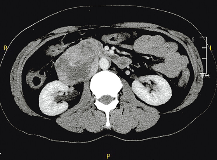 Fig. 1
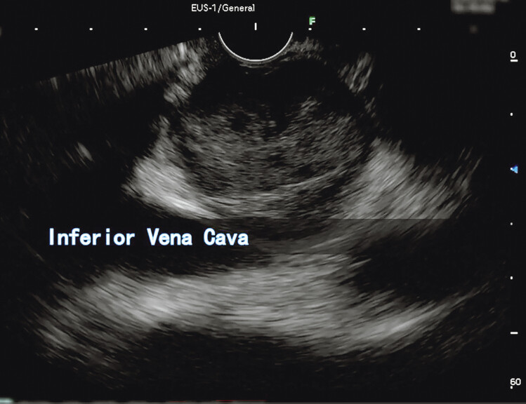 Fig. 2
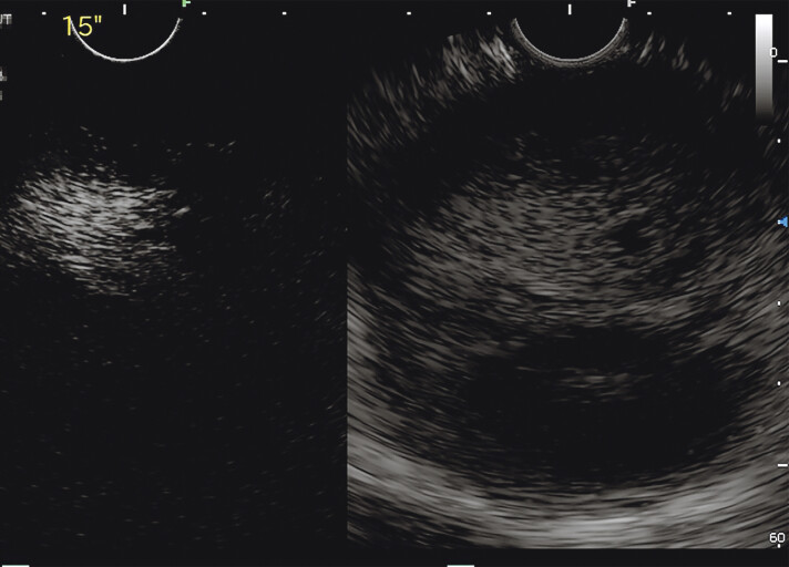 Fig. 3
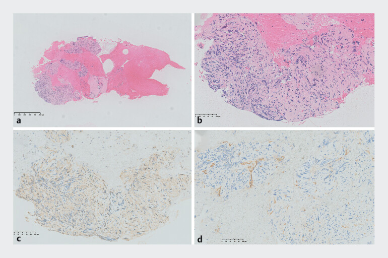 Fig. 4
Peer Reviews
No public reviews on file for this paper yet. If you reviewed it on a platform where reviews are public (OpenReview, ICLR, NeurIPS, ICML), you can paste yours below so the community can read it here.
Videos
No videos yet. Explain this paper in a talk, walkthrough, or lecture? Add one.
Taxonomy
TopicsSarcoma Diagnosis and Treatment · Soft tissue tumors and treatment · Cardiac tumors and thrombi
Endoscopic ultrasound (EUS) has become an important diagnostic tool for various diseases 1 . Contrast-enhanced (CE)-EUS has emerged as an effective technique that is complementary to conventional EUS and allows visualization of microvessels and parenchymal perfusion, and more accurate characterization of the lesion 2 .
We report the case of a 46-year-old woman with abdominal pain. On a contrast-enhanced computed tomography scan, a mass of approximately 7 cm in diameter was discovered in the right posterior peritoneum, with compression of the duodenum and inferior vena cava (IVC) ( Fig. 1 ). We decided to perform CE-EUS for this patient ( Video 1 ).
Contrast-enhanced computed tomography scan showed a mass of approximately 7 cm in diameter in the right retroperitoneum.
Contrast-enhanced endoscopic ultrasonography for diagnosis of leiomyosarcoma of the inferior vena cava.Video 1
On CE-EUS, we discovered a solid hypoechoic lesion originating from the IVC wall, independent of the duodenum ( Fig. 2 ). After injection of contrast reagent (Sonovue; Bracco, Milan, Italy), the lesion showed heterogeneous hyper-enhancement into the lesion ( Fig. 3 ). For a pathological diagnosis, EUS-guided fine-needle aspiration (EUS-FNA) of the lesion was performed with a 22-gauge needle (SharkCore; Medtronic, Minneapolis, Minnesota, USA).
Endoscopic ultrasound showed a 7-cm hypoechoic mass originating from the inferior vena cava.
Contrast-enhanced endoscopic ultrasonography showed a heterogeneous hyper-enhancing pattern into the lesion.
Histological examination revealed an abnormal high proliferation of spindle cells, obvious nuclear atypia, and mitotic activity. The immunohistochemical stains revealed positivity for B-cat, EMA, and SMA, and negativity for CD34, CD117, CgA, S100, SOX11, and SYN ( Fig. 4 ). We suspected a malignant spindle cell tumor originating from the IVC, most compatible with leiomyosarcoma. The patient accepted surgical resection and vascular reconstruction.
Histopathology of tissue mass samples by endoscopic ultrasound-guided fine-needle aspiration. a Original magnification of hematoxylin and eosin stain (×5). b Local magnification showed large areas of spindle cells (×200). c Tumor cells showed diffuse SMA expression (×200). d Tumor cells showed negativity for CD34 (×200).
Finally, the specimen confirmed the diagnosis of leiomyosarcoma. Immunostains showed diffuse positivity for caldesmon, calponin, desmin, EMA, and SMA but negativity for CD10, CD34, CD117, Muc-4, S100, and STAT6.
Primary leiomyosarcoma of the IVC is a rare soft tissue sarcoma 3 . This is the first instance where CE-EUS was applied in the preoperative diagnosis of IVC leiomyosarcoma. The essential role of CE-EUS and EUS-FNA in evaluating IVC leiomyosarcoma preoperatively is remarkable.
Endoscopy_UCTN_Code_CCL_1AF_2AG_3AD
The reference list from the paper itself. Each links out to its DOI / PubMed record.
- 1Itonaga M Ashida R Kitano M Endoscopic ultrasound-guided fine-needle aspiration (EUS-FNA) with image enhancement Diagnostics (Basel)20201088810.3390/diagnostics 1011088833143258 PMC 7692599 · doi ↗ · pubmed ↗
- 2Kitano M Sakamoto H Kudo M Contrast-enhanced endoscopic ultrasound Dig Endosc 201426798510.1111/den.1217924118242 · doi ↗ · pubmed ↗
- 3Wang MX Menias CO Elsherif SB Current update on IVC leiomyosarcoma Abdom Radiol (NY)2021465284529610.1007/s 00261-021-03256-934415408 · doi ↗ · pubmed ↗