Phenotypic and genetic heterogeneity of Acinetobacter baumannii in the course of an animal chronic infection
Léa Bednarczuk, Alexandre Chassard, Julie Plantade, Xavier Charpentier, Maria-Halima Laaberki

TL;DR
This study examines how Acinetobacter baumannii evolves genetically and phenotypically during a 5-year urinary tract infection in an animal patient after antibiotic treatment was stopped.
Contribution
The study reveals genetic and phenotypic adaptations of A. baumannii during a chronic infection without antibiotic pressure.
Findings
A. baumannii strains showed genome reduction through IS recombination, phage excision, and plasmid curing.
Genetic variations were observed in biofilm formation and adhesion genes, but metabolism remained stable.
Colistin resistance was preserved due to a pmrB mutation despite overall decreased antibiotic resistance.
Abstract
Acinetobacter baumannii is a nosocomial pathogen associated with various infections, including urinary tract infections (UTIs). In the course of an infection, A. baumannii is known to rapidly become resistant to antibiotic therapy, but much less is known about possible adaptation without antibiotic pressure. Through a retrospective study, we investigated within-host genetic diversity during a subclinical 5-year UTI in an animal–patient after withdrawal of colistin treatment. We conducted whole-genome sequencing and phenotypic assays on 17 clonally related isolates from the Sequence Type 25 lineage. Phylogenomic analysis revealed their proximity with animal and human strains from the same country suggesting zoonotic transmission (France). In this case study, the clonally related strains presented variations in genome sizes and nucleotide sequences. Over the course of the infection, A.…
Genes, proteins, chemicals, diseases, species, mutations and cell lines named across the full text — each resolved to its canonical identifier and authoritative record.
Click any figure to enlarge with its caption.
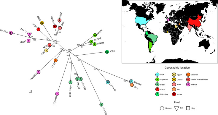 Fig. 1
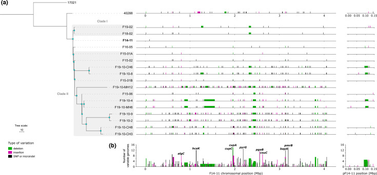 Fig. 2
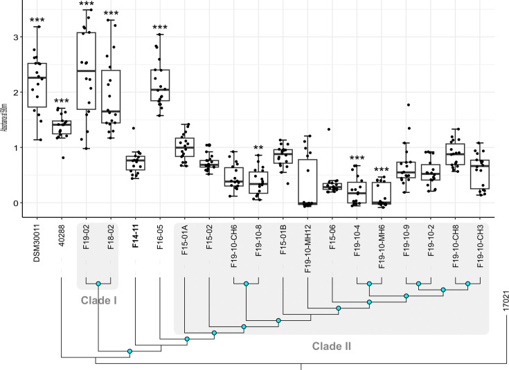 Fig. 3
 Fig. 4
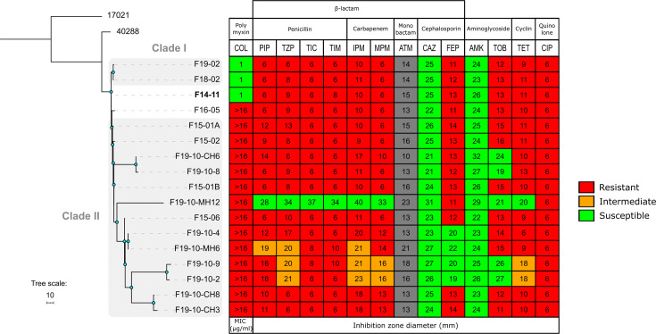 Fig. 5
- —http://dx.doi.org/10.13039/501100011073 VetAgro Sup
- —Fondation pour la Recherche Médicale (FR)
- —Agence Nationale de la Recherche (FR)
- —Agence Nationale de la Recherche (FR)
Peer Reviews
No public reviews on file for this paper yet. If you reviewed it on a platform where reviews are public (OpenReview, ICLR, NeurIPS, ICML), you can paste yours below so the community can read it here.
Videos
No videos yet. Explain this paper in a talk, walkthrough, or lecture? Add one.
Taxonomy
TopicsAntibiotic Resistance in Bacteria · Vibrio bacteria research studies · Escherichia coli research studies
Data Summary
The newly sequenced genomes are listed in Table S4; these genomes are available in GenBank (National Center for Biotechnology Information) and their BioProject number is PRJNA1100485. The publicly available genomes of bacteria from human and animal origin used in this study are listed in Table S5. All the supplementary tables are available in the Supplementary Material.
Introduction
Acinetobacter baumannii is a non-fermentative Gram-negative pathogenic bacterium responsible for nosocomial infections in humans and animals, particularly in intensive care units [12]. A. baumannii represents a major threat to public health due to the rapid emergence of multidrug-resistant isolates increasingly difficult to eradicate [3]. Indeed, few antibiotics remain effective against this pathogen, such as carbapenems, and the carbapenem resistance of clinical isolates may require poorly tolerated antibiotics such as colistin [4]. The emergence of carbapenem resistance in A. baumannii, therefore, limits treatment options and led the World Health Organization to classify carbapenem-resistant strains of A. baumannii as a priority for research and development of new drugs [5]. This pathogen is also described in animal infections, especially in pets with strains belonging to clones common with human medicine with concerning resistance to human-restricted antibiotics such as carbapenems [16].
This bacterium has no particular tropism, causing mainly pulmonary infections, but is also capable of skin, blood, bone and urinary tract infections (UTIs) [7]. The existence of extra-hospital reservoirs has long been disputed, but recent studies demonstrate that A. baumannii is a ubiquitous bacterium found both in diverse environments and carried by healthy individuals – humans and animals [814]. This bacterium is therefore capable of thriving in highly contrasted environments, demonstrating a great capacity for adaptation; antibiotic resistance would be just one facet of its versatility applied to the healthcare context.
Several within-host studies of A. baumannii infection describe antibiotic resistance emergence in response to treatment due to non-synonymous SNP or horizontal gene transfer [1516]. However, little is known about host adaptation, because brief timeframes of infection limit observation of bacterial evolution within a host. Additionally, these investigations focused on human infections, typically involving extended antibiotic treatments, making it difficult to decipher bacteria’s adaptation to the antibiotic from host adaptation. They also involve mostly single-isolate approaches hindering the understanding of within-host diversity. Consequently, there remains a limited understanding of the fate of antibiotic resistance genes and more broadly of gene evolution post-antibiotic exposure and during host colonization. Seminal within-host microevolution studies on A. baumannii observed the accumulation of variations in genes involved surface structures such as motility, adhesion, biofilm formation or cell wall and capsule biogenesis, suggesting a consistent selective pressure during antibiotic-treated infections, but phenotypic validation is usually missing [1719]. Phenotypically, antibiotic exposure prompts bacteria to adopt a biofilm-like lifestyle, enhancing their resistance [20]. These biofilms pose challenges for infection treatment and promote recurrence, notably in UTIs [21]. Despite the over-representation of pulmonary and bloodstream infections in A. baumannii studies, the urinary tract is a major source of A. baumannii isolates in humans [22]. Tissue tropism may result from mobile genetic element (MGE) regulation as exemplified by plasmid-driven regulation of chromosomal genes modifying strain adhesion patterns [62324]. Therefore, A. baumannii within-host microevolution may involve a crosstalk of MGE and core genome determinant.
In the following case study, a pet infection emerged from a subcutaneous ureteral bypass placement which was particularly described as a risk factor for the development of A. baumannii UTI in cats [25]. We used whole-genome sequencing and phenotype assays on 17 A. baumannii strains isolated from the same cat with UTI over a 5-year period without antibiotic treatment. We sought to investigate the fate of antibiotic resistance and explore evolutionary strategies upon infection.
Methods
Strains
A. baumannii strains identified in the Laboratoire Vétérinaire Départemental du Rhône, France (LVD69), were stored in an archive since 2014. Nine strains originated from distinct urine samples collected between 2014 and 2019 and were referred to as the longitudinal strains, while all others were derived from a single urine sample collected later in October 2019, referred to as the synchronic isolates. Samples were obtained not for the purpose of this study or research but rather for diagnostic purposes. The longitudinal strains were initially isolated on a blood agar plate, and the synchronic strains were isolated using three different media: lysogeny broth (LB, Sigma) agar, Mueller–Hinton agar (Sigma) and CHROMagar Acinetobacter (CHROMagar). All the strains used in this study are listed in Table S1 (available in the Supplementary Material).
Metabolic assays
Metabolic assays were conducted using Biolog Gen III Microplates (Hayward, CA) following the manufacturer’s instructions. In brief, the strains were streaked on LB agar and incubated overnight at 30 °C. For each strain, a single colony was resuspended in inoculating fluid A (IFA, Biolog) using a cotton-tipped Inoculatorz swab (Biolog) to reach a cell density of 90–98%. Each well of the Gen III MicroPlate, containing either a specific carbon source or a chemical sensitivity testing agent, was filled with 100 µl of the bacterial suspensions and incubated at 33 °C for 48 h in the OmniLog incubator/reader measuring the reduction of tetrazolium redox dye (forming a purple colour) due to bacterial growth.
Genome sequencing and annotation
Bacteria were grown in a liquid LB medium, and DNA was purified using a Wizard Genomic DNA kit (Promega). DNA quantifications were performed using Qubit™ dsDNA HS Assay Kit (Thermo Fisher Scientific). For each isolate, Oxford Nanopore and Illumina DNA sequencings were both performed. Nanopore sequencing was performed using MinIon flow cells (FLO-MIN106) using the SQK-RBK004 barcoding kit. Nanopore sequencing data were then basecalled using ONT Guppy base-calling software (2.22-r1101), which automatically removed reads with a quality score Q below 10. Median read quality and read length N50 were analysed with NanoPlot (1.42.0) [26], confirming that no additional filtering was required (Table S2). Illumina sequencing was performed on Illumina HiSeq 2000 with paired 150-base sequence reads (Novogene). Quality check of Illumina sequencing data was performed using fastQC (0.11.9) [27]. The analysis of distributions of per base sequence quality (Table S2) was performed using R functions mean() (package base 4.3.1) and sd() (package stats 4.3.1) and showed that no trimming or filtering was necessary. Each read set was assembled individually using Unicycler (version 0.4.9, [28]) in hybrid mode and annotated using Prokka (version 1.14.5,[29]) on the Galaxy pipeline using as reference AB5075-UW protein sequences (GenBank accession number: CP008706.1, [30]). For analysis purposes, plasmid pF14-11 was annotated using pA297-3 sequence (GenBank accession number: KU744946)[31] except for the tetR genes that were named after pAB04 (Genbank accession number: CP012007, 19, 23–26). Sequence typing was assigned using the PubMLST database and the Pasteur scheme [3233]. Putative prophages were predicted using PHASTER [34]. Putative resistance genes were detected using Resfinder (version 4.5.0, [3536]) and insertion sequences using ISfinder [37]. All genomes are available at National Center for Biotechnology Information (NCBI) under BioProject PRJNA1100485 with quality performed by the NCBI platform using CheckM (v1.2.2) with completeness between 98.46 and 99.75% and contamination ranging from 0.27 to 0.53%.
Construction of bacterial strains
All the oligonucleotides used in this study for genetic modification are listed in Table S1. Gene disruptions were performed using a scarless genome editing strategy described previously [38]. For plasmid curing of strain F14-11, its ∆higA-higB::sacB-aac4 derivative was grown overnight in LB (2 ml) at 37 °C; the next day, 100 µl was spread on M63 with 10% sucrose for 24 h growth at 37 °C and then purified on the same medium. Approximately 150 sucrose-resistant c.f.u. values were then patched on LB agar, LB tetracycline 30 µg ml^−1^ and LB apramycin 30 µg ml^−1^. Tetracycline- and apramycin-resistant clones were screened by PCR and plasmid extraction for plasmid loss.
Phylogenetic analysis of the study strains
All chromosomes were aligned to the F14-11 chromosome, set as a reference, using the Gubbins script generate_ska_alignment.py, which creates an alignment using split k-mer analysis version 2 (SKA2, version 0.3.7, [39]) (GitHub link: https://github.com/nickjcroucher/gubbins/blob/master/python/scripts/generate_ska_alignment.py). The resulting alignment measured 4 061 760 bp with 337 variable positions. The strain 17021 was additionally included to root the tree (accession number: GCA_022241165; Table S5). This strain is a suitable root as it lies outside the other study strains while still being phylogenetically close. Maximum likelihood tree reconstruction was performed with Gubbins (version 3.2.1, [40]) relying on RAxML using the GTRGAMMA model (version 8.2.12, [41]) for constructing the phylogeny in each iteration to mitigate the bias of horizontal sequence transfer mechanisms. Statistical support for internal branches of the tree was evaluated by bootstrapping with 10 000 iterations. Tree visualization was conducted using iTOL (version 6.9, [42]).
Phylogenetic analysis with other closely related ST25
Given the close genetic relationships among the study strains, only the first isolated one, F14-11, was used for the selection of closely related strains from other studies.
The topgenome (-t) feature of WhatsGNU was used to identify the ten closest strains from a database comprising 4325 genomes of A. baumannii available on GenBank [43] (‘top-ten strains’). The database was constructed by downloading all publicly accessible genomes of A. baumannii from NCBI in February 2023 (Genome database, search criteria: assembly level = {Chromosome, Complete, Contig}, exclude partial, exclude anomalous).
To expand the diversity of the strains set, we also added the closest genomes of each of the ‘top-ten strains’ as well as four additional ST25 strains (OIFC143, CI79, Naval18 and 107m) bringing a total of 24 strains. To prevent erroneous conclusions regarding geographic proximity, only one strain per study was included. Incomplete sequenced genomes were excluded. All the genomes were mapped to the F14-11 chromosome set as a reference using the Gubbins script generate_ska_alignment.py, which creates an alignment using SKA2 (version 0.3.7). The resulting alignment measured 4 061 760 bp with 1511 variable positions. The output was then analysed by the Gubbins algorithm relying on RAxML using the GTRGAMMA model (version 8.2.12) for constructing the phylogeny in each iteration to generate an unbiased phylogeny. The reproducibility of the node positions in the tree topology was assessed using bootstrap analysis with 10^4^ replicates. No root was assigned because strains known to be outgroups were genetically too distant. Including such distant outgroups would result in insufficient sequence alignment and, therefore, insufficient information for accurate phylogenetic analysis. Finally, the tree was visualized, incorporating host and isolation location data, using the R package ggtree (version 3.12.0, [44]).
Variant detection analysis
Primary Illumina sequence reads (fastq) of monophyletic isolates were aligned to the earliest (F14-11), using the tool Snippy using default parameters (Galaxy version 4.6.0, [45]). The primary Illumina reads of the F14-11 strain were also subjected to the analysis as control and returned no SNPs. Deletions, insertions and rearrangements were identified using the software package MAUVE (version 2.4.0) [46]. Specifically, we developed a script to automatically extract regions present or absent in the strain genome compared to the F14-11 genome by analysing the backbone files, which lists the aligned genomic regions. The list of the identified genetic variations is provided in Table S3.
Statistical analysis
All statistical analyses were performed using RStudio 4.1.2 (www.rstudio.com). The medians of the biofilm formation levels of the different strains were compared using the non-parametric Kruskal–Wallis test, followed by a Bonferroni–Holm correction for pairwise comparisons with strain F14-11. The potential correlation between chromosome size and the number of months elapsed between isolation dates was evaluated using Spearman’s correlation test. For months with multiple strains isolated, the mean chromosome size of those strains was used.
Antibiotic susceptibility testing
Susceptibility of the strains to a panel of antibiotics was evaluated by disc diffusion on Mueller–Hinton agar (Biorad, France) following the CA-SFM 2013 recommendations (https://www.sfm-microbiologie.org/wp-content/uploads/2020/07/CASFM_2013.pdf). Inhibition values were interpreted according to CA-SFM 2013 breakpoints for all antibiotics but aztreonam, for which Pseudomonas spp. breakpoints are given. The strain Pseudomonas aeruginosa CIP 7110 was used as a control for all the antibiotic susceptibility testing. Minimum inhibitory concentration (MIC) experiments were also conducted to evaluate susceptibility to colistin and piperacillin according to EUCAST guidelines [47].
Biofilm formation assay
Biofilm-forming capacities of the isolates were evaluated using the crystal violet staining method, with slight modifications to the protocol previously described [48]. Briefly, exponential phase cells were inoculated into 96-well polystyrene plates in 200 µl of BM2 minimal medium [49] supplemented with 10 mM potassium glutamate (BM2G) at starting OD_600_ of 0.1 and incubated at 37 °C in the absence of light for 48 h, without shaking. The medium was replaced with fresh BM2G medium after 24 h of incubation. After 48 h, the cells in suspension were removed, and the adherent cells were fixed with 3.7% formaldehyde for 10 min. The wells were then washed twice with distilled water, and 200 µl of 0.1% crystal violet was added to each well, incubated for 10 min and washed three times with distilled water. To solubilize the crystal violet, 200 µl of 95% ethanol was added, and the absorbance was measured at 590 nm with Infinite M200 PRO (Tecan) and Magellan 7.1 SP1 (Tecan).
Results
An ST25 monophyletic infection related to human and animal cases
A retrospective study was performed on A. baumannii isolates cultured from a single animal patient (cat) over a 5-year period (2014–2019). This patient–animal developed a urinary A. baumannii infection following the placement of a urinary invasive medical device. The earliest isolate was identified as multidrug-resistant, including to human-restricted antibiotic carbapenems, but not to colistin. However, two colistin treatments failed to clear the infection. Given the limited antibiotic arsenal authorized in veterinary medicine, and the absence of invalidating clinical signs, no further antibiotic treatment was attempted. During medical follow-ups, eight strains (referred to as longitudinal strains) were sequentially isolated and stored by the hospital laboratory from November 2014 to February 2019 (Fig. S1 and Table S4). In addition, nine strains (referred to as synchronic strains) were subsequently isolated from a single complete urine sample collected in October 2019.
Whole-genome sequencing and hybrid assembly of the 17 study strains revealed clonally related strains belonging to sequence type (ST) 25 clone, ST associated with UTI in humans and animals [623]. To identify the closest genomes to F14-11, the earliest isolate, we used the Similar Genome Finder utility of the WhatsGNU tool to query an A. baumannii genome database retrieved from the NCBI (5234 genomes, downloaded on 17 February 2023) [43]. Most strains were from human origin as publicly available A. baumannii genomes are mostly related to human medicine. The phylogenetic tree constructed from 23 ST25 strains brings evidence of geographical clustering for Eurasian and American isolates. However, both North American strains were potentially military imports from overseas explaining their clustering with an Iraqi strain [50] (Fig. 1). This analysis also indicated a clustering of clones independently of their host suggestive of zoonotic transfer and highlighting the relevance of investigating animal infections for human health [5152]. The closest human strain, 16A1524, was indeed isolated in France in 2016. Interestingly, the closest strain, 40288, was also isolated in France from a dog UTI in 2015 [38]. F14-11 and 40288 exhibit only 31 SNPs across their chromosomes and 1 SNP between their ~145 Kb-length plasmids p40288 and pF14-11 (Table S5). The pF14-11 backbone was also detected in 22 of the closest genomes (coverage from 65 to 100%; Table S5). Despite its reduced size, pF14-11 shares 100% identity with larger plasmids pVB82 (215 Kb) and pHWBA8 (195 Kb) carried by close eponymous strains (Fig. S2 and Table S5). This size reduction may explain the inability of pF14-11 to undergo conjugation (Fig. S2).
Phylogenetic proximity of F14-11 to other strains isolated from human and animal infections worldwide. (a) Unrooted maximum likelihood phylogeny constructed using Gubbins based on RAxML. Tips are coloured by geographic origin, labelled with the country’s initial and shaped by host type. Bootstrap values are shown at nodes. The scale indicates branch length in base substitutions. (b) World map with countries highlighted according to the specified colour scheme.
Genome reduction and IS dynamics
To further investigate within-host strain diversity, we a performed whole-genome comparison of the 16 study strains to the early one F14-11, set as a reference (Fig. S1 and Table S4). This analysis revealed a low number of SNPs scattered throughout the genome in comparison to F14-11, with a mean of 31 SNPs per genome ranging from 3 SNPs for the closest genomes to 78 SNPs for the most distant one (Table S4).
Phylogenetic analysis suggests an absence of correlation between isolation date and phylogenetic relationships (Fig. 2). Notably, longitudinal isolates F18-02 and F19-02 form an independent clade, referred to as clade I, but share a hypothetical common ancestor with all other strains. In contrast, late synchronic strains isolated in 2019 cluster with strains isolated in 2015 implying the coexistence of different lineages during the infection. A correlation is observed between the chromosome size of strains and the time elapsed (R²=0.86, P=6.7×10^−3^, Spearman test) indicating a reduction in size during infection (Table S4). Genome reduction can operate on MGE such as prophages. Indeed, most strains show independent excisions of one to three of the four prophages identified in strain F14-11. Close strains F18-02 and F19-02 lost one and two prophages, respectively (Fig. 2, large deletions highlighted in green, and Fig. S3A, prophage regions 2 and 4). The endogenous plasmid underwent further size reduction in four of the synchronic isolates and lacks a 12–18 kb plasmid region corresponding to the BREX phage resistance system, while three have lost the plasmid entirely (Figs 2 and S3B, C) [53].
Phylogenic tree of the study strains and 40288 associated with the distribution of the genetic variations. (a) Left: maximum likelihood phylogeny constructed with Gubbins based on RAxML. The tree was rooted with strain 17021. The scale indicates branch length in base substitutions. Blue nodes indicate bootstrap values >80. Right: Representation of SNPs and microindels in black, insertions in pink and deletions in green along the genomes in comparison to F14-11 (reference genome highlighted in bold). (b) Cumulative distribution of the genetic variations along the F14-11 genome with the y-axis indicating the number of study strains harbouring the genetic variation. The genes most prone to variation are shown.
Biofilm formation by the study strains and the dog UTI strain 40288 associated with their phylogenetic relationships. Blue nodes indicate bootstrap values >80. Biofilm formation was assessed by measuring the OD of the crystal violet stained matrix and compared to environmental strain DSM30011. Error bars are generated from 12 replicas from four independent experiments. Statistically significant differences with F14-11 (Kruskal–Wallis rank-sum tests with Bonferroni–Holm correction) are highlighted with asterisks: () if the P-value<0.05, (* ) if the P-value≤0.01 and ( * ) if the P-value≤0.001. The first isolated strain (F14-11) is highlighted in bold.
IS recombination led to chromosome deletions. The most striking evidence of this phenomenon is the deletion of a 36.6 kb region, fixed in a subclade of eight strains within clade II, which is flanked by two identical ISAba34 in F14-11 (Figs23). In addition, in comparison to the F14-11 chromosome, we observed multiple new chromosomal insertions by an IS originating from the plasmid (Fig. S4). Indeed, IS17, an IS5 family IS, is originally found only in the plasmid pF14-11 of the earliest strain F14-11. While a single instance of IS17 was found in the chromosomes of strains isolated in 2015, it significantly multiplied in synchronic strains, climaxing with 75 occurrences in strain F19-10-MH12. Interestingly, IS17 is also present on p40288 of the related 40288 strain although restrained to this replicon.
Recurrent variation of adhesion and biofilm genes
Over 82% of SNPs (497 SNPs) and microindels of the strains studied are in coding sequences, with 80% being non-synonymous SNPs. Several of these genes are involved in adhesion and biofilm formation – genes that are also affected by insertion and deletion events (Fig. 2; cup [54], csu [55], pga [56], bapA [57] and algC [58] genes). All clade II strains exhibit at least one mutation in one of these genes, while clade I strains retain genetic sequences identical to those of F14-11. Strikingly, genes involved in the biogenesis of archaic and classical types of chaperone-usher pili involved in the adhesion step of biofilm formation (csu and cup operon, respectively) were subjected to IS insertion, deletion and frameshift. Specifically, the csu operon is affected by insertions by distinct IS in strains F15-01A (IS17) and F15-06 (ISAba33), this latter insertion being conserved in most synchronic isolates (seven out of nine) (Fig. S5). Genes of the classical pili Abp1 (formerly Cup), cupA and cupC, were also affected by large deletions and indel leading to a frameshift in most strains of the clade II. Additionally, genes involved in biofilm formation such as algC and pgaABCD but also bapA gene were also disrupted by IS insertion or subjected to deletion events (Fig. 2).
Hence, during the infection, strains may coexist with varying adhesion and biofilm abilities. To bring experimental evidence to this hypothesis, the biofilm-forming capacities of the study strains were measured and compared with those of the environmental DSM30011 strain described as a strong biofilm producer (Fig. 3) [49]. As observed for human clinical strains, the animal clinical strain F14-11 forms less biofilm than DSM30011 although having similar growth in the biofilm medium (Fig. 3, threefold; P-value: 0.00143, Fig. S6A). However, F14-11 carries a plasmid closely related to plasmid pAB5 that has been demonstrated to downregulate biofilm pga and adhesion cup genes in strain UPAB1 [2324]. We, therefore, tested the hypothesis that pF14-11 may restrain biofilm production. pF14-11 curing did not increase adhesion, biofilm or PNAG production (Fig. S6B, C). We also tested if the residual biofilm capacity of F14-11 could involve type IV pili or prp type I pili but failed to identify its adhesion mechanism (Fig. S6B, D).
Surprisingly, the F16-05 isolate is a strong biofilm producer despite a frameshift in the cupC gene and a non-synonymous SNP in pgaB. However, its adhesion capacity is indeed greatly affected (Fig. S6D). Moreover, despite the genome proximity between F14-11, F18-02 and F19-02, the latter two strains produce biofilm amount comparable to DSM30011 (P-value=1) (Fig. 3). However, none of the SNPs, mainly found in intergenic regions, nor the phage deletions of these strains are common between F16-05 and F18-02 and F19-02 isolates and could account for this phenotype. In contrast, strains from the clade II exhibit comparable or reduced biofilm production in comparison to F14-11. However, considering the numerous adverse variations in their adhesion and biofilm genes, a more drastic reduction in biofilm formation was expected. In addition, F14-11, despite identical biofilm and adhesion genes (cup, csu, pga and algC genes), produces less biofilm than dog UTI strain 40288 (P-value: 1.3e−05). These results indicate that the F14-11 strain isolated at an early stage of infection exhibits low biofilm production. One hypothesis would be that biofilm production is repressed in the F14-11 strain by a chromosomally encoded factor. Biofilm genes were subjected to recurrent mutation in subsequently isolated strains though not with statistically significant differences in biofilm production compared with F14-11.
Limited conserved metabolism modification
Bacterial metabolism versatility is a crucial factor for the successful colonization of a new environment. Similarly to humans, a cat’s urine composition is highly variable and complex with thousands of unique compounds [5960]. The variations occur both among different individuals and within the same individual over time, under the influence of environmental factors, particularly the type of diet [59]. Interestingly, genetic variations in study strains mainly occur in metabolic genes (Fig. S7).
However, only two metabolic entities appear recurrently affected in the isolated strains: the pyrG gene and the hca operon. Most strains (13/17) have a substitution (1 SNP) in the pyrG gene encoding the putative PyrG enzyme catalysing amination of UTP (Uridine triphosphate) to CTP (Cytidine triphosphate), thereby regulating intracellular CTP levels (Fig. 2) [61]. In the non-synonymous substitution (H193R, 1 SNP) being in the vicinity of the conserved putative UTP binding site, we hypothesized that it increases the enzyme affinity for its substrate. We also observed repeated gene variation (for 14 strains out of 17) for aminobenzoate utilization (hca operon, Fig. 2). Strains presented either a non-synonymous substitution (G269V, 1 SNP) in the putative membrane transporter HcaK or a stop codon mutation in the gene encoding its putative repressor HcaR, indicating a possible increase in aminobenzoate utilization. This family of compounds can be found in human and animal food, where it is used as a food preservative, and excreted in urine [62]. Metabolic capacities may also be affected by deletion events. For instance, the 36.6 kb deletion mentioned previously encompasses 27 genes including 19 metabolism genes, whereas only 40.8% of F14-11 genes are of this category (1533/3758). This conserved large deletion suggests a dispensability of associated metabolic functions for growth in the bladder.
We further explored the metabolism capacities of study strains using Biolog metabolic phenotypic microarray (Fig. 4). Overall strains demonstrated a poor ability to grow in carbohydrates but were able to utilize most amino acids as sole nutrient source. This shared metabolic profile is advantageous in the context of urinary infection as urine usually lacks carbohydrates, whereas amino acids and small peptides are abundant [5960]. The metabolic profiles of the different strains were mostly similar, with the exception of the F19-10-MH12 strain whose metabolic capacities may have been profoundly affected by numerous IS17 insertions (Figs 4 and S4). For the set of nutrient sources tested, metabolic capacities were inconsistently altered and could be explained by allelic variation and transposition. For instance, a P51H substitution (1 SNP) likely abrogated GABA permease function resulting in a growth defect in GABA in strains F19-10-8 and F19-10-CH6. ISAba53 insertion upstream an amino acid transporter and downstream an arginine N-succinyltransferase may account for slower growth on l-arginine, with approximately fivefold lower area under the curve over 48 h (AUC_48h_) of strains F19-10-2 and F19-10-9 compared to F14-11. More surprisingly, seven strains exhibit a more efficient growth with d-malic acid with up to approximately fivefold higher AUC_48h_ compared to F14-11 (F19-10-2, F19-10-9, F19-10-CH3, F19-10-CH6 and F19-10-CH8). This gain of function for utilization of an artificial amino acid may reflect an increased efficiency of enzymes from the tricarboxylic acid cycle and the requirement of this metabolism pathway for growth in the urinary tract as observed for uropathogenic Escherichia coli [63]. An alternative explanation would be an increased metabolization of d-malic acid which is a food preservative authorized in animal feed [64]. This unexpected gain in function, may, therefore, reflect adaptation to the host’s urine composition.
Heatmap of AUC48h for growth either in 70 different nutrient sources or in two pH and three salinity conditions. Amino acids are highlighted in bold blue. The AUC48h parameter was extracted from kinetic data over 48 h in Biolog Gen III MicroPlate. The highest and lowest AUC48h are indicated by green and red colours, respectively. The x-axis lists the tested compounds and conditions, while the y-axis lists the study strains, which are displayed in the order corresponding to the phylogenetic tree presented in Fig. 2a. Mean data of two independent experiments are shown. The first isolated strain (F14-11) is highlighted in bold.
Overall resistance loss contrasts with colistin resistance fixation
The first isolate F14-11 is resistant to penicillins, cephalosporins (but ceftazidime), fluoroquinolones, aminoglycosides (but amikacin) and carbapenems. Following colistin treatments, colistin resistance was developed in 2015 isolates (Fig. 5). Colistin resistance was mainly maintained after cessation of treatment with the exception of two sensitive isolates (F18-02 and F19-02). Colistin-resistant isolates share a PmrB(P170T) substitution (1 SNP). We ensured that PmrB(P170T) transfer to F14-11 resulted in colistin resistance (MIC>16 µg ml^−1^). In contrast to PmrB(P170L), which has been associated with a growth defect in laboratory define medium [65], F14-11 derivatives PmrB(P170T) and parental F14-11 have comparable growth rates (Fig. S8). Isolation of colistin-sensitive strains with wt pmrB sequence suggests the coexistence of both resistant and sensitive strains (Fig. 5, F18-02 and F19-02).
Heatmap of antibiotic susceptibility of the study strains associated with their phylogenetic relationships. Blue nodes indicate bootstrap values >80. Abbreviations. AMK, amikacin; GEN, gentamicin; TOB, tobramycin; CIP, ciprofloxacin; PIP, piperacillin; TZP, piperacillin+tazobactam; TIC, ticarcillin; TIM, TIC+clavulanic acid; CAZ, ceftazidime; FEP, cefepime; IMP, imipenem; MEM, meropenem; ATM, aztreonam; TET, tetracycline; COL, colistin. Antibiotic susceptibilities were either measured using MIC (Minimum Inhibitory Concentration) or by the disc diffusion methods. The antibiotics tested are listed on the x-axis and the study strains on the y-axis. The first isolated strain (F14-11) is highlighted in bold.
In contrast to colistin resistance, several antibiotic resistances were lost in the clade II in comparison to F14-11. Most susceptibilities emerged due to plasmid loss. Early isolate plasmid pF14-11 harbours genes conferring resistance to tetracycline (tetA(B)), aminoglycosides (strA/strB, aac [3]-IIa, aac(6′)-Ian) and sulfonamides (sul2) frequently associated with this family of plasmid [51]. Plasmid loss results as expected in susceptibility to tetracycline and tobramycin as observed for strains F19-10-MH12, F19-10-2 and F19-10-9 (Fig. 5). However, plasmid loss alone may not explain recovered susceptibility to antibiotics as two tobramycin-sensitive strains still carry a plasmid (Figs25; F19-10-CH6 and F19-10-8). Their intact resistance gene sequences led us to hypothesize that plasmid-specific deletion events encompassing a putative DNA primase-encoding gene may lower plasmid copy number and decrease associated resistance to tobramycin.
Chromosomal resistance located in MGEs was also subjected to curing. F14-11 strain carries two copies of Tn2006 carrying the blaOXA-23 gene accounting for its carbapenem resistance. Closely related strain 40288 also bears two Tn2006 copies located either in an AbaR4 resistance island inserted in the comM gene or interrupting the vgrG gene [38]. Compared to 40288, F14-11 presents a chromosomal inversion between Tn2006 copies leading to a split AbaR4 (Fig. S9). Deletions of one or both copies of Tn2006 found in several strains may explain the increased susceptibility to carbapenems (Fig. 5, respectively, in F19-10-4, F19-10-CH3, F19-10-CH8 F19-10-MH6 and F19-10-MH12). Additionally, resistance decreases without antibiotic gene loss may be related to compromised growth in laboratory medium associated with large chromosomal deletion events (Figs 5 and S10, F19-10-2, F19-10-9 and F19-10-MH6). Taken together, these results indicate an overall decline in antibiotic resistance mediated by the curing of MGEs. They also suggest the coexistence of strains with varying resistance levels within a population, hence illustrating within-host heteroresistance in the absence of antibiotic pressure.
Discussion
The high versatility of certain clones of A. baumannii may explain their ability to colonize various hosts and dominate the global epidemiology of this pathogen. In this study, we examined within-host genomic evolution during prolonged urinary infection of an ST25 strain in an animal host. The genetic proximity between this animal strain and human strains favours the hypothesis of zoonotic transmission, with a potential transmission from human to animal hosts (a cat for the F14-11 strain and a dog for the 40288 strain). A relatedness between human and animal isolates of the ST25 lineage was also suggested by a recent study [51]. Remarkably, most animal ST25 strains analysed in this latter study were isolated from urinary infection to a greater extent than human strains, suggesting a host-specific tropism. Following transmission, strains may further evolve by mutations, insertions and deletions presumably mediated by MGE-driven intragenomic rearrangements, particularly through IS dynamics known to promote evolution in vitro [6667]. In our study, the single plasmid copy of IS17 was subsequently detected in the chromosomes of strains collected during infection and extensively propagated in the genomes of late isolates, leading to the pseudogenization of accessory genes. Additionally, recombination of ISAba34 led to the loss of a large chromosomal fragment encompassing genes mainly involved in metabolism. While IS element expansion and subsequent recombination events are traditionally associated with the restricted lifestyle of endosymbionts and their dependence on the host, genome reductions have recently been linked with pathogenicity [68]. These results bring additional evidence of IS dynamics shaping bacterial evolution to adapt to a new environment, as recently described for the plasmid-mediated IS1 transposition in clinical Enterobacteriaceae [69].
Genetic variations are mainly observed in genes associated with biofilm formation and metabolism. Strikingly, we found that two of three type I pili structures encoded by these ST25 isolates were profoundly affected by mutations (csu and cup genes). Interestingly, several studies described recurrent downregulation or mutation affecting the csu operon following infection by human clinical isolates [186570]. Besides type I pili, other biofilm formation genes present genetic variations such as algC and pga operon. The cup and pga genes were also described as downregulated by a large conjugative plasmid of a urinary strain involving plasmid-encoded transcriptional regulators that directly or indirectly repressed their expression [23]. For the present study urinary strain, curing the related large plasmid did not modify the adhesion pattern suggesting that a strong repression of these genes may also involve chromosomal determinants. Repression of biofilm and adhesion gene expression indicates a dispensability for infection and led to their evolution by mutation. This hypothesis is strongly sustained by the recurrent and varying types of gene inactivation observed in those loci. Adhesive structures such as pili are highly immunogenic and are even exploited for vaccine development [71]. Loss of these appendages would favour immune escape.
Furthermore, our data suggest a coexistence of strong and poor biofilm producers rather than a homogeneous population. This coexistence was also described in aother urinary pathogen during a long-term human urinary infection with Klebsiella pneumoniae [72]. These observations may indicate the heterogeneous occupation of the urinary tract with spatially distinct niches. Alternatively, strong and poor biofilm producers could collaborate, with sufficient extracellular matrix to allow poor biofilm-producer strains to embed themselves in the biofilm. The metabolic genes have undergone the strongest evolution within the host during this long-term infection. This trend is also observed in longitudinal within-host studies conducted on other bacterial species indicating common survival strategies [7376]. However, strains display an unexpectedly stable metabolism profile. This observation may reflect our technical limitation as only 70 nutrient sources were tested. Nevertheless, the reduced genomes may be sufficient to efficiently grow on nutrients available in their host environment, such as amino acids, and even to adapt to this specific medium with a near-neutral pH, low sodium and chloride, and even the presence of diet-associated compound (d-malic acid metabolism). This latter observation brings new perspectives to the management of patients suffering from A. baumannii infection and the potential role of host diet in patient care. Our study also present divergences with previous studies on human infections. Indeed, we observed only a few mutations in translational machinery genes or in genes related to replication, recombination and repair observed in strains isolated from human patients [1970]. Other studies identified mutations in porins and cell wall-associated genes [77]. However, these evolutions may have been shaped by alternative antibiotic treatments performed on human patients. Moreover, contrary to the previous report, colistin resistance was maintained in the study strains. Snitkin et al. isolated colistin-sensitive strains from human patients after the removal of this antibiotic [70]. However, this study used a single-isolate approach randomly isolated that may not reflect the overall population. Alternatively, the PmrB(P170T) described in the present study may represent a low-cost mutation or even be beneficial in the context of urine infection. Indeed, recent findings on PmrAB indicate that this two-component system responds to low pH (5.8) and metal ions (Fe^2+^, Zn^2+^ and Al^3+^) [78]. Therefore, the PmrB(P170T) mutation could have been maintained to mitigate urine component toxicity.
Our results also unveiled a hidden phenotypic and genetic diversity within a clinical sample. Rather than restricting the conclusion to a single isolate with defined characteristics, the advent of new sequencing technology such as single-cell sequencing should enable a comprehensive understanding of pathogen genomic dynamics during infection [79].
supplementary material
10.1099/mgen.0.001352Supplementary Material 1.
10.1099/mgen.0.001352Supplementary Material 2.
The reference list from the paper itself. Each links out to its DOI / PubMed record.
- 1Nocera FP Attili A-R De Martino L Acinetobacter baumannii: Its clinical significance in human and veterinary medicine Pathogens 20211012710.3390/pathogens 1002012733513701 PMC 7911418 · doi ↗ · pubmed ↗
- 2van der Kolk JH Endimiani A Graubner C Gerber V Perreten V Acinetobacter in veterinary medicine, with an emphasis on Acinetobacter baumannii J Glob Antimicrob Resist 201916597110.1016/j.jgar.2018.08.01130144636 · doi ↗ · pubmed ↗
- 3Sun R-X Song P Walline J Wang H Xu Y-C et al Morbidity and mortality risk factors in emergency department patients with Acinetobacter baumannii bacteremia World J Emerg Med 20201116416810.5847/wjem.j.1920-8642.2020.03.00632351649 PMC 7183915 · doi ↗ · pubmed ↗
- 4Iacono M Villa L Fortini D Bordoni R Imperi F et al Whole-genome pyrosequencing of an epidemic multidrug-resistant Acinetobacter baumannii strain belonging to the European clone II group Antimicrob Agents Chemother 2008522616262510.1128/AAC.01643-0718411315 PMC 2443898 · doi ↗ · pubmed ↗
- 5Tacconelli E Carrara E Savoldi A Harbarth S Mendelson M et al Discovery, research, and development of new antibiotics: the WHO priority list of antibiotic-resistant bacteria and tuberculosis Lancet Infect Dis 20181831832710.1016/S 1473-3099(17)30753-329276051 · doi ↗ · pubmed ↗
- 6Lupo A Châtre P Ponsin C Saras E Boulouis H-J et al Clonal spread of Acinetobacter baumannii sequence type 25 carrying blaoxa-23 in companion animals in france Antimicrob Agents Chemother 201661018811610.1128/AAC.01881-16PMC 519210327799214 · doi ↗ · pubmed ↗
- 7Maragakis LL Perl TM Acinetobacter baumannii: epidemiology, antimicrobial resistance, and treatment options Clin Infect Dis 2008461254126310.1086/52919818444865 · doi ↗ · pubmed ↗
- 8Bordenave S Goñi-Urriza MS Caumette P Duran R Effects of heavy fuel oil on the bacterial community structure of a pristine microbial mat Appl Environ Microbiol 2007736089609710.1128/AEM.01352-0717704271 PMC 2075027 · doi ↗ · pubmed ↗