Evaluation of the variations of mandibular molars and the distance from root apex to the inferior alveolar nerve in Saudi Sub-population: Three-dimensional radiographic evaluation
Tariq Mohammed Aqili, Esam Sami Almuzaini, Abdulbari Saleh Aljohani, Ahmed Khaled Al Saeedi, Hassan Abdulmuti Hammudah, Muath Alassaf, Muhannad M. Hakeem

TL;DR
This study examines the root and canal variations in lower mandibular molars of a Saudi population using 3D imaging and measures distances to a key nerve for safer dental procedures.
Contribution
The study provides new insights into mandibular molar anatomy and root-nerve distances specific to the Saudi subpopulation using 3D radiographic analysis.
Findings
Radix molaris prevalence ranged from 0.7% to 3.4% in lower first and second molars.
Most molars had three canals, with 2% of first molars and 9.2% of second molars having two canals.
Age-related variations in distances from root apices to the inferior alveolar canal were observed.
Abstract
To investigate the prevalence of various morphological variations in the roots and canals of lower mandibular molar teeth in the Saudi subpopulation and measure the distance from the root apices to the inferior alveolar canal (IAC). A cross-sectional analysis was conducted on 149 CBCT scans from Taibah University the College of Dentistry (TUCD). Three evaluators independently reviewed scans for anatomical features such as the number of canals, the presence of radix molaris (RM), and root-to-IANC distances. Teeth observed from the medullary cavity to the root apical layers on the coronal, sagittal and cross-section views. Data was analyzed using SPSS 21.0 software. Statistically significant differences were defined at p < 0.05. The prevalence of RM ranged between 0.7%-3.4% in lower first and second molars. The number of the canals in the apex ranged between 2–4 canals, with most molars…
Genes, proteins, chemicals, diseases, species, mutations and cell lines named across the full text — each resolved to its canonical identifier and authoritative record.
Click any figure to enlarge with its caption.
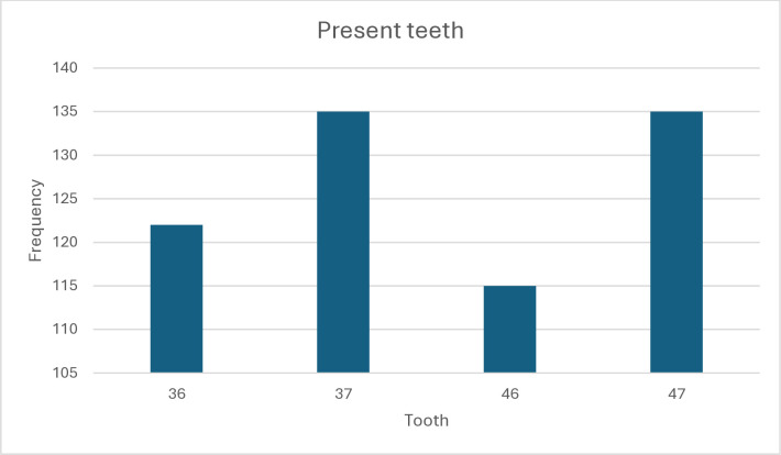 Figure 1
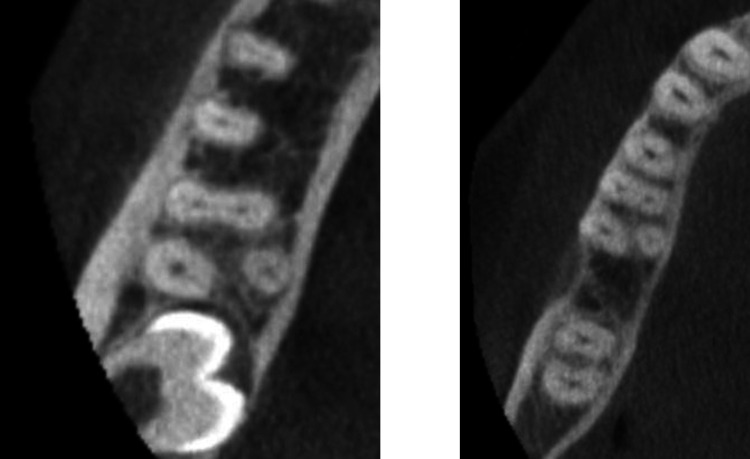 Figure 2
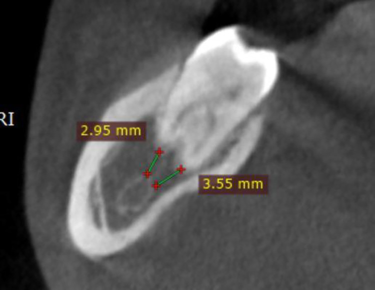 Figure 3
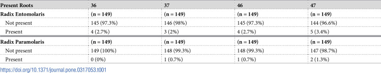 Figure 4
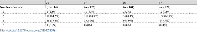 Figure 5
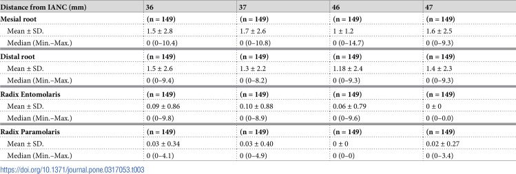 Figure 6
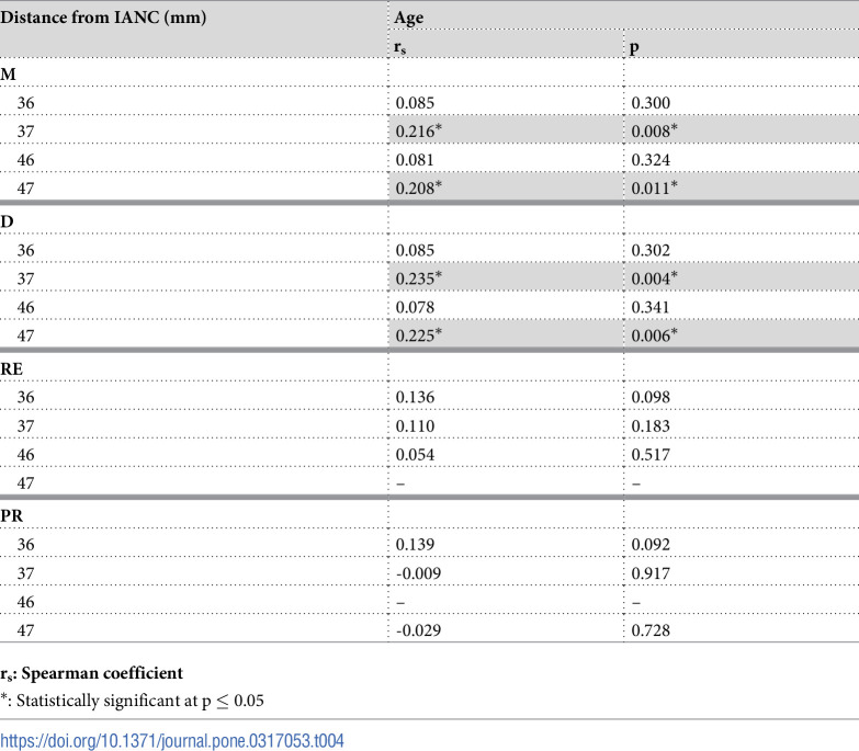 Figure 7
Peer Reviews
No public reviews on file for this paper yet. If you reviewed it on a platform where reviews are public (OpenReview, ICLR, NeurIPS, ICML), you can paste yours below so the community can read it here.
Videos
No videos yet. Explain this paper in a talk, walkthrough, or lecture? Add one.
Taxonomy
TopicsEndodontics and Root Canal Treatments · Dental Radiography and Imaging