Learning neuroimaging models from health system-scale data
Yiwei Lyu, Samir Harake, Asadur Chowdury, Soumyanil Banerjee, Rachel Gologorsky, Shixuan Liu, Anna-Katharina Meissner, Akshay Rao, Akhil Kondepudi, Cheng Jiang, Xinhai Hou, Rushikesh Joshi, Volker Neuschmelting, Ashok Srinivasan, Dawn Kleindorfer, Brian Athey, Vikas Gulani

TL;DR
A new AI model called Prima is developed using a large health system dataset to improve neuroimaging diagnosis and reduce healthcare system strain.
Contribution
Prima is the first vision language model for neuroimaging trained on health system-scale data and tested in real-world clinical settings.
Findings
Prima achieved a mean diagnostic area under the ROC curve of 90.1 ± 5.0% across 52 radiologic diagnoses.
Prima outperformed existing general and medical AI models in neuroimaging tasks.
Prima provides explainable diagnoses, worklist prioritization, and referral recommendations while demonstrating algorithmic fairness.
Abstract
Neuroimaging is a ubiquitous tool for evaluating patients with neurological diseases. The global demand for magnetic resonance imaging (MRI) studies has risen steadily, placing significant strain on health systems, prolonging turnaround times, and intensifying physician burnout [1, 2]. These challenges disproportionately impact patients in low-resource and rural settings [3]. Here, we utilized a large academic health system as a data engine to develop Prima, the first vision language model (VLM) serving as an AI foundation for neuroimaging that supports real-world, clinical MRI studies as input. Trained on over 220,000 MRI studies, Prima uses a hierarchical vision architecture that provides general and transferable MRI features. Prima was tested in a 1-year, prospective, health system-wide study that included 30K MRI studies. Across 52 radiologic diagnoses from the major neurologic…
Genes, proteins, chemicals, diseases, species, mutations and cell lines named across the full text — each resolved to its canonical identifier and authoritative record.
Click any figure to enlarge with its caption.
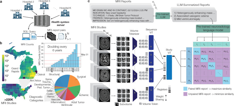 Figure 1
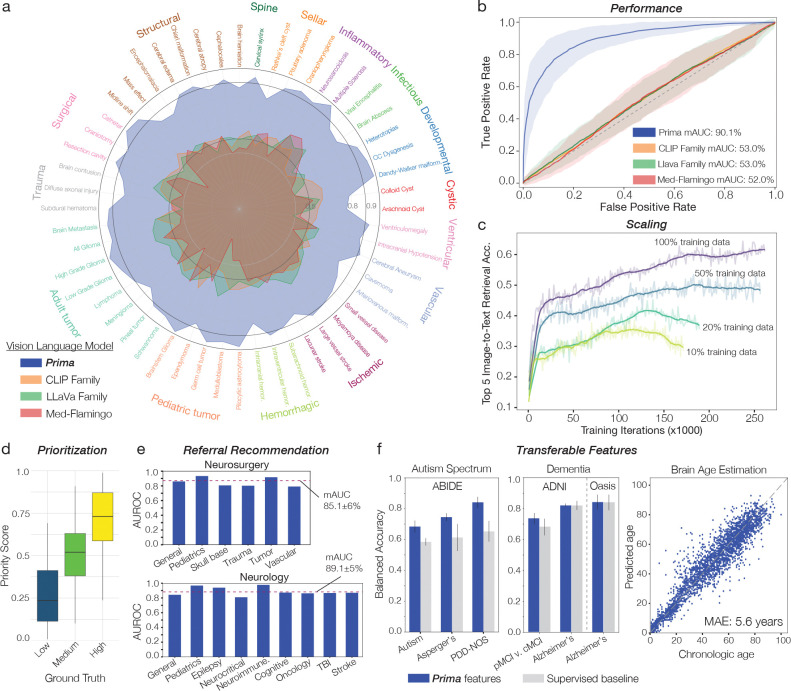 Figure 2
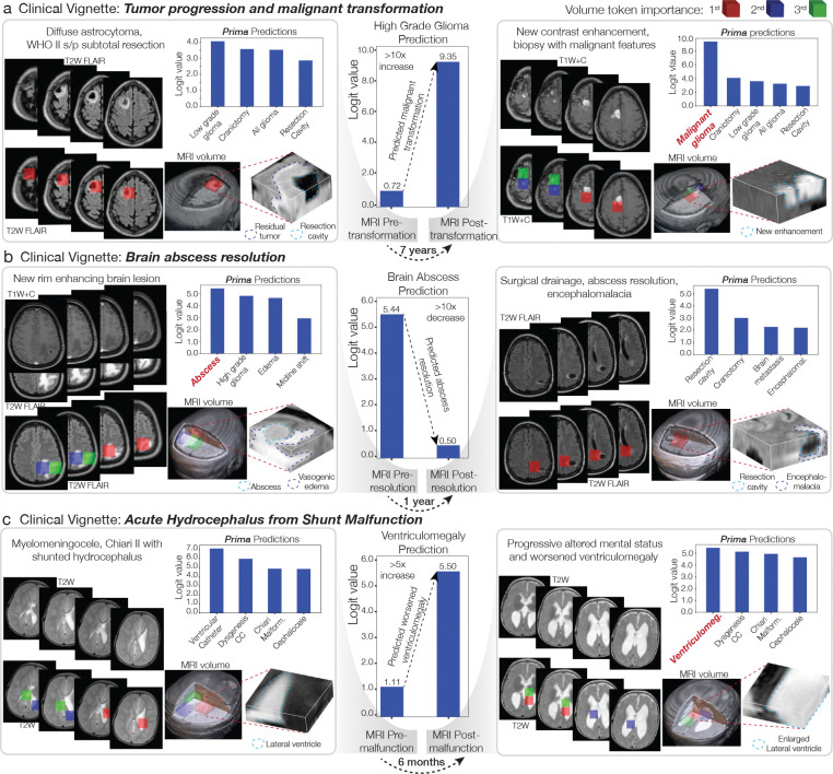 Figure 3
 Figure 4
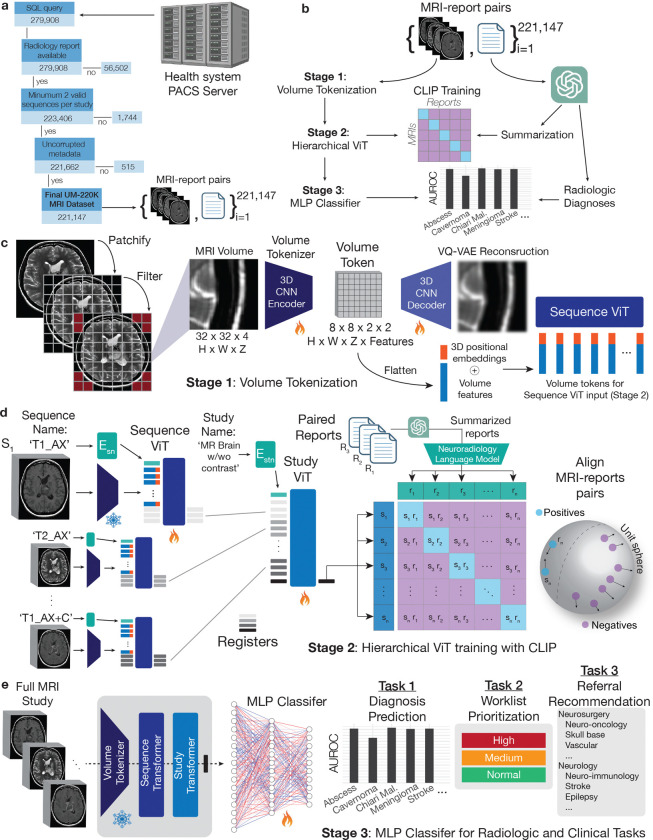 Figure 5
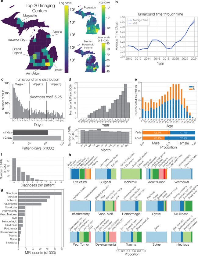 Figure 6
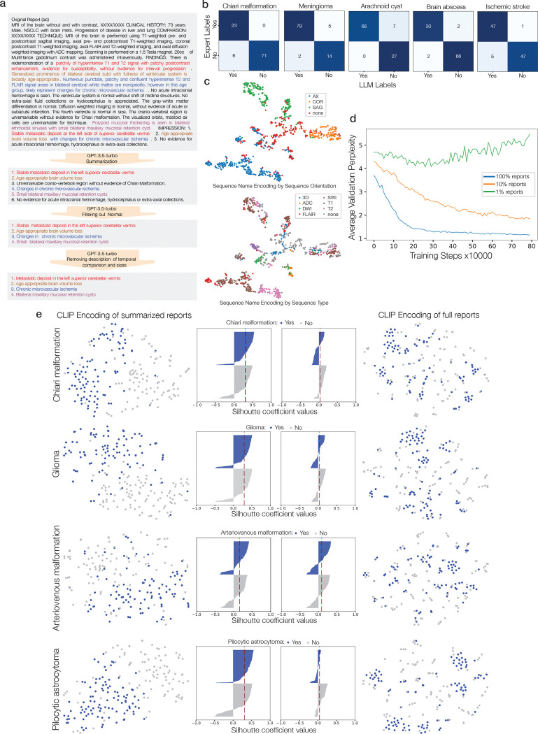 Figure 7
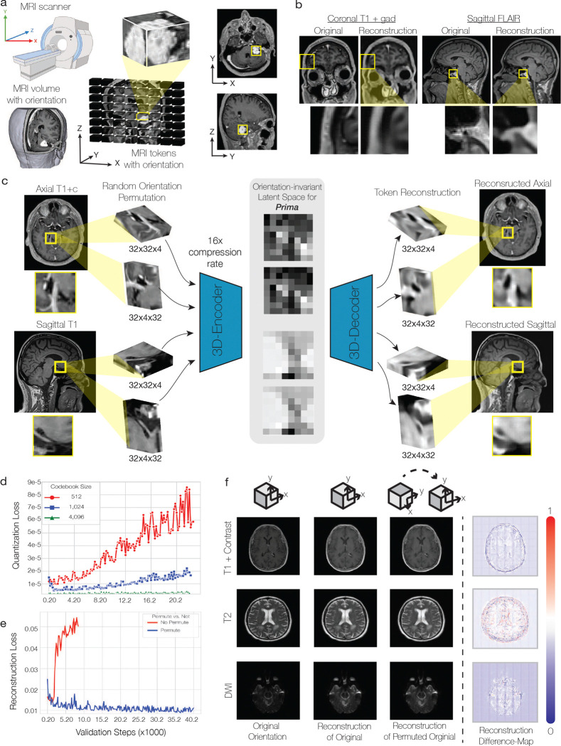 Figure 8
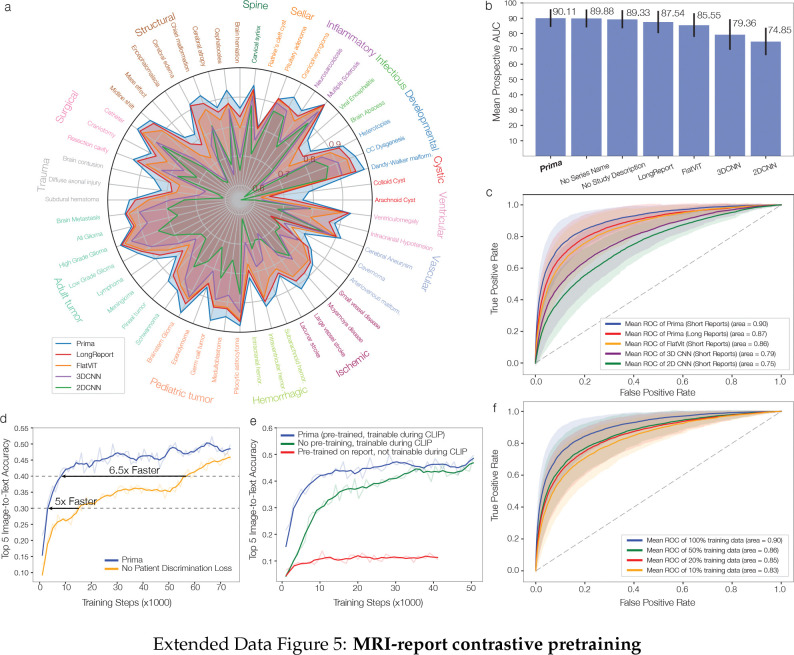 Figure 9
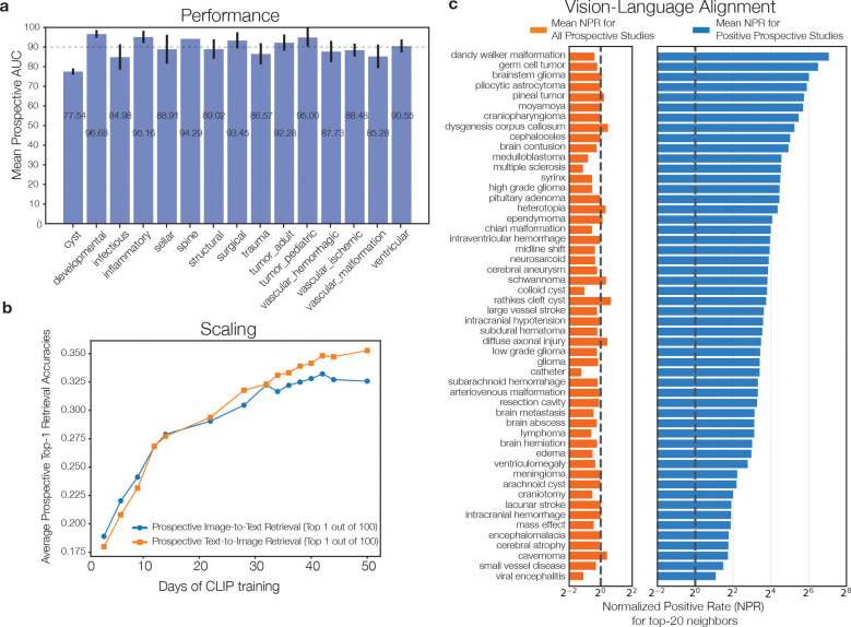 Figure 10
 Figure 11
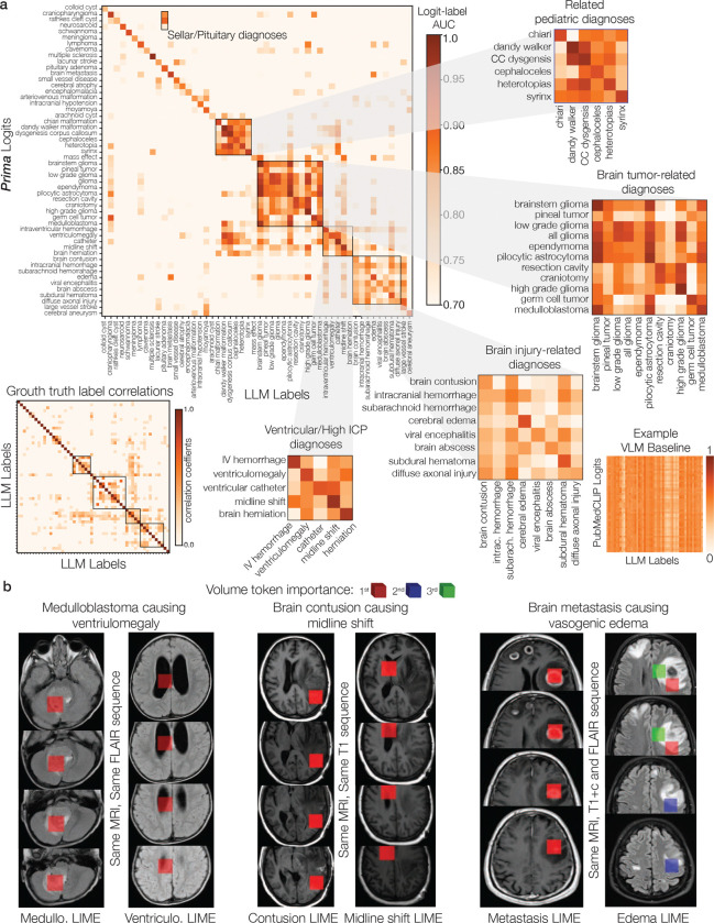 Figure 12
 Figure 13
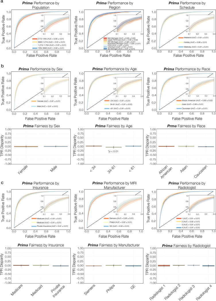 Figure 14
Peer Reviews
No public reviews on file for this paper yet. If you reviewed it on a platform where reviews are public (OpenReview, ICLR, NeurIPS, ICML), you can paste yours below so the community can read it here.
Videos
No videos yet. Explain this paper in a talk, walkthrough, or lecture? Add one.
Taxonomy
TopicsRadiomics and Machine Learning in Medical Imaging · Artificial Intelligence in Healthcare and Education · Glioma Diagnosis and Treatment