Cholangioscopy-guided lithotripsy and extraction of gallbladder stones through the natural lumen
Tong Xiao, Changqin Xu, Hongwei Xu, Yuemin Feng, Tong Su, Lechang Zhang, Shulei Zhao

Abstract
Genes, proteins, chemicals, diseases, species, mutations and cell lines named across the full text — each resolved to its canonical identifier and authoritative record.
Click any figure to enlarge with its caption.
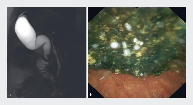 Fig. 1
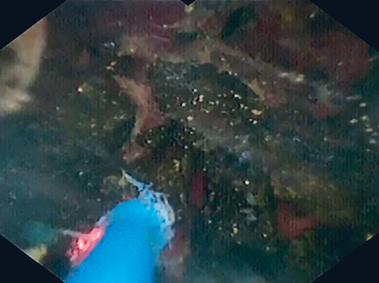 Fig. 2
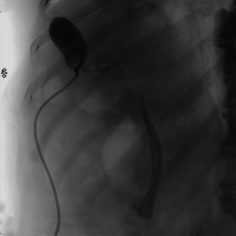 Fig. 3
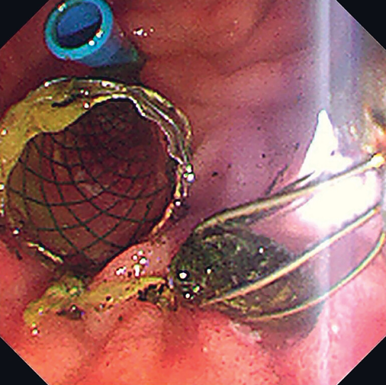 Fig. 4
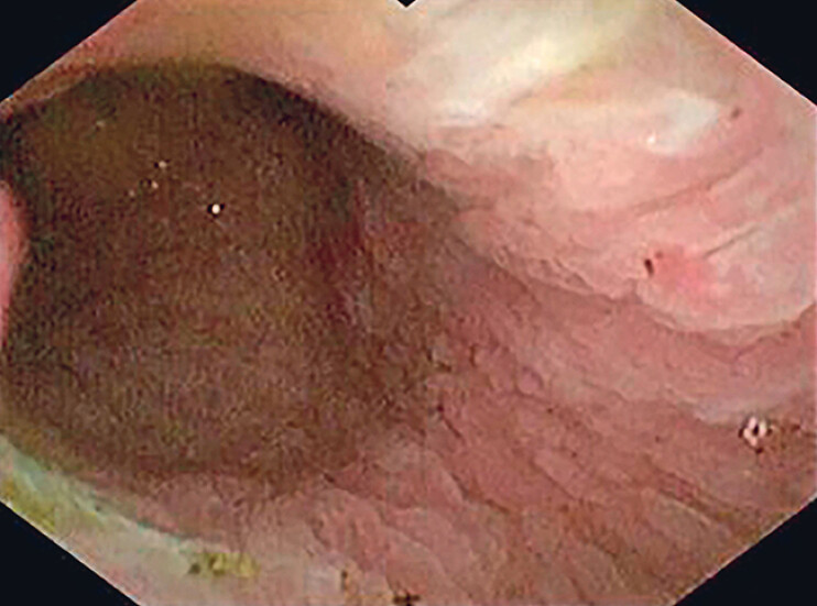 Fig. 5
Peer Reviews
No public reviews on file for this paper yet. If you reviewed it on a platform where reviews are public (OpenReview, ICLR, NeurIPS, ICML), you can paste yours below so the community can read it here.
Videos
No videos yet. Explain this paper in a talk, walkthrough, or lecture? Add one.
Taxonomy
TopicsGallbladder and Bile Duct Disorders · Pediatric Hepatobiliary Diseases and Treatments · Biliary and Gastrointestinal Fistulas
In recent years, the integration of endoscopic retrograde cholangiopancreatography (ERCP) and cholangioscopy has emerged as a prominent method for managing complex cholelithiasis 1 2 3 . We present a case demonstrating an innovative and effective approach to gallbladder stone extraction via the natural lumen ( Video 1 ).
Cholangioscopy-guided lithotripsy is performed to fragment a large stone lodged in the neck of the gallbladder, allowing subsequent complete stone clearance through the natural lumen.Video 1
A 52-year-old man presented with intermittent abdominal pain persisting over 2 months. Magnetic resonance cholangiopancreatography revealed an enlarged gallbladder with a stone lodged in its neck and additional stones in the common bile duct (CBD), a finding subsequently confirmed through direct observation with a cholangioscope ( Fig. 1 ). Given the functional status of the gallbladder and the patient's history of two prior abdominal surgeries, we opted for a natural lumen stone extraction strategy. The procedure commenced with gallbladder puncture and drainage to alleviate pressure, followed by ERCP. Two CBD stones were initially extracted using a balloon technique. Subsequently, a 9-Fr cholangioscope was navigated along the guidewire, to enable laser lithotripsy under direct visualization. Following multiple applications, the stone was successfully fragmented into smaller pieces ( Fig. 2 ).
Images showing a stone of approximately 1.0 cm in size lodged in the neck of the gallbladder on: a magnetic resonance cholangiopancreatography; and b cholangioscopy.
Cholangioscopic view showing the stone being fragmented into smaller pieces using laser lithotripsy.
A 10-mm × 10-cm fully coated metal stent was then positioned, with its upper part located at the gallbladder neck and the lower part exiting at the duodenal papilla. To prevent bile duct obstruction, an 8.5-Fr ×7-cm plastic stent was inserted ( Fig. 3 ). The patient experienced mild abdominal discomfort and transient amylase elevation post-ERCP, which promptly resolved with symptomatic measures.
Fluoroscopic image showing the metal and plastic stents in position in the bile duct.
The gallbladder was re-accessed 4 days later using a choledochoscope passed through the metal stent. Under direct visualization, all of the remaining gallstones were extracted using a mini stone-retrieval basket ( Fig. 4 ). After complete stone clearance had been confirmed ( Fig. 5 ), both stents were removed. The patient was kept fasted for 1 day, before being discharged. A subsequent ultrasound examination 3 months later revealed no evidence of residual stones.
Endoscopic image 4 days after the initial procedure showing the gallstones being removed using a mini stone-retrieval basket.
Cholangioscopic view showing no evidence of residual stones in the gallbladder.
Endoscopy_UCTN_Code_TTT_1AR_2AH
The reference list from the paper itself. Each links out to its DOI / PubMed record.
- 1Troncone E Mossa M De Vico P Difficult biliary stones: A comprehensive review of new and old lithotripsy techniques Medicina 20225812010.3390/medicina 5801012035056428 PMC 8779004 · doi ↗ · pubmed ↗
- 2Oh CH Dong SH Recent advances in the management of difficult bile-duct stones: A focus on single-operator cholangioscopy-guided lithotripsy Korean J Intern Med 20213623524610.3904/kjim.2020.42532972127 PMC 7969058 · doi ↗ · pubmed ↗
- 3Zhang W-L Ji R Cystic duct stump stone removal by retrieval basket under direct visualization using a novel peroral choledochoscope Endoscopy 202255 E 100E 10110.1055/a-1938-808136216264 PMC 9829795 · doi ↗ · pubmed ↗