Exploratory high b value diffusion-weighted MR for quantitative differentiation of ileocecal inflammatory conditions and tumors
Hao Yu, Yucheng Hai, Jingyu Lu

TL;DR
This study shows that high b value diffusion-weighted MRI can better distinguish between bowel tumors and inflammatory conditions than conventional MRI methods.
Contribution
The study introduces high b value (2000 s/mm²) diffusion-weighted imaging as a more effective quantitative tool for differentiating ileocecal tumors from inflammatory conditions.
Findings
ADC values and SIR values from high b value DWI showed significantly better diagnostic accuracy than conventional b value DWI.
SIR values from high b value DWI achieved the highest area under the curve (AUC) for differentiating tumors from inflammatory conditions.
Quantitative DWI parameters, especially SIR from high b value imaging, are promising non-invasive biomarkers for diagnostic differentiation.
Abstract
To explore the quantitative analysis of high b value (2000 s/mm2) diffusion-weighted imaging (DWI) for the differentiation of ileocecal inflammatory conditions and tumors, compared with conventional b value (800 s/mm2) DWI. Sixty-six patients with 30 tumors and 36 inflammatory conditions underwent MR enterography with conventional and high b values DWI. Quantitative apparent diffusion coefficient (ADC) values and signal intensity ratios (SIRs) of lesions of the psoas muscle were measured from the two b value DWIs. The receiver operating characteristic (ROC) curve was applied to determine the diagnostic value of ADC and SIR for differentiating tumors from inflammatory conditions. The ADC values of tumors were significantly lower than those of inflammatory conditions in 800 s/mm2 (p = 0.001) and 2000 s/mm2 (p < 0.001) DWI. In addition, tumors exhibited significantly higher SIR values…
Genes, proteins, chemicals, diseases, species, mutations and cell lines named across the full text — each resolved to its canonical identifier and authoritative record.
Click any figure to enlarge with its caption.
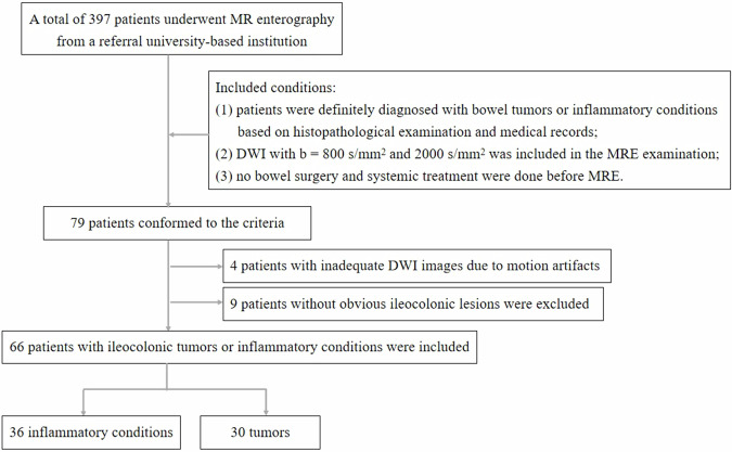 Figure 1
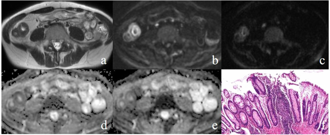 Figure 2
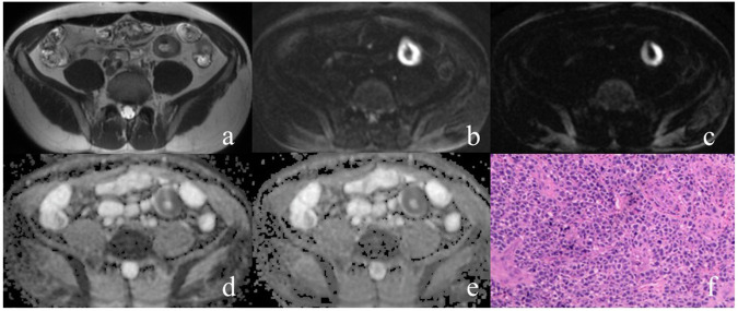 Figure 3
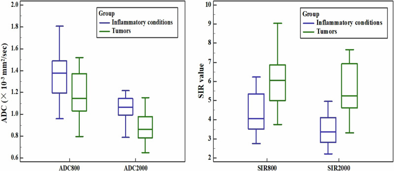 Figure 4
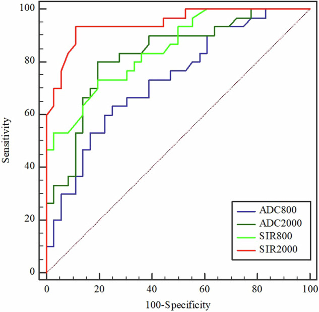 Figure 5
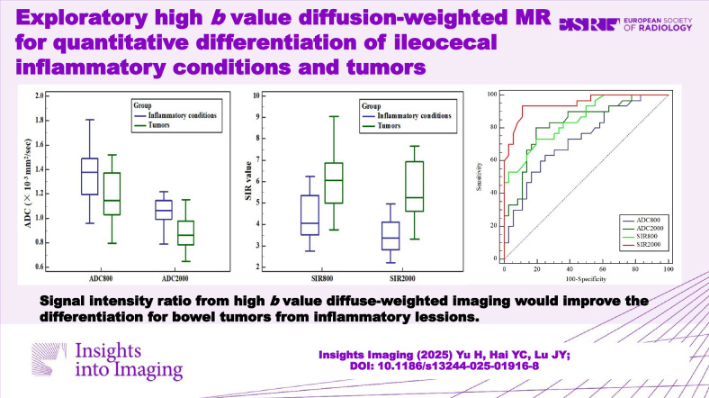 Figure 6
Peer Reviews
No public reviews on file for this paper yet. If you reviewed it on a platform where reviews are public (OpenReview, ICLR, NeurIPS, ICML), you can paste yours below so the community can read it here.
Videos
No videos yet. Explain this paper in a talk, walkthrough, or lecture? Add one.
Taxonomy
TopicsMRI in cancer diagnosis · Radiomics and Machine Learning in Medical Imaging · Inflammatory Biomarkers in Disease Prognosis