Author Correction: SGF29 nuclear condensates reinforce cellular aging
Kaowen Yan, Qianzhao Ji, Dongxin Zhao, Mingheng Li, Xiaoyan Sun, Zehua Wang, Xiaoqian Liu, Zunpeng Liu, Hongyu Li, Yingjie Ding, Si Wang, Juan Carlos Izpisua Belmonte, Jing Qu, Weiqi Zhang, Guang-Hui Liu

Abstract
Genes, proteins, chemicals, diseases, species, mutations and cell lines named across the full text — each resolved to its canonical identifier and authoritative record.
Click any figure to enlarge with its caption.
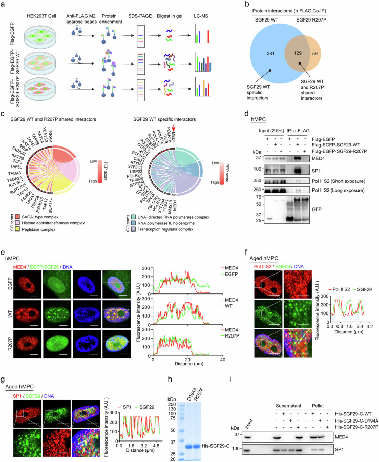 Figure 1
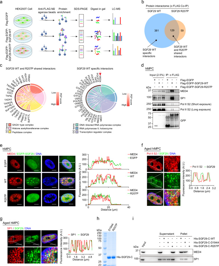 Figure 2
Peer Reviews
No public reviews on file for this paper yet. If you reviewed it on a platform where reviews are public (OpenReview, ICLR, NeurIPS, ICML), you can paste yours below so the community can read it here.
Videos
No videos yet. Explain this paper in a talk, walkthrough, or lecture? Add one.
Taxonomy
TopicsRNA Research and Splicing · RNA modifications and cancer · Genomics and Chromatin Dynamics
Correction to: Cell Discovery 10.1038/s41421-023-00602-7, published online 07 November 2023
In this article, we identified an oversight in Fig. 5g^1^, where the red and green channel images were inadvertently reversed. Also, Supplementary Fig. S7d contained an oversight with the selection of an atypical nucleus for EGFP-SGF29-WT group.
Incorrect Figure 5. Fig. 5Identification of SGF29 interacting proteins sensitive to condensate perturbation.a Flow chart of the mass spectrometry strategy for the identification of EGFP-SGF29-WT (WT) and EGFP-SGF29-R207P (R207P) interacting proteins. Flag-EGFP was used as a control. b Shared and specific interacting proteins of EGFP-SGF29-WT (WT) and EGFP-SGF29-R207P (R207P) identified by mass spectrometry. c Chord diagrams showing the enriched pathways of shared interaction partners of EGFP-SGF29-WT (WT) and EGFP-SGF29-R207P (R207P) (left) and those of specific protein interaction partners of EGFP-SGF29-WT (right). d Co-IP analysis showing the interaction between indicated proteins and EGFP-SGF29-WT (WT) and EGFP-SGF29-R207P (R207P) in hMPCs. e Immunofluorescence staining of MED4 in hMPCs transduced with lentiviruses expressing either EGFP, EGFPSGF29-WT (WT) or EGFP-SGF29-R207P (R207P). Left, representative images. Scale bar, 10 μm. Right, quantification of the fluorescence intensity along the line embedded the image following the arrow direction. f Immunofluorescence staining of Pol II S2 and SGF29 in senescent hMPCs. Left, representative images. Scale bars, 10 μm and 5 μm (zoomed-in image). Right, quantification of the fluorescence intensity along the line embedded the image following the arrow direction. g Immunofluorescence staining of SP1 and SGF29 in senescent hMPCs. Left, representative images. Scale bars, 10 μm and 5 μm (zoomed-in image). Right, quantification of the fluorescence intensity along the line embedded the image following the arrow direction. h Coomassie blue staining of purified recombinant SGF29-C-D194A and SGF29-C-R207P after being resolved on SDS-PAGE. i Pelleting assay show that SGF29-C-D194A and SGF29-C-R207P interact with MED4 and SP1.
Correct Figure 5. Fig. 5Identification of SGF29 interacting proteins sensitive to condensate perturbation.a Flow chart of the mass spectrometry strategy for the identification of EGFP-SGF29-WT (WT) and EGFP-SGF29-R207P (R207P) interacting proteins. Flag-EGFP was used as a control. b Shared and specific interacting proteins of EGFP-SGF29-WT (WT) and EGFP-SGF29-R207P (R207P) identified by mass spectrometry. c Chord diagrams showing the enriched pathways of shared interaction partners of EGFP-SGF29-WT (WT) and EGFP-SGF29-R207P (R207P) (left) and those of specific protein interaction partners of EGFP-SGF29-WT (right). d Co-IP analysis showing the interaction between indicated proteins and EGFP-SGF29-WT (WT) and EGFP-SGF29-R207P (R207P) in hMPCs. e Immunofluorescence staining of MED4 in hMPCs transduced with lentiviruses expressing either EGFP, EGFPSGF29-WT (WT) or EGFP-SGF29-R207P (R207P). Left, representative images. Scale bar, 10 μm. Right, quantification of the fluorescence intensity along the line embedded the image following the arrow direction. f Immunofluorescence staining of Pol II S2 and SGF29 in senescent hMPCs. Left, representative images. Scale bars, 10 μm and 5 μm (zoomed-in image). Right, quantification of the fluorescence intensity along the line embedded the image following the arrow direction. g Immunofluorescence staining of SP1 and SGF29 in senescent hMPCs. Left, representative images. Scale bars, 10 μm and 5 μm (zoomed-in image). Right, quantification of the fluorescence intensity along the line embedded the image following the arrow direction. h Coomassie blue staining of purified recombinant SGF29-C-D194A and SGF29-C-R207P after being resolved on SDS-PAGE. i Pelleting assay show that SGF29-C-D194A and SGF29-C-R207P interact with MED4 and SP1.
While these issues do not impact the results or conclusions, we are committed to the highest standards of publication integrity. The original figure and Supplementary figures have been corrected. We apologize for any inconvenience that it may have caused.
Supplementary information
Supplementary information