Comparing the postoperative refractive predictability of Pentacam HR and IOLMaster 500 after a multifocal intraocular lens implantation
Newton Andrade Junior, Wilson Takashi Hida, André Marcio Vieira Messias, João Marcelo Lyra, Carlos André Mont’Alverne Silva, Milton Ruiz Alves

TL;DR
This study compares the accuracy of two devices, Pentacam HR and IOLMaster 500, in predicting postoperative vision outcomes after multifocal lens implantation.
Contribution
The study introduces a comparison of refractive predictability using different biometric measurements from two devices in multifocal intraocular lens implantation.
Findings
Pentacam HR and IOLMaster 500 showed significant differences in keratometry measurements.
The anterior chamber depth measurements were equally accurate between the two devices.
Using IOLMaster 500 for anterior chamber depth resulted in smaller prediction differences.
Abstract
To compare the postoperative refractive predictability of IOLMaster 500 and Pentacam HR on the basis of keratometry and anterior chamber depth values in eyes with an indication for multifocal intraocular lens (IOL) implantation. This was a retrospective study conducted on 118 eyes treated with phacoemulsification and multifocal intraocular lens implantation. Only the eyes that achieved emmetropia in the dynamic refraction performed on postoperative day 30 were included. Haigis’ formula was used in each case to calculate the intraocular lens power, and the intraocular lens with the target refraction closest to emmetropia was implanted. Four lens calculation scenarios were tested by combining keratometry and anterior chamber depth measurements obtained using the two devices. IOLMaster 500 and Pentacam HR differed with regard to mean keratometry (∆ 0.07 ± 0.03 D; p=0.0065) and anterior…
Genes, proteins, chemicals, diseases, species, mutations and cell lines named across the full text — each resolved to its canonical identifier and authoritative record.
Click any figure to enlarge with its caption.
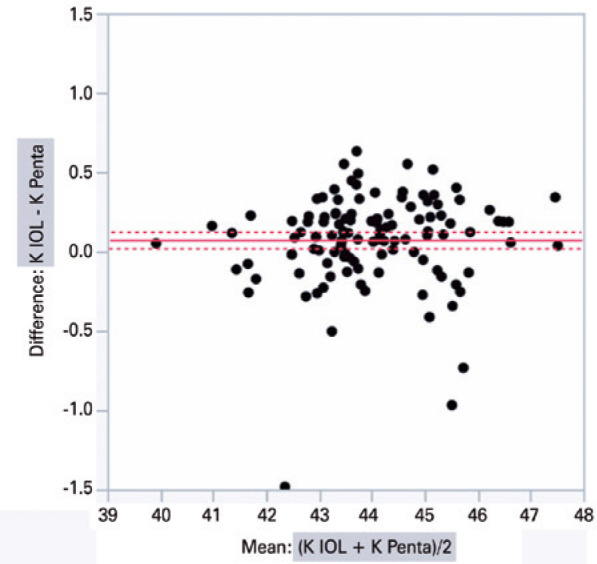 Figure 1
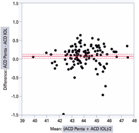 Figure 2
| Lens calculation scenario | ACD IOL | ACD penta | Best IOL | Ideal best | Ideal IOL | K IOL | K penta |
|---|---|---|---|---|---|---|---|
| K IOL × ACD IOL | 3.11 ± 0.03 | 3.19 ± 0.03 | 21.73 ± 0.24 | 0.18 ± 0.01 | 21.56 ± 0.24 | 44.03 ± 0.12 | 43.95 ± 0.12 |
| K IOL × ACD Penta | 3.11 ± 0.03 | 3.19 ± 0.03 | 21.73 ± 0.24 | 0.15 ± 0.01 | 21.59 ± 0.24 | 44.03 ± 0.12 | 43.95 ± 0.12 |
| K Penta × ACD IOL | 3.11 ± 0.03 | 3.19 ± 0.03 | 21.73 ± 0.24 | 0.28 ± 0.03 | 21.66 ± 0.24 | 44.03 ± 0.12 | 43.95 ± 0.12 |
| K Penta × ACD Penta | 3.11 ± 0.03 | 3.19 ± 0.03 | 21.73 ± 0.24 | 0.28 ± 0.03 | 21.69 ± 0.24 | 44.03 ± 0.12 | 43.95 ± 0.12 |
| Mean K/ACD | Mean K/ ACD | Difference | SE | Lower Upper | ||
|---|---|---|---|---|---|---|
| Penta/IOL | IOL/Penta | 0.14 | 0.03 | 0.06 | 0.21 | <0.0001 |
| Penta/Penta | IOL/Penta | 0.13 | 0.03 | 0.06 | 0.21 | <0.0001 |
| Penta/IOL | IOL/IOL | 0.11 | 0.03 | 0.04 | 0.19 | 0.0012 |
| Penta/Penta | IOL/IOL | 0.11 | 0.03 | 0.04 | 0.19 | 0.0016 |
| IOL/IOL | IOL/Penta | 0.03 | 0.03 | -0.06 | 0.10 | 0.8654 |
| Penta/IOL | Penta/Penta | 0.01 | 0.03 | -0.08 | 0.08 | 0.9967 |
Peer Reviews
No public reviews on file for this paper yet. If you reviewed it on a platform where reviews are public (OpenReview, ICLR, NeurIPS, ICML), you can paste yours below so the community can read it here.
Videos
No videos yet. Explain this paper in a talk, walkthrough, or lecture? Add one.
Taxonomy
TopicsIntraocular Surgery and Lenses · Corneal surgery and disorders · Ophthalmology and Visual Impairment Studies
INTRODUCTION
Over the past 30 years, several formulas and devices have been proposed to improve the refractive predictability and reduce refractive errors after a cataract sur gery^(1-3)^. As calculations are based on preoperative eye dimensions, such as axial length (AL), keratometry (K), and anterior chamber depth (ACD), careful eye mea surements should be performed for accuracy. The refractive outcome is predicted based on three main factors: i) uneventful surgery with a well-centered in-the-bag implanted intraocular lens (IOL); ii) accuracy of preoperative biometric data (AL, ACD, and K); and iii) predictability of the formula used to calculate IOL power, using optimized IOL constants^(4-13)^. For example, a 1-mm deviation in the corneal diameter, axial diameter, or ACD has been reported to result in a postoperative refractive error of 5.7 D, 2.7 D, or 1.5 D, respectively^(11)^.
Postoperative refraction predictability is even more important when implanting multifocal IOLs. IOLMaster 500 (Carl Zeiss Meditec AG, Jena, Germany) is the gold standard for biometric measurements and calculations; however, some studies have questioned the accuracy of its generated K measurements (using data from six light reflections at a 2.3-mm diameter), especially when compared to Pentacam HR (Oculus Optikgeräte GmbH, Wetzlar, Germany), which uses a Scheimpflug camera (180°) and a monochromatic slit-light source combined with a static camera^(14)^. Reitblat et al.^(15)^ recently compared the accuracy of IOLMaster and Lenstar in patients undergoing multifocal IOL implantation (SN6AD1; Alcon Laboratories, Inc., Fort Worth, TX, USA) and concluded that both devices were highly accurate, when using similar measurement methods.
Therefore, this study aimed to compare the postoperative refractive predictability of IOLMaster 500 and Pentacam HR based on K and ACD values in the eyes implanted with multifocal IOLs.
METHODS
The study was conducted at the Cataract Division of Hospital Oftalmológico de Brasilia, Brazil, with the study protocol complying the tenets of the Declaration of Helsinki and approved by the institutional ethics committee.
Patients and contraindications for multifocal IOL
Medical records of all eyes submitted for cataract surgery with multifocal IOL implantation (AcrySof IQ ReSTOR SN6AD1, Alcon, USA) between January 2014 and October 2015 were retrospectively reviewed. Eligible participants were all patients aged 45-65 years with bilateral senile cataract, corneal astigmatism of <1.00 diopter in both eyes; pupil diameter of at least 3.5 mm under mesopic conditions; and absence of other eye diseases, topical hypotensive medication use, and previous eye surgery. Intraoperative and postoperative exclusion criteria were doubts about IOL implantation within the capsular bag or capsulorhexis described as >0.5 mm as verified by the slit-lamp examination, and patients who did not achieve emmetropia in the dynamic refraction performed on postoperative day 30. Because postoperative ACD was not measured, the study was designed to analyze only the eyes that achieved emmetropia in the dynamic refraction performed on postoperative day 30.
The main contraindications for multifocal IOL implantation in this study were:
History of ocular surgery.Systemic changes capable of interfering with postoperative healing (e.g., diabetes mellitus, autoimmune conditions, connective tissue disorders).Preexisting ocular disease compromising visual acuity (e.g., herpetic ocular disease, moderate or severe dry eye syndrome, uveitis, glaucoma, retinal disorders).Incomplete records with regard to the study parametersMacular changes indicating imminent central vision loss (age-related macular degeneration, macular edema, macular hole, epiretinal membrane).Corneal changes interfering with K (pterygium, scars, other opacities).
Surgical procedure
All surgical procedures were performed by a single experienced surgeon (WTH) at a surgical center following the standardized surgical technique. Under topical anesthesia, a clear self-sealing corneal 2.75-mm incision was made in the steep meridian, followed by continuous circular capsulorhexis and hydrodissection with 1% lidocaine without preservatives diluted in 10 mL of balanced saline solution. Then, the soft-shell technique was performed using Celoftal^®^ (hydroxypropyl methylcellulose; Alcon Laboratories, Fort Worth, TX, USA) and cohesive Provisc^®^ (sodium hyaluronate 1%; Alcon Laboratories, Fort Worth, TX, USA), whereas conventional phacoemulsification was performed using an Infiniti^®^ system (Alcon Laboratories, Fort Worth, TX, USA) with an IOL implanted in the capsular bag using a Royale^®^ injector (ASICO, Westmont, IL, USA).
Postoperatively, a fluoroquinolone (moxifloxacin 0.5%, Vigamox^®^; Alcon Laboratories, Fort Worth, TX, USA) was topically administered every 6 h for 7 days along with a topical corticosteroid (dexamethasone 1%, Maxidex^®^; Alcon Laboratories, Fort Worth, TX, USA), initially 1 drop every 4 h, and gradually tapered over 30 days.
Measurements and calculations
The analysis included visual acuity with and without correction, biomicroscopy, specular microscopy, retinal mapping, and preoperative measurements obtained with IOLMaster 500 (Zeiss, Germany) and Pentacam HR (Oculus, Germany). As only the eyes that achieved emmetropia on postoperative day 30 were analyzed, the dynamic refraction performed on postoperative day 30 was used as a reference when comparing the postoperative refractive predictability of IOLMaster 500 and Pentacam HR based on K and ACD values. Haigis’ formula was used in each case to calculate the IOL power, and the IOL with the target refraction closest to emmetropia was implanted. Four lens calculation scenarios were tested by combining K and ACD measurements obtained using the two devices: K and ACD measured with IOLMaster 500; K and ACD measured with Pentacam HR; K measured with IOLMaster 500/ACD measured with Pentacam HR; and K measured with Pentacam HR/ACD measured with IOLMaster 500 (Table 1).
Table 1: Calculation of intraocular lens power using Haigis’ formula (n = 118 eyes)
ACD was measured from the corneal epithelium to the anterior lens capsule and from the corneal endothelium to the anterior lens capsule^(16)^. To ensure comparability between the measurements obtained with the two devices, the central corneal thickness was measured from the epithelium to the endothelium when using Pentacam HR, and this value was added to the ACD endothelium-to-lens value (“AD” on the display). This result is equivalent to the ACD epithelium-to-lens value calculated by IOLMaster 500.
Statistical analysis
Paired t-test and Bland-Altman plot analysis were used to compare the K and ACD measured with the two devices. The analysis of covariance was used to determine the influence of AL and each measuring device in order to include all effects in the model, and then the Tukey’s HSD test was subsequently performed. The level of statistical significance was set at p<0.05.
RESULTS
A total of 118 operated eyes (M=55/F=63) that achieved emmetropia in the dynamic refraction on postoperative day 30 were included in this study. The mean patient age, nuclear classification, and preoperative visual acuity were 62.3 years, 2 (N2), and 0.49 without correction and 0.89 with correction, respectively (expressed in logMAR and measured using the Early Treatment Diab etic Retinopathy Study table). No intraoperative com plications were observed.
IOLMaster 500 and Pentacam HR significantly differed with regard to the mean K (44.03 ± 1.34 D vs. 43.95 ± 1.32 D; intra-individual difference of 0.07 ± 0.02 D; p<0.001) and ACD (3.11 ± 0.35 mm vs. 3.19 ± 0.35 mm; intra-individual difference of 0.08 ± 0.01 mm; p<0.001), respectively. The graphical analysis of paired differences in K measurements obtained with IOLMaster 500 and Pentacam HR is shown in figure 1. Likewise, the graphical analysis of paired differences in ACD measurements obtained with the two devices is shown in figure 2.
Figure 1. Graphical analysis of paired differences in keratometry (K) measurements obtained with IOLMaster 500 (IOL) and Pentacam HR (Penta).
Figure 2. Graphical analysis of paired differences in the anterior chamber depth (ACD) measurements obtained with IOLMaster 500 (IOL) and Pentacam HR (Penta).
The analysis of covariance produced the following values when comparing the biometric results of different combinations of K and ACD from Pentacam HR and IOLMaster 500, respectively, inserted according to the Haigis’ formula: PENTA/IOL × IOL/PENTA (0.13 ± 0.03, p<0.0001); PENTA/PENTA × IOL/PENTA (0.13 ± 0.03, p<0.0001); PENTA/IOL × IOL/IOL (0.11 ± 0.03, p=0.001); PENTA/PENTA × IOL/IOL (0.11 ± 0.03, p=0.002); IOL/ IOL × IOL/PENTA (0.02 ± 0.03, p=0.865); and PENTA/ IOL × PENTA/PENTA (0.002 ± 0.03, p=0.99) (Table 2). The two columns in the left show the device used to measure K and ACD.
Table 2: Differences between mean K and ACD values from Pentacam HR and IOLMaster 500 based on analysis of covariance (n=118 eyes)
The difference was smaller when ACD was measured with IOLMaster 500, regardless of which device used to measure K.
DISCUSSION
To improve the accuracy of biometric calculations, fourth-generation formulas, such as Haigis’ formula^(1,17)^, include not only K and AL but also ACD^(17)^. The more accurately these variables are measured, the greater the postoperative refractive predictability provided in the formula. Several measuring methods and devices are available; however, systematic differences have been observed between their results^(17,18)^. Currently, Pentacam HR significantly differed from IOLMaster 500 when calculating K. As regards ACD, the two devices were equally accurate.
Several authors have shown that coherence tomography generates higher ACD values than IOLMaster^(9,19,20)^. Previous studies also demonstrated that ACD values were significantly greater with Pentacam than with IOLMaster or Orbscan^(12,14,18,21,22)^. This is supported by our finding of a positive difference of 0.08 ± 0.01 mm in ACD when using Pentacam HR. As for K, IOLMaster and Pentacam are reported to generate similar values in the central 4.5 mm; however, the two devices differed by 0.07 ± 0.02 D in this study.
Haigis’ formula was developed to measure with IOLMaster, suggesting that this is the most appropriate technology for biometric calculations. However, after introducing the Pentacam HR technology (rotational Scheimpflug camera with controlled fixation making a detailed 3D scan of the anterior segment), anterior chamber and corneal measurements were expected to become more accurate, positively impacting the postoperative refractive predictability^(1,9,17,18,22-25)^.
This study has several limitations that should be addressed. Eighty eligible eyes submitted for cataract surgery with multifocal IOL implantation during the study period were not included in the study because they did not achieve emmetropia in the dynamic refraction on postoperative day 30. To determine the lens power in the IOL plane, postoperative ACD should have been considered. However, postoperative ACD was not measured to make this correction and achieve the accuracy required in this study. Therefore, only 118 eyes that achieved emmetropia on postoperative day 30 were included. The 118 analyzed eyes were also included based on K and ACD measurements of the IOLMaster 500, which can be considered bias in the present study. Therefore, further studies should be conducted to correlate patients who had spherical equivalent was different from 0 D in the dynamic refraction on postoperative day 30. The importance of correlating the effective measurement of postoperative lens with this residual refraction should also be emphasized in future studies.
In conclusion, within the limitations in this study, the biometric calculations obtained from K measurements with Pentacam HR and IOLMaster 500 had a disagreement. However, for ACD measurements, the two devices were equally accurate.
The reference list from the paper itself. Each links out to its DOI / PubMed record.
- 1Miraftab M Hashemi H Fotouhi A Khabazkhoob M Rezvan F Asgari S. Effect of anterior chamber depth on the choice of intraocular lens calculation formula in patients with normal axial length Middle East Afr J Ophthalmol 20142143073112537163510.4103/0974-9233.142266 PMC 4219221 · doi ↗ · pubmed ↗
- 2Sahin A Hamrah P. Clinically relevant biometry Curr Opin Ophthalmol 201223147532208103210.1097/ICU.0b 013e 32834 cd 63e PMC 3299090 · doi ↗ · pubmed ↗
- 3Tappeiner C Rohrer K Frueh BE Waelti R Goldblum D. Clinical comparison of biometry using the non-contact optical low coher ence reflectometer (Lenstar LS 900) and contact ultrasound biometer (Tomey AL-3000) in cataract eyes Br J Ophthalmol 20109456666672044797610.1136/bjo.2009.167700 · doi ↗ · pubmed ↗
- 4Behndig A Montan P Lundström M Zetterström C Kugelberg M. Gender differences in biometry prediction error and intra-ocular lens power calculation formula Acta Ophthalmol 20149287597632493080610.1111/aos.12475 · doi ↗ · pubmed ↗
- 5Rönbeck M Lundström M Kugelberg M. Study of possible predictors associated with self-assessed visual function after cataract surgery Ophthalmology 2011 Sep 1189173217382171501310.1016/j.ophtha.2011.04.013 · doi ↗ · pubmed ↗
- 6Norrby S. Sources of error in intraocular lens power calculation J Cataract Refract Surg 20083433683761829905910.1016/j.jcrs.2007.10.031 · doi ↗ · pubmed ↗
- 7Preussner PR Olsen T Hoffmann P Findl O. Intraocular lens calculation accuracy limits in normal eyes J Cataract Refract Surg 20083458028081847163610.1016/j.jcrs.2008.01.015 · doi ↗ · pubmed ↗
- 8Aristodemou P Knox Cartwright NE Sparrow JM Johnston RL. Formula choice: hoffer Q, Holladay 1, or SRK/T and refractive outcomes in 8108 eyes after cataract surgery with biometry by partial coherence interferometry J Cataract Refract Surg 201137163712118310010.1016/j.jcrs.2010.07.032 · doi ↗ · pubmed ↗