Third case of gallbladder hemangioma associated with gallstones: a comprehensive literature review
Imane Boujguenna, Soumia Moujane, Fatima Boukis, Soufiane Abdouh, Ahmed Elguazzar

TL;DR
This paper reports the third known case of a rare gallbladder tumor linked to gallstones and reviews all previously documented cases.
Contribution
The paper adds a new case and emphasizes the underdiagnosis of gallbladder hemangiomas.
Findings
Only three cases of gallbladder hemangioma associated with gallstones have been reported.
Gallbladder hemangiomas are likely underdiagnosed and require careful examination of removed gallbladder tissue.
Abstract
Gallbladder hemangiomas are extremely rare benign tumors. Eighteen cases have been reported in the literature, with only two associated with gallstones. We report the third case of a gallbladder hemangioma associated with gallstones in a 54-year-old Moroccan woman, along with a comprehensive review of the literature. Gallbladder hemangiomas are likely underdiagnosed, underscoring the need for careful examination of cholecystectomy specimens.
Genes, proteins, chemicals, diseases, species, mutations and cell lines named across the full text — each resolved to its canonical identifier and authoritative record.
Click any figure to enlarge with its caption.
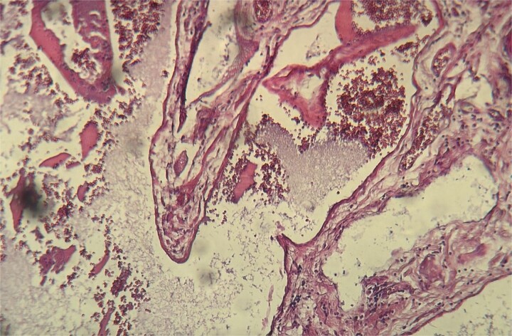 Figure 1
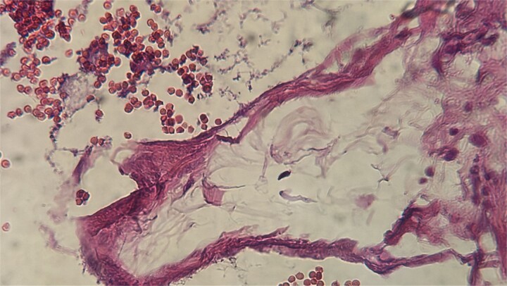 Figure 2
| Case | Year | Country | Age | Gender |
|---|---|---|---|---|
| [ | 2022 | Italy | 76 | M |
| [ | 2022 | India | 45 | F |
| Our case | 2024 | Morocco | 54 | F |
| Case | Year | Country | Age | Gender |
|---|---|---|---|---|
| [ | 1969 | |||
| [ | 1973 | |||
| [ | 1987 | 11 | F | |
| [ | 1997 | |||
| [ | 1997 | Spain | 50 | F |
| [ | 2005 | Italy | 49 | F |
| [ | 2016 | 51 | F | |
| [ | 2017 | China | 44 | M |
| [ | 2018 | USA | ||
| [ | 2018 | France | ||
| [ | 2018 | USA | ||
| [ | 2019 | USA | 80 | F |
| [ | 2019 | Korea | 53 | M |
| [ | 2019 | Japan | 75 | M |
| [ | 2021 | India | 62 | M |
| [ | 2024 | Turkey | 49 | F |
Peer Reviews
No public reviews on file for this paper yet. If you reviewed it on a platform where reviews are public (OpenReview, ICLR, NeurIPS, ICML), you can paste yours below so the community can read it here.
Videos
No videos yet. Explain this paper in a talk, walkthrough, or lecture? Add one.
Taxonomy
TopicsCholangiocarcinoma and Gallbladder Cancer Studies · Genetic and Kidney Cyst Diseases
Introduction
Hemangiomas are benign vascular tumors primarily located in the skin. Gallbladder hemangiomas are exceptionally rare, with only 18 cases reported in the literature, including 2 associated with gallstones [1]. We present the third case of gallbladder hemangioma associated with gallstones in a 54-year-old Moroccan woman.
Case report
A 54-year-old Moroccan woman with no significant medical history presented with right upper quadrant pain lasting for two months, accompanied by nausea but no fever, weight loss, or other systemic or digestive symptoms. Abdominal ultrasonography revealed gallstones. The patient underwent laparoscopic cholecystectomy. Macroscopic examination revealed a gallbladder measuring 8.6 cm in length and 2.3 cm in width, with a slightly thickened wall and the presence of gallstones. Microscopic examination showed chronic diverticular cholecystitis and a vascular proliferation measuring 10 mm, composed of variably sized, often ectatic blood vessels containing red blood cells (Fig. 1). The vessels were lined by a single layer of regular endothelium (Fig. 2). The patient’s postoperative course was uneventful.
Histopathological examination showing ectatic blood vessels containing red blood cells.
The vessels were lined by a single layer of regular endothelium.
Discussion
Hemangiomas are predominantly located in the skin, and gallbladder hemangiomas are exceedingly rare. We identified 18 cases in the literature, 2 of which were associated with gallstones (Tables 1 and 2), with our case being the third. Clinically, gallbladder hemangiomas are usually asymptomatic but can present with abdominal pain [1, 2], hemoperitoneum [17], or jaundice [18]. Abdominal ultrasonography, the initial imaging modality for hepatobiliary diseases, may fail to detect hemangiomas, particularly when gallstones are present, as seen in our case. Gallstones can explain the symptoms or pose a differential diagnostic challenge with neoplastic pathologies [18]. Macroscopically, gallbladder hemangiomas can be asymptomatic or appear as violaceous hemorrhagic tumors [1]. Microscopically, they resemble hemangiomas in other locations, showing vascular proliferation with ectatic vessels containing red blood cells and lined by regular endothelium. The histopathological diagnosis is generally straightforward, but in cases of endothelial detachment, immunohistochemistry using endothelial markers can aid diagnosis. The treatment is surgical, consisting of cholecystectomy. In cases with bleeding risk or suspected malignancy, laparotomy exploration is recommended [18]. Our case represents the third reported instance of gallbladder hemangioma associated with gallstones. Gallbladder hemangioma is an extremely rare benign tumor that is likely underdiagnosed. Careful macroscopic and microscopic examination of cholecystectomy specimens is essential for accurate diagnosis.
The reference list from the paper itself. Each links out to its DOI / PubMed record.
- 1Trucco G, Chiusa L, Tandoi F, et al. First report of a gallbladder hemangioma coexisting with gallstones: a case report and literature review of a rare finding. BMC Surg 2022;22:128. 10.1186/s 12893-022-01554-7.35382806 PMC 8985283 · doi ↗ · pubmed ↗
- 2Mohan S, Sudhanshu A, Badyal S, et al. Gallbladder masking haemangioma at the gallbladder fossa. Int J Med Sci Curr Res 2022;5:781–6.
- 3Sewell JH, Miron MA. Benign cavernous hemangioma of the gallbladder. Arch Pathol 1969;88:30–1.5793686 · pubmed ↗
- 4Moffat JH . Cavernous hemangioma of the gallbladder and liver. Can J Surg 1973;16:172–5.4689894 · pubmed ↗
- 5Jones WP, Keller FS, Odrezin GT, et al. Venous hemangioma of the gallbladder. Gastrointest Radiol 1987;12:319–21. 10.1007/BF 01885171.3305129 · doi ↗ · pubmed ↗
- 6Furukawa H, Kanai Y, Mukai K, et al. Arteriovenous hemangioma of the gallbladder: CT and pathologic findings. AJR Am J Roentgenol 1997;168:1383. 10.2214/ajr.168.5.9129455.9129455 · doi ↗ · pubmed ↗
- 7Mayorga M, Hernando M, Val-Bernal JF. Diffuse expansive cavernous hemangioma of the gallbladder. Gen Diagn Pathol 1997;142:211–5.9065585 · pubmed ↗
- 8Crucitti A, La Greca A, Antinori A, et al. Cavernous hemangioma of the gallbladder: case report and review of the literature. Tumori J 2005;91:432–5. 10.1177/030089160509100511.16459643 · doi ↗ · pubmed ↗