Polymicrobial Osteomyelitis in a Patient With Isolation of Trueperella bernardiae: A Case Report and Literature Review
Marco Antonio Delaye-Martínez, Edgar Samuel Vanegas-Rodríguez, Braulio Mendez-Sotelo, María de Lourdes García-Hernández, Claudia Adriana Colín-Castro, Rafael Franco-Cendejas, Luis Esaú López-Jácome

TL;DR
A 24-year-old woman with a chronic wound and multiple antibiotic treatments developed polymicrobial osteomyelitis involving Trueperella bernardiae, highlighting the role of bacterial interactions in such infections.
Contribution
This case report adds to the limited literature on Trueperella bernardiae in polymicrobial osteomyelitis and emphasizes the need for standardized susceptibility testing.
Findings
Trueperella bernardiae was isolated alongside multiple other bacteria in a case of polymicrobial osteomyelitis.
The patient's chronic wound and prior antibiotic use likely contributed to the infection's progression.
Successful treatment required a combination of antibiotics after initial monotherapy failed.
Abstract
Background: Trueperella bernardiae is a Gram-positive rod that has been described as an opportunistic pathogen in immunocompromised patients. In a significant number of documented cases, infections with Trueperella bernardiae have been associated with polymicrobial infections, which highlight the fact that important bacteria–bacteria relations might be involved in the natural course of these infections, especially in patients with chronic disease courses and a history of multiple antibiotic treatments. Case Presentation: We present a case of a 24-year-old woman with a 3-year history of a chronic pressure ulcer on the right foot associated with varus and cavus deformity. As per relevant medical history, she was positive for multiple wound healing sessions with wound debridement and a large number of antibiotic treatments with minimal improvement. Microbiological cultures were taken…
Genes, proteins, chemicals, diseases, species, mutations and cell lines named across the full text — each resolved to its canonical identifier and authoritative record.
Click any figure to enlarge with its caption.
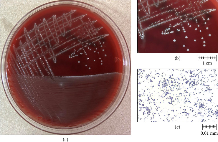 Figure 1
Peer Reviews
No public reviews on file for this paper yet. If you reviewed it on a platform where reviews are public (OpenReview, ICLR, NeurIPS, ICML), you can paste yours below so the community can read it here.
Videos
No videos yet. Explain this paper in a talk, walkthrough, or lecture? Add one.
Taxonomy
TopicsBacterial Identification and Susceptibility Testing · Diphtheria, Corynebacterium, and Tetanus · Streptococcal Infections and Treatments
1. Background
Trueperella bernardiae is a Gram-positive rod, predominantly coccobacilli, non-spore-forming, non-motile, facultatively anaerobic, catalase-negative, oxidase-negative, and indole-negative bacterium, with colony diameters ranging from 0.5 mm to 1.5 mm [1–4]. It can be found as a commensal in the human skin [3–6], oropharynx [3–5], and urogenital tract microbiota [2, 3]. The pathogenic role of T. bernardiae in human disease has not been established due to difficulties related to its isolation [2, 6, 7], its consideration as a contaminant in cultures [2, 7], and its low frequency as a part of a pathogenic process as well [4, 8]. However, the use of matrix-assisted laser desorption/ionization time-of-flight mass spectrometry (MALDI-TOF MS) technology has allowed clinical microbiologists to identify it. Therefore, it is increasingly documented in the literature [3].
Some authors have classified this microorganism as an opportunistic pathogen [1, 9], in which the presence of comorbidities related to an immunocompromised state, such as chronic degenerative diseases [3, 10–15] or reduced mobility [7, 8, 16–18], is frequently reported. In some cases, a history of multiple surgical procedures [7, 8, 19, 20] or prolonged antibiotic treatment courses [10, 21, 22] appears to be a common factor for infection with this pathogen, especially for polymicrobial infections. Therefore, the clinical context seems to be the most relevant component of T. bernardiae infection.
To our knowledge, only 39 cases associated with human infection have been reported in the medical literature, which consist of 4 cases of urinary tract infection [2, 10, 19, 20], 8 cases of joint and prosthesis infection (hip and knee) [2, 7, 11, 12, 23–25], 10 cases of soft tissue and wound infection [1, 2, 4–6, 8, 13–15], 13 cases of bacteremia or sepsis [2, 3, 9, 16, 17, 21, 26–28], 2 cases of cerebral abscess [22, 29], 1 case of endocarditis [18], and 1 case of Lemièrre syndrome [30].
A notable number of articles have documented cases of Trueperella bernardiae infection where more than one microorganism has been involved, in which patients have long antibiotic treatments due to the presence of chronic infections. These observations suggest a potential association between Trueperella bernardiae and the establishment of interspecies relationships that may contribute to its pathogenicity. Nevertheless, the precise nature of these relationships remains to be established in the context of this infection.
Here, we present a case of a young woman with a history of chronic soft tissue infection that later evolved into polymicrobial osteomyelitis caused by Trueperella bernardiae infection. Written informed consent was obtained from the patient to publish this case report.
2. Case Report
A 24-year-old woman was referred to our institution because of a 3-year history of soft tissue infection in the right lower limb associated with varus foot deformity. She had received multiple antibiotic treatments and wound-healing sessions with minimal improvement. As part of her relevant medical history, the patient underwent myelomeningocele correction at birth and numerous correcting surgeries in both feet for varus and cavus deformity. The last correction surgeries were in 2002 for a tenotomy in the left lower limb and instrumentation of the right foot with placement of the osteosynthesis material in 2017. Otherwise, her past medical history was unremarkable.
Physical examination revealed varus of the right lower limb with cavus deformity of the foot. A 7 × 2 × 3 cm painful ulcer was noted in the lateral aspect of the right heel with well-defined margins, a clean bottom, and no evidence of purulent exudate. Sensitivity and range of motion were preserved without vascular involvement. The patient denied any history of fever, dizziness, nausea, or vomiting. The laboratory included a blood leukocyte and platelet count of 7700/mm^3^ and 639,000/mm^3^, respectively, C-reactive protein (CRP) levels of 2.95 mg/L, and an erythrocyte sedimentation rate (ESR) of 20 mm/hr. Due to multiple antibiotic courses, clinical history, and adverse wound evolution, it was decided to obtain a biopsy for microbiological cultures. The specimens were inoculated onto 5% blood sheep agar (BD DIFCO, Beckton, Dickinson and Company, US), MacConkey agar (BBL, Beckton, Dickinson and Company, US), both under aerobic conditions, and phenylethyl alcohol agar (PEA) (TM Media, Bhiwadi), under anaerobic conditions, at 37°C. Preliminary Gram staining of the exudate revealed abundant Gram-negative bacilli and Gram-positive cocci. A primary diagnosis of soft-tissue infection was made. The wound was initially treated locally with Prontosan (B|Braun) solution washings with surgical debridement.
Three Gram-negative bacilli were isolated from aerobic MacConkey agar culture, and one Gram-positive cocci was isolated from 5% blood sheep agar two days after culture incubation. Species identification with MALDI-TOF MS, Vitek MS (BioMérieux, France) technology revealed the presence of Providencia stuartii, Pseudomonas aeruginosa, Proteus penneri, and Streptococcus agalactiae. On PEA, small, whitish, nonhemolytic, and round colonies were observed (Figures 1(a) and 1(b)). Gram staining revealed Gram-positive coccobacilli (Figure 1(c)), which later were identified as Trueperella bernardiae by Vitek MS [31]. The identification was confirmed by Sanger sequencing targeting the 16S rRNA gene.
Minimum inhibitory concentration (MIC) for susceptibility tests was performed by the broth microdilution technique Vitek2 System (BioMérieux, MarciÈtoile, France) in all isolates, with the exception of Trueperella bernardiae. Providencia stuartii showed resistance to ampicillin-sulbactam (> 32 μg/mL) and ciprofloxacin (2 μg/mL), and Proteus penneri showed intermediate resistance to imipenem (2 μg/mL). Pseudomonas aeruginosa and Streptococcus agalactiae were pansusceptible.
Trueperella bernardiae MIC was determined by the agar dilution method in Brucella agar supplemented with hemin (5 μg/mL), vitamin K_1_ (1 μg/mL), and 5% sheep blood. As no specific clinical breakpoints have been established for resistance to Trueperella bernardiae, the “MIC breakpoints for Anaerobes” of the Clinical and Laboratory Standards Institute (CLSI) guides were used [32]. Trueperella bernardiae MIC values are shown in Table 1. Considering microbiological isolations, a severe polymicrobial soft tissue infection diagnosis was made, and empiric antibiotic treatment with levofloxacin (750 mg of PO daily for 7 days) was initiated.
Three weeks later, a medical assessment by the Infectious Diseases staff showed a purulent exudate and a foul-smelling of the wound with a positive probe-to-bone test. The antibiotic therapy was changed to ciprofloxacin (500 mg of PO q8h) and trimethoprim-sulfamethoxazole (160/800 mg of PO q8h) for 4 weeks as polymicrobial osteomyelitis was established.
Fourteen days later, the patient showed symptoms of dyspepsia and decided to discontinue treatment on a personal basis. On clinical assessment, a significant wound improvement was noted, with a reduction in size to 4 × 3 × 2 cm, cleaned margins, and no purulent exudate. The importance of continuing treatment after 1 month was emphasized to the patient. Estericide solution (ESTERIPHARMA) washings were performed with wound debridement, and KitosCell gel (Cell Therapy and Technology) and Sorbact dressings (BSN medical) were applied.
The treatment plan was accomplished, and total wound closure was observed. On physical examination, there was no evidence of exudates or fistulous tracts.
Eight months after the suspension of antibiotic treatment, the patient's progress continued to be satisfactory. The computed tomography (CT) scan had documented the absence of data suggesting infection, and the orthopedic surgeon scheduled her for right tibiotalocalcaneal arthrodesis with a retrograde intramedullary nail for cavus deformity completion of treatment.
3. Discussion
Trueperella bernardiae is described as part of the normal microbiota of the skin, oropharynx, and urinary tract [3]. Since its description in 1995, its classification has been widely changed with the emergence of next-generation sequencing technologies (NGS) [33]. Originally, the Centers for Disease Control and Prevention (CDC) classified it as “Coryneform group 2” bacteria [34], given their differences from the group 1 and group 4A-Coryneform bacteria. They tested negative for catalase, esculin hydrolysis, nitrogen reduction, triple sugar iron, and gelatin hydrolysis. Then, in 1995, Funke et al. [35] performed the phenotypic characterization that led to their integration into the genus Actinomyces. Two years later, Ramos et al. [33] analyzed the hypervariable regions V1-V4 of the 16S ribosomal gene and reclassified Actinomyces bernardiae into the Arcanobacterium genus. It was Yassin et al. [36] who in 2011 finally proposed the conformation of the Trueperella genus where Arcanobacterium bernardiae was included.
Clinical presentations of Trueperella bernardiae infection have been observed to vary considerably, encompassing a spectrum of manifestations ranging from severe conditions, such as necrotizing fasciitis, brain abscesses, or bacteremia, to less severe forms, including soft tissue infections, bone and prosthetic joint infections, or urinary tract infections (Table 2). Some others have reported cases of endocarditis or Lemièrre syndrome; however, these presentations are rare. On the other hand, it seems that Trueperella bernardiae infection occurs in an opportunistic manner, frequently associated with comorbidities, such as obesity [13, 17], type 2 diabetes mellitus [9, 13, 26], multiple surgical interventions [6–8, 23, 24], cancer [3, 5, 12, 15, 18], decreased functional status [27, 28], and history of past infections [21–29].
The methods used to identify Trueperella bernardiae have undergone significant developments in the last years. NGS has proven invaluable in differentiating between various Trueperella species, establishing itself as the gold standard [37]. However, the utilization of methodologies such as MALDI-TOF has proven to be beneficial, as they facilitate identification in a shorter time [31]. Following the work of Hijazin et al., the validation of databases for identifying Trueperella bernardiae has commenced since 2012. From this moment, almost all documented cases of Trueperella bernardiae infection after 2015 have employed this technology for identification with good concordance when confirming with 16S sequencing. Overall, it appears that mass spectrometry is an effective method for the identification of Trueperella bernardiae and should be considered for the initial approach.
Trueperella bernardiae natural course infection is complicated to establish, given the coexistence with polymicrobial infections. However, in patients with osteoarticular and periprosthetic infection, it appears to be associated with infection courses of more chronicity. For example, Bemer et al. [8] reported in 2009 a case of a 63-year-old man who presented for almost 30 years with recurrent episodes of infection and knee swelling. In his last relapse, both Staphylococcus aureus and Trueperella bernardiae were isolated and required surgical debridement and antibiotic therapy. Another case was reported by Otto et al. [17], involving a 78-year-old woman with a history of a pressure ulcer in the sacral region. Clinically relevant history was positive for diabetes mellitus, obesity, and multiple episodes of superinfected ulcers in both lower limbs. Three strains were isolated from the wound: Bacteroides fragilis, Enterococcus avium, and Trueperella bernardiae. The patient developed bacteremia secondary to Bacteroides fragilis dissemination, which later resolved with amoxicillin and clavulanic acid for 10 days. In our case, the patient had a chronic ulcer with a protracted evolution, which was previously treated on multiple occasions with antibiotic therapy with no improvement. It was also presented in the context of a polymicrobial infection. Considering the similarity to the previously mentioned cases, it is expected that Trueperella bernardiae infection represents a long-standing infection that might coexist with other microorganisms at the time of the diagnosis, where an immunocompromising state plays an important role in the host's susceptibility. The relationship between Trueperella bernardiae and anaerobic bacteria needs to be considered. In most of the cases where a polymicrobial infection was reported, other bacteria, such as Bacteroides fragilis [17], Bacteroides thetaiotaomicron [28], Fusobacterium gonidiaformans [2], Peptoniphilus harei [22], Peptostreptococcus lacrimalis [9], or Actynomices spp. [2], were also isolated. This particularity may correlate with the chronicity observed in clinical presentations that include isolates of anaerobic bacteria and the presence of Trueperella bernardiae, where, as noted above, it is frequently reported as part of chronic infections. Another important observation that might contribute to the polymicrobial nature of these infections is the relation between Trueperella bernardiae and Staphylococcus aureus. Almost 3 cases have been reported in the literature where both microorganisms were isolated as part of a soft tissue infection that later evolved into bacteremia. It is known that Staphylococcus aureus establishes relations with other microorganisms such as Pseudomonas aeruginosa. For example, in cases of pressure ulcers, P. aeruginosa is found to grow in the basal layers of the ulcer, and S. aureus grows more superficially [38]. By this, whether Trueperella bernardiae might benefit from S. aureus or P. aeruginosa warrants further investigations. Some potential mechanisms that could be involved in the development of this polymicrobial infection include the following: (i) the formation of bacterial biofilms, (ii) the presence of commensal interactions between the bacteria involved in the infection, and (iii) interspecies genetic exchange promoting antimicrobial resistance [38].
Antimicrobial treatment of Trueperella bernardiae infections remains a discussion topic, mainly because of the lack of consensus for antibiotic therapeutic regimens. According to literature-reported cases, the pharmacological approaches used in the susceptibility tests with the diffusion gradient epsilometry method (E-test) had been widely variable. This has resulted in difficulty to standardize MICs that are useful to establish clinical breakpoints for therapeutic decisions. Resistance to at least 16 drugs has been reported in the literature; among those that stand out are erythromycin [1, 2, 4, 7, 26], clindamycin [1, 4, 7], penicillin G [23, 26], cefotaxime [30], sulfamethoxazole-trimethoprim [4, 23, 27], amikacin [23], norfloxacin [17], daptomycin [28], phosphomycin [17], ciprofloxacin [15], levofloxacin [27], meropenem, imipenem, moxifloxacin [5], gentamicin [30], and metronidazole [11] (Supporting Table 1). This interpretation must be taken with caution because, to date, neither the EUCAST nor the CLSI guidelines have yet established specific clinical breakpoints for Trueperella bernardiae. Furthermore, there is no consensus regarding the most appropriate methodology for testing susceptibility. Considering this, dilution methods such as macrodilution, microdilution, or agar dilution represent a good approach for the determination of antimicrobial susceptibility for unknown clinical breakpoint bacteria, as in the case of Trueperella bernardiae [39]. These methods offer an advantage over epsilometry, a diffusion method, as they permit a more precise control of the inoculum size. In this sense, the number of colony-forming units (CFU) determined by McFarland densitometry and the volume to be used during the inoculation can be more accurate with dilution methods than diffusion methods, where a swab is used to spread the inoculum on the agar plate. For this reason, diffusion methods for susceptibility testing reports might be a better approach for clinical breakpoint standardization.
Despite the lack of clinical breakpoints, the therapeutic approach to Trueperella bernardiae infection should prioritize the selection of an antimicrobial regimen that demonstrates optimal penetration into the affected tissue and provides good coverage against other microorganisms that may accompany the infection.
All treatments used to manage infections involving Trueperella bernardiae have been effective. In general, the antibiotic most frequently prescribed is amoxicillin with clavulanic acid for 7–14 days, depending on the clinical context of each patient. There have been reported cases where the treatment was extended for over a month. For example, Loïez et al. [23] used a 12-week therapeutic scheme for a hip prosthetic joint infection. Pan et al. [29] used a 6-month scheme with amoxicillin to treat a 5-year-old child with a cerebral abscess, and Lawrance et al. [2] reported a 4-week scheme with amoxicillin to treat a patient with bacteremia. The treatment of Trueperella bernardiae infections will typically depend on the location of the infection, the presence of a monomicrobial or polymicrobial infection, and the susceptibility of the isolates. In consideration of the cases that have been published, the recommended treatment duration for bone and periprosthetic joint infections is between six and eight weeks [1, 7, 11, 23–25]. The use of quinolones [7], clindamycin [11], tetracyclines [24], or carbapenems [25] has been demonstrated to be adequate for the treatment of these infections. In contrast, the use of beta-lactams and beta-lactam inhibitors such as amoxicillin with clavulanic acid has been demonstrated to be an effective approach for the treatment of soft tissue infections [1, 2, 4–6, 8, 14, 15], urinary tract infections [2, 10, 19, 20, 27], and bacteremia [2, 3, 9, 12, 16, 21]. The recommended duration of treatment for these infections is 2–4 weeks, respectively. In the case of central nervous system infections, the use of 3^rd^ generation cephalosporins and amoxicillin has been effective with a treatment duration of 3–6 months [22, 29]. Just one case of abdominal necrotizing fasciitis has been documented, in which treatment with piperacillin-tazobactam was effective [13].
In our case, Trueperella bernardiae showed resistance to clindamycin (MIC ≥ 8 μg/mL) and metronidazole (MIC ≥ 32 μg/mL), according to the CLSI clinical breakpoints for anaerobes, and resistance to levofloxacin (MIC 2 μg/mL) and ciprofloxacin (MIC 4 μg/mL), given the EUCAST PK/PD for nonrelated species (Table 1). With this in mind, the patient initially received levofloxacin for 7 days. Then, because of the worsening of symptoms, the treatment was switched to ciprofloxacin and sulfamethoxazole-trimethoprim for 4 weeks until complete remission was achieved. It is well established that fluoroquinolones are recommended for the treatment of pressure ulcers or ulcers associated with venous insufficiency [40], as they provide good coverage against Staphylococcus aureus and gram-negative bacilli. Furthermore, in the context of bone involvement, they are an excellent option due to their ability to penetrate in such tissue [41]. The initial failure in our case was likely attributed to the polymicrobial nature of the infection. We opted for ciprofloxacin switching because of its well-known antipseudomonal activity and its effectiveness against Proteus species, while trimethoprim-sulfamethoxazole was selected for its efficacy against both Streptococcus and Proteus species. In addition to levofloxacin, antibiotics from the macrolide family, such as azithromycin, and first-generation cephalosporins, such as cephalothin, teicoplanin, or gentamicin, can also be useful.
Despite the resistance observed in the Trueperella bernardiae isolate, treatment was sufficient to achieve the complete remission of symptoms up to 8 months after treatment discontinuation.
4. Conclusion
Trueperella bernardiae is a variable hemolytic, facultatively anaerobic, Gram-positive, rod-shaped bacterium that can be found in patients with an immunocompromised state. Its isolation in the context of a polymicrobial infection, is a common finding among the medical literature, and a long course of treatment might be necessary for complete remission of the symptoms. The use of the MALDI-TOF MS system for bacterial identification appears to be effective. There is a lack of consensus on the best method to determine the susceptibility testing for Trueperella bernardiae, and this can be seen reflected in the lack of standardized clinical breakpoints for clinical decisions. So, it is important to define which method is better to homogenize the data reported in similar cases. Moreover, the antimicrobials to be tested in this type of assay are still needed, which might be solved if intrinsic resistance mechanisms are evaluated experimentally.
The reference list from the paper itself. Each links out to its DOI / PubMed record.
- 1Rattes A. L. R. Araujo M. R. Federico M. P. Magnoni C. D. Neto P. A. M. Furtado G. H. Trueperella Bernardiae: First Report of Wound Infection Post Laparoscopic Surgery Clinical Case Reports 20164881281510.1002/ccr 3.60027525092 PMC 4974436 · doi ↗ · pubmed ↗
- 2Lawrence C. H. D. Waseem S. Newsholme W. Klein J. L. Trueperella Bernardiae: an Unusual Cause of Septic Thrombophlebitis in an Injection Drug User New Microbes and New Infections 201826899110.1016/j.nmni.2018.09.0012-s 2.0-8505445899530310680 PMC 6178208 · doi ↗ · pubmed ↗
- 3Casale R. Bianco G. Cosma S. Trueperella Bernardiae Bloodstream Infection Following Onco-Gynaecologic Surgery and Literature Review Informe Medico 202230112412810.53854/liim-3001-15PMC 892973835350261 · doi ↗ · pubmed ↗
- 4Calatrava E. Borrego J. Cobo F. Breast Abscess Due to Trueperella Bernardiae and Actinotignum Sanguinis Revista Española de Quimioterapia 201932220020230847460 PMC 6441983 · pubmed ↗
- 5Casanovas I. Foronda C. Calatrava E. Cobo F. Abscessed Wound Resulting from Trueperella Bernardiae Salud(i)Ciencia. 2019234348350
- 6Tang W. Desai K. A Case of Prosthetic Hip Infection and Abscess Caused by Trueperella Bernardiae New Microbes and New Infections 202141 p. 10088510.1016/j.nmni.2021.100885 PMC 816692434094580 · doi ↗ · pubmed ↗
- 7Gilarranz R. Chamizo F. Horcajada I. Bordes-Benítez A. Prosthetic Joint Infection Caused by Trueperella Bernardiae Journal of Infection and Chemotherapy 201622964264410.1016/j.jiac.2016.02.0032-s 2.0-8495945258626964529 · doi ↗ · pubmed ↗
- 8Bemer P. Eveillard M. Touchais S. Redon H. Corvec S. A Case of Osteitis Due to Staphylococcus aureus and Arcanobacterium Bernardiae Coinfection Diagnostic Microbiology and Infectious Disease 200963332732910.1016/j.diagmicrobio.2008.10.0162-s 2.0-5974908393819097838 · doi ↗ · pubmed ↗