Sebaceous Carcinoma as a Presentation of Muir-Torre Syndrome
Emily Saurborn, Bukola Adeshina, Isabella G Stuart, Shane Cook

TL;DR
A case of sebaceous carcinoma is presented as a sign of Muir-Torre Syndrome, a rare genetic condition linked to Lynch syndrome and multiple cancers.
Contribution
The paper highlights sebaceous carcinoma as a rare but important clinical presentation of Muir-Torre Syndrome.
Findings
A sebaceous adenocarcinoma was identified in a patient with Muir-Torre Syndrome.
Immunohistochemistry showed loss of MSH2 and MLH1, supporting the diagnosis.
The case underscores the importance of recognizing sebaceous tumors in MTS evaluation.
Abstract
Muir-Torre syndrome (MTS) is a rare, autosomal dominant condition that is within the spectrum of Lynch syndrome (hereditary nonpolyposis colorectal cancer (HNPCC)). Sebaceous adenomas are among the most specific manifestations of MTS. Other malignancies include tumors of the colon, rectum, and genitourinary systems, such as endometrial, ovarian, urothelial, and prostate cancer. Individuals at risk for MTS are identified using the Mayo score, which assesses risk based on family history of Lynch syndrome-associated cancers, personal history of these cancers, and age at diagnosis of a sebaceous adenoma or visceral malignancy. We present a case of a firm, red-yellow papule on the upper extremity, which was revealed by a biopsy to be a sebaceous adenocarcinoma. Immunohistochemistry was significant for the loss of MSH2 and MLH1.
Genes, proteins, chemicals, diseases, species, mutations and cell lines named across the full text — each resolved to its canonical identifier and authoritative record.
Click any figure to enlarge with its caption.
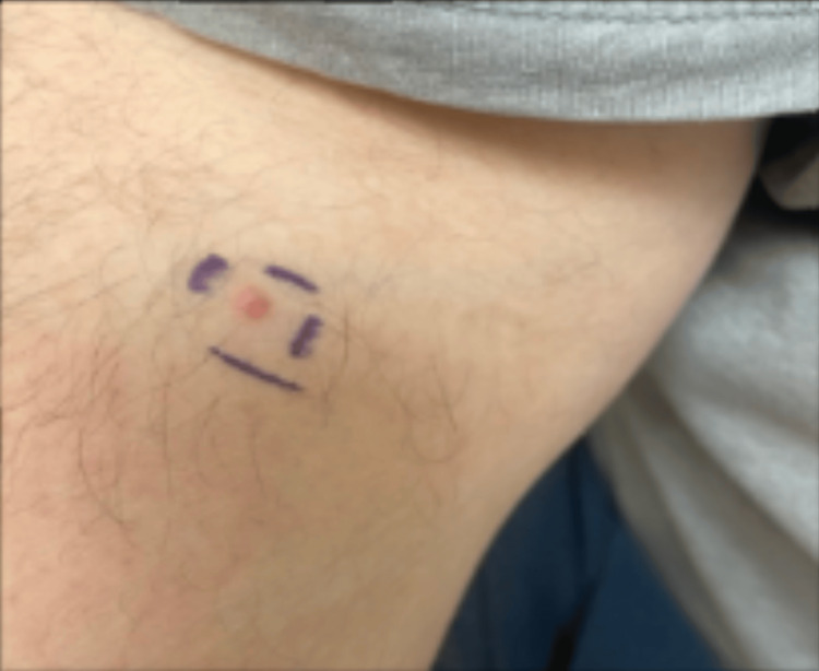 Figure 1
| Criteria | Points |
| Age at presentation of initial sebaceous neoplasm | 1 point if under 60 years old |
| Total number of sebaceous neoplasms or keratoacanthomas | 2 points if two or more |
| Personal history of Lynch syndrome-associated internal malignancies | 1 point |
| Personal history of Lynch syndrome-associated cancers | 1 point |
| Screening recommendation | |
| Skin | Annually |
| Colonoscopy | Start at 20-25 years, and then every 1-2 years |
| Pelvic exams | Start at 30-35 years, and then annually |
| Upper endoscopy | Start at 30-35 years and then every 2-3 years |
| Urinalysis and cytologic examination | Start at 30-35 years, and then annually |
Peer Reviews
No public reviews on file for this paper yet. If you reviewed it on a platform where reviews are public (OpenReview, ICLR, NeurIPS, ICML), you can paste yours below so the community can read it here.
Videos
No videos yet. Explain this paper in a talk, walkthrough, or lecture? Add one.
Taxonomy
TopicsGenetic factors in colorectal cancer · Cancer Genomics and Diagnostics · Colorectal Cancer Treatments and Studies
Introduction
Muir-Torre syndrome (MTS) is a rare autosomal dominant genetic variant of hereditary non-polyposis colorectal cancer, or Lynch syndrome, with both visceral and cutaneous involvement [1]. The syndrome was first described by Muir in 1967 and again by Torre in 1968 [1]. Occurrence is due to mutations in DNA mismatch repair (MMR) genes, MLH1, MSH2, MSH6, and PMS2, resulting in microsatellite instability [1,2]. Hallmark dermatological manifestations of MTS include sebaceous adenomas, sebaceous epitheliomas, sebaceous carcinomas, and keratoacanthomas that may or may not include sebaceous differentiation, typically presenting on the head and neck [1,2]. Often, benign cutaneous lesions are the initial presentation of MTS [3]. On physical exam, sebaceous adenomas and carcinomas present as yellowish or skin-colored papules. Visceral malignancies most commonly include colorectal cancers but may also be comprised of cancers of the endometrium, ovaries, cervix, breast, uroepithelium, brain, blood, lung, small bowel, pancreas, hepatobiliary tract, and gastric organs [1,2,4].
Early detection of the various presentations of the MTS is important for the workup of other visceral malignancies to ensure that patients receive integrative care across a multitude of specialties. Thus, we present a case of a patient with a small papule with a biopsy consistent with sebaceous adenocarcinoma. This case underscores the importance of providers being aware of presentations of various syndromes in order to ensure appropriate workup and timely referrals for patients.
Case presentation
We present a case of a Caucasian male patient with a past medical history significant for nonalcoholic steatohepatitis (NASH) and intestinal polyps who presented to the dermatology clinic with a red-yellow papule present on the upper extremity, depicted in Figure 1. The lesion was biopsied, and pathology revealed a diagnosis of sebaceous adenocarcinoma. The sample underwent immunohistochemistry (IHC) staining and was found to be negative for MLH1, MSH2, MSH6, and PMS2. The patient was treated with wide local excision and was referred to gastroenterology for further workup, including a scheduled fibrosure test, unrelated to his diagnosis of MTS. Urology consultation included urine cytology, which was negative for any evidence of hematuria or urothelial carcinoma. The patient has continuous follow-up with gastroenterology and urology services as a part of a multi-disciplinary approach.
Red-yellow papule present on the upper extremity
Discussion
MTS is a clinical diagnosis based on the presence of at least one sebaceous gland tumor (benign or malignant) and at least one Lynch syndrome-associated malignancy [2,4]. Additional diagnostics include histopathology consistent with sebaceous differentiation, germline genetic testing for MMR gene mutations, and microsatellite instability testing - the hallmark of MMR gene deficiency. IHC testing for MMR proteins remains a widely used screening method, though it does not distinguish somatic and germline mutations [2]. If both IHC and genetic testing are consistent with MTS, screening for internal malignancies is warranted [1]. IHC is used to detect loss-of-function mutations (LOF) in MMR genes, for instance, MLH1, MSH2, MSH6, and PMS2. LOF mutations result in the lack of MMR protein expression, which presents as absent staining on IHC, a delineating feature of MTS tumors [5]. It has also been suggested to perform germline testing for MMR gene defects as part of the initial workup of all immunocompromised patients who develop an MTS-associated neoplasm; especially if they have a current or past history of malignancy [4]. Individuals at risk for MTS may be identified using the Mayo MTS risk score, one of several diagnostic tools, as outlined in Table 1.
Management of MTS requires a multidisciplinary approach with interprofessional collaboration. Treatment of sebaceous tumors may involve local excision or cryotherapy while continuing to monitor for new lesions [1]. Sebaceous carcinomas can spread locally and to distant sites, necessitating more aggressive treatment with wide local excision or Mohs micrographic surgery [1]. Radiation therapy may be used as an adjunct after excision [1]. Additionally, combining interferon-alpha with oral isotretinoin has been shown to reduce the occurrence of cutaneous and visceral cancers [1]. Due to the high risk of internal malignancies, patients require regular screening and imaging studies tailored to the individual’s risk profile as outlined in Table 2 [1].
Although MTS is a rare condition, there are other reports of similar presentations within the literature. Pancholi et al. reported a case of a 57-year-old female presenting with a 4 cm lesion on the left buttock. Histopathology revealed a sebaceous adenoma. The colonoscopy performed was significant for a malignant lesion at the ileocecal region, and biopsy results from the hemicolectomy were positive for a poorly differentiated signet ring adenocarcinoma with lymph node involvement [6]. Further, Shaker et al. reported a case of a 47-year-old woman who presented with multiple skin lesions involving the face, back, flanks, and scalp. Pathology results were significant for a sebaceous adenoma, and staining revealed a loss of MSH2 and MSH6. Continued workup revealed multiple tumors throughout the colon and rectum, with biopsy results significant for adenocarcinoma [2].
Conclusions
Recognizing the cutaneous manifestations of MTS can lead to early identification and management of internal malignancies, as highlighted by this case. A multidisciplinary approach encompassing dermatology, gastroenterology, urology, and oncology is vital in early detection, management, and overall patient outcomes. Providers should maintain a high index of suspicion when encountering sebaceous tumors, especially in patients under 60 years old with a relevant personal or family history of Lynch syndrome-associated malignancies.
The reference list from the paper itself. Each links out to its DOI / PubMed record.
- 1Muir-Torre syndrome Stat Pearls [Internet] Gay JT Troxell T Gross GP Treasure Island (FL)Stat Pearls Publishing 2024 http://www.ncbi.nlm.nih.gov/books/NBK 513271/30020643 · pubmed ↗
- 2Muir-Torre syndrome and recent updates on screening guidelines: the link between colorectal tumors and sebaceous adenomas in unusual locations J Surg Oncol Shaker N Shaker N Abid A Shah S Shakra RA Sangueza OP 138013841282023 https://doi.org/10.1002/jso.274403770660710.1002/jso.27440 · doi ↗ · pubmed ↗
- 3Looking beyond the surface: Muir Torre syndrome Arch Clin Cases Bagga E Innes D Leung E 1191221020233773659610.22551/2023.40.1003.10255 PMC 10510334 · doi ↗ · pubmed ↗
- 4Patients with a new-onset cutaneous sebaceous neoplasm following immunosuppression should be evaluated for Muir-Torre syndrome with germline mismatch repair gene mutation analysis: case reports Dermatol Online J Cohen PR Kurzrock R 302024 https://pubmed.ncbi.nlm.nih.gov/38762859/10.5070/D 33016328738762859 · doi ↗ · pubmed ↗
- 5Screening for Muir-Torre syndrome using mismatch repair protein immunohistochemistry of sebaceous neoplasms J Genet Couns Roberts ME Riegert-Johnson DL Thomas BC 393405222013 https://doi.org/10.1007/s 10897-012-9552-42321217610.1007/s 10897-012-9552-4 · doi ↗ · pubmed ↗
- 6Muir-Torre syndrome: a case report and screening recommendations Ann R Coll Surg Engl Pancholi A Collins D Lindley R Gandhi P 010902008 https://pubmed.ncbi.nlm.nih.gov/18990276/10.1308/147870808 X 360387 PMC 272781718990276 · doi ↗ · pubmed ↗