Cardiac CT Perfusion Imaging of Pericoronary Adipose Tissue (PCAT) Highlighting Potential Confounds in CTA Analysis
Hao Wu, Yingnan Song, Ammar Hoori, Juhwan Lee, Sadeer G. Al-Kindi, Wei-Ming Huang, Chun-Ho Yun, Chung-Lieh Hung, Sanjay Rajagopalan, David L. Wilson

TL;DR
This study uses cardiac CT perfusion to show that pericoronary adipose tissue (PCAT) features in CT scans can be affected by blood flow and timing, which may lead to misinterpretation of cardiovascular risk.
Contribution
The study introduces dynamic cardiac CT perfusion to investigate PCAT perfusion and its impact on PCAT assessment in coronary CT angiography.
Findings
PCAT Hounsfield units (HU) increased more than other adipose depots and showed a 7 HU swing with a two-second timing offset.
PCAT blood flow was about 23% of contiguous myocardium, and radiomic features changed by over 10% relative to initial scans.
PCAT enhancement and time-to-peak were reduced distal to significant coronary stenoses.
Abstract
Background: Features of pericoronary adipose tissue (PCAT) from coronary computed tomography angiography (CCTA) are associated with inflammation and cardiovascular risk. As PCAT is vascularly connected with coronary vasculature, the presence of iodine is a potential confounding factor on PCAT HU and textures that has not been adequately investigated. We aim to use dynamic cardiac CT perfusion (CCTP) to understand the perfusion of PCAT and determine its effects on PCAT assessment. Methods: From CCTP, we analyzed HU dynamics of territory-specific PCAT, the myocardium, and other adipose depots in patients with coronary artery disease. HU, blood flow, and radiomics were assessed over time. Changes from peak aorta time, Pa, chosen to model the acquisition time of CCTA, were obtained. Results: HU in PCAT increased more than in other adipose depots. Blood flow in PCAT was ~23% of that in the…
Genes, proteins, chemicals, diseases, species, mutations and cell lines named across the full text — each resolved to its canonical identifier and authoritative record.
Click any figure to enlarge with its caption.
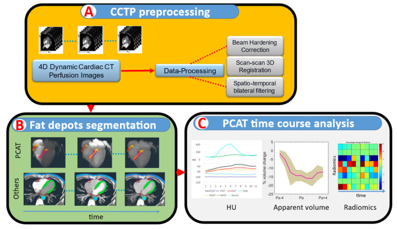 Figure 1
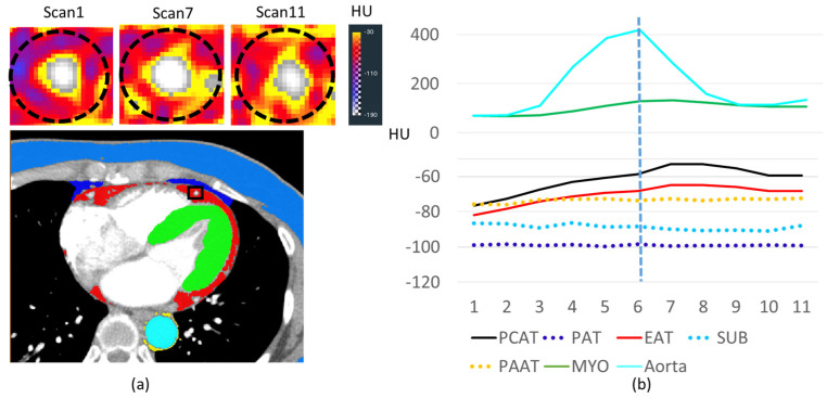 Figure 2
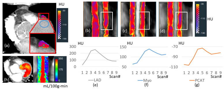 Figure 3
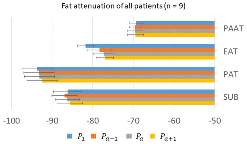 Figure 4
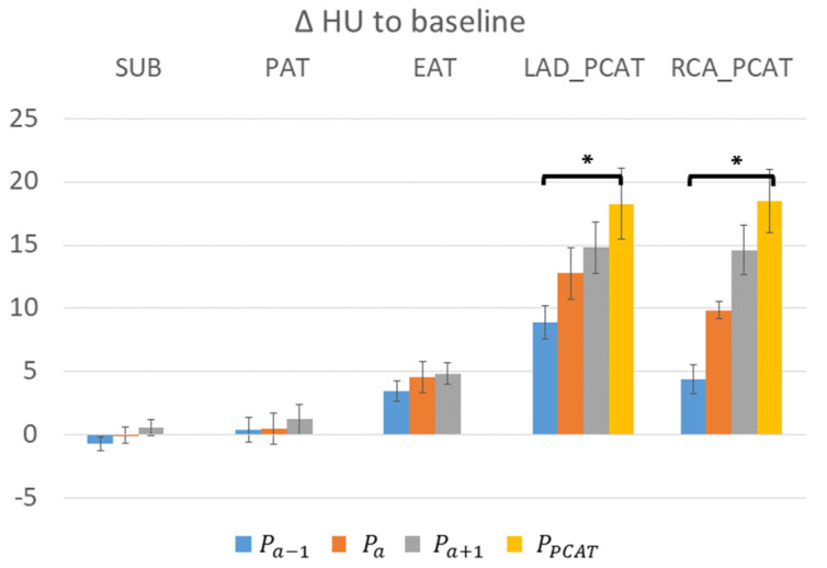 Figure 5
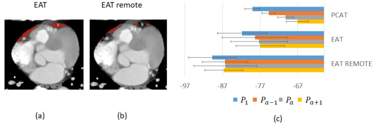 Figure 6
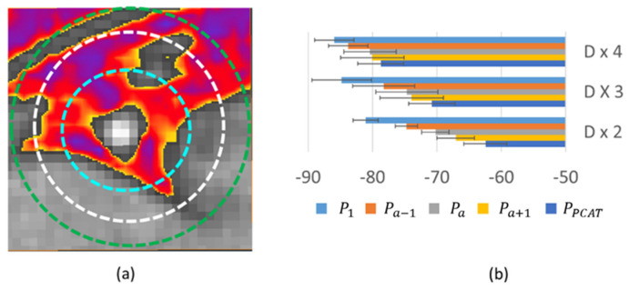 Figure 7
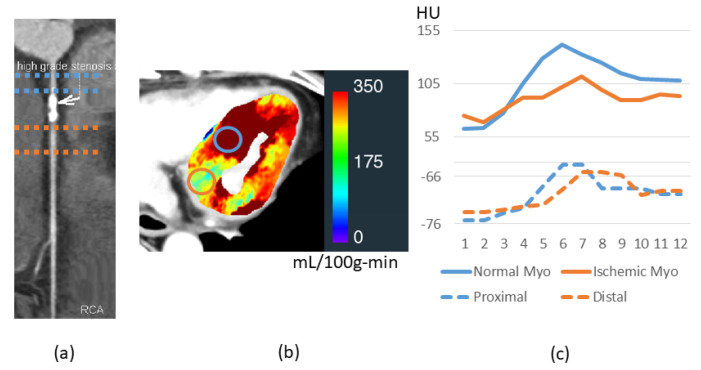 Figure 8
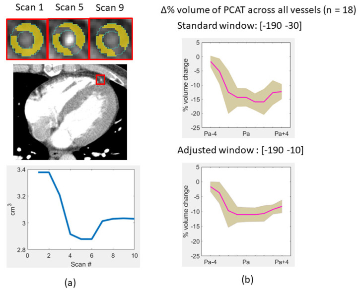 Figure 9
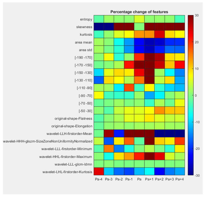 Figure 10
Peer Reviews
No public reviews on file for this paper yet. If you reviewed it on a platform where reviews are public (OpenReview, ICLR, NeurIPS, ICML), you can paste yours below so the community can read it here.
Videos
No videos yet. Explain this paper in a talk, walkthrough, or lecture? Add one.
Taxonomy
TopicsCardiovascular Disease and Adiposity · Cardiac Imaging and Diagnostics · Cardiovascular Function and Risk Factors