Throwing light on night blindness associated with rare manifestation of vitamin A deficiency
Avadhesh Oli, Sujata Upadhyay, Agrima Bhatia, Bhavaraj Veerabhadhra Rao

TL;DR
A 38-year-old man with vitamin A deficiency showed a rare eye condition called the Mizuo phenomenon, highlighting the link between low vitamin A and night blindness.
Contribution
This is the first reported case of the Mizuo phenomenon associated with vitamin A deficiency and normal ERG but abnormal EOG.
Findings
The patient exhibited a metallic sheen in the retina, which disappeared after dark adaptation, consistent with the Mizuo phenomenon.
Multimodal imaging showed retinal thinning and hyperautofluorescent flecks, but ERG was normal while EOG was abnormal.
Vitamin A supplementation did not significantly improve serum levels, suggesting a need for further study on treatment efficacy.
Abstract
This case report describes a rare presentation of the Mizuo phenomenon in a vitamin A deficiency-associated patient. A 38-year-old male presented with nyctalopia for 10 years and a fresh onset of dry eyes. He was a known case of alcoholic liver disease with cirrhosis and Oguchi disease. Anterior segment evaluation revealed moderate dry eyes, and fundus examination revealed metallic sheen OU, the latter extinguished after dark adaptation via a modified patch test suggestive of the Mizuo Nakamura phenomenon. Multimodal imaging modalities like SD-OCT, short-wave autofluorescence, and electrophysiological tests were performed. Findings suggested a diagnosis other than Oguchi’s disease. The gene panel comprising SAG and GRK1 mutations was negative for Oguchi. Serum vitamin A levels were significantly low (0.03 mg/l). The patient was started on topical lubricants and oral vitamin A…
Genes, proteins, chemicals, diseases, species, mutations and cell lines named across the full text — each resolved to its canonical identifier and authoritative record.
Click any figure to enlarge with its caption.
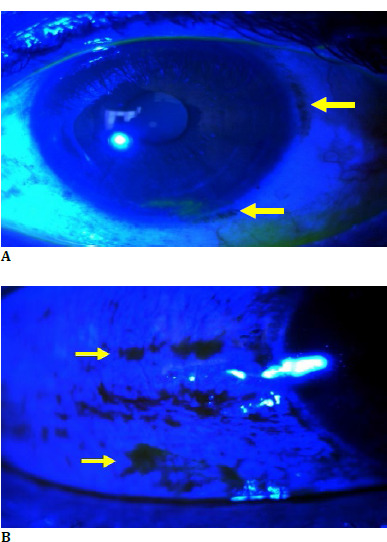 Fig. 1
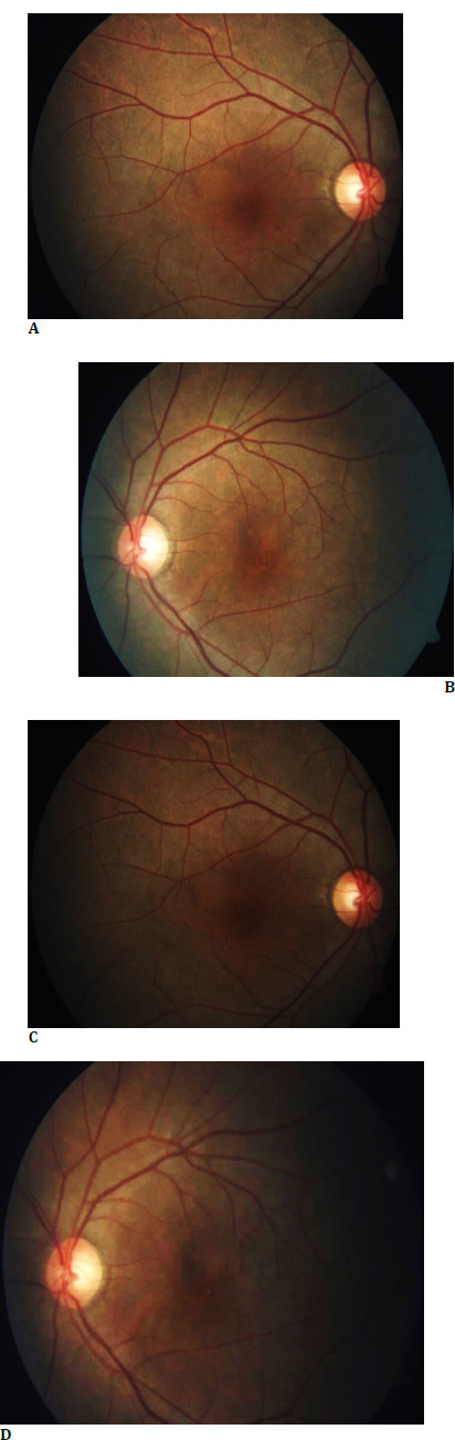 Fig. 2
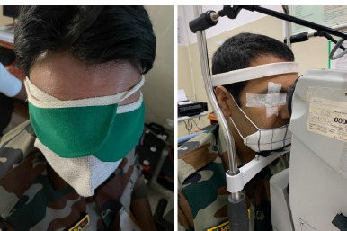 Fig. 3
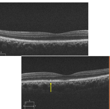 Fig. 4
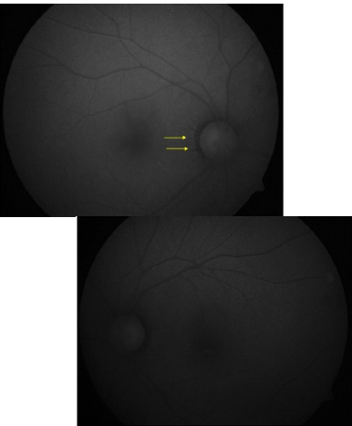 Fig. 5
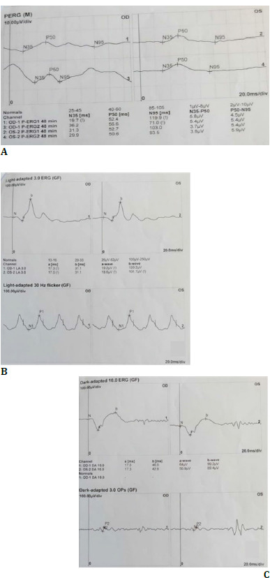 Fig. 6
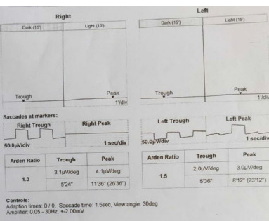 Fig. 7
Peer Reviews
No public reviews on file for this paper yet. If you reviewed it on a platform where reviews are public (OpenReview, ICLR, NeurIPS, ICML), you can paste yours below so the community can read it here.
Videos
No videos yet. Explain this paper in a talk, walkthrough, or lecture? Add one.
Taxonomy
TopicsRetinoids in leukemia and cellular processes · Antioxidant Activity and Oxidative Stress
Introduction
Vitamin A deficiency is rare in developed and nutrient-rich countries but is still prevalent in developing countries. Vitamin A deficiency-associated ocular symptoms have been shown to occur at concentrations of less than ten micrograms/dL [1]. Besides nutritional deficit, pancreatic, liver, and intestinal pathology are the leading causes of vitamin A deficiency. Chronic liver disease of any type has been associated with vitamin A deficiency. It is an acquired form of rod photoreceptor disease leading to night blindness. Vitamin A deficiency can lead to night blindness due to poor visual pigment regeneration in retinal rods [2]. Many inherited retinal pathologies manifest as night blindness (spectrum of congenital stationary night blindness). One is Oguchi disease with characteristic retinal findings, called the Mizou-Nakamura phenomenon, in which golden-yellow fundus discoloration disappears after prolonged dark adaptation.
We present a case of Severe Vitamin A deficiency in a patient with alcoholic liver cirrhosis with portal hypertension who had presented with nyctalopia for the past 10 years and dry eye symptoms for the past few months.
Case report
A 38-year-old male patient presented to us with complaints of nyctalopia for the past 10 years and foreign body sensation in both eyes for the past few months. There was no family history of consanguinity or any ocular disorder. He was a known case of alcoholic liver disease with cirrhosis and Oguchi disease. The patient’s best-corrected visual acuity was 20/40 in the Right Eye and 20/20 in the Left eye; color vision, as determined with pseudoisochromatic Ishihara color plates, was expected in each eye. Anterior segment evaluation suggested moderate dry eyes with a tear break time of 5 seconds OU (Fig. 1 A, B).
A, B. Anterior segment evaluation suggestive of conjunctival xerosis and moderate dry eyes
Fundus examination revealed metallic sheen in both eyes (Fig. 2 A, B), which disappeared after dark adaptation for 1 hour (Fig. 2 C, D). Due to time and space constraints, we chose a modified dark adaptation test wherein instead of making the patient sit in a dark room for 3-4 hours, we patched both his eyes for 1 hour with a cloth patch to obstruct the light completely (Fig. 3).
A, B. Fundus examination revealed metallic sheen in both eyes; C, D. Extinguished metallic sheen in both eyes post-dark adaptation of 1 hour
Modified dark adaptation (Patch) test using a cloth patch
The golden sheen of the fundus, which was extinguished after dark adaptation, was suggestive of the Mizou Nakamura phenomena. Interestingly, the patient had been diagnosed with Oguchi disease ten years back at another institution, underscoring the importance of re-evaluation in cases with overlapping clinical features. We, however, chose to perform a meticulous examination protocol as the symptoms of nyctalopia were persistent with the fresh onset of features of dry eyes. He showed increased dark and light adaptation time (approx. 45 minutes). Multimodal imaging modalities like Spectral Domain Optical Coherence tomography cross sections demonstrated thinning of the ellipsoid region with multiple hyperreflective excrescences in the outer retinal layers (Fig. 4) colocalized with hyperautofluorescent flecks on short-wave autofluorescence imaging (Fig. 5).
Spectral Domain Optical Coherence tomography cross sections demonstrated thinning of the ellipsoid region with multiple hyperreflective excrescences in the outer retinal layers
Hyperautofluorescent flecks on short-wave autofluorescence imaging
The electrophysiological tests performed according to ISCEV standards were utilized to navigate the diagnosis objectively. Pattern, scotopic, and photopic ERG were normal bilaterally (Fig. 6 A-C). Electrooculogram showed a significantly decreased Arden ratio (OD 1.3 and OS 1.5) (Fig. 7). This raised suspicion of an underlying additional retinal pathology or a diagnosis other than Oguchi disease. To aid in unraveling this perplexity, a gene panel comprising SAG and GRK1 mutations was conducted, which yielded negative results for Oguchi. Serum vitamin A levels, however, were detected to be significantly low (0.03 mg/l).
A-C. Pattern, Scotopic, and Photopic ERG were regular bilaterally
Electrooculogram showed a significantly decreased Arden ratio in both eyes
The significantly low vitamin A levels, slowly progressive night blindness, and gradually worsening dry eye symptoms favored Vitamin A deficiency-associated retinal pathology with associated Mizou Nakamura phenomena. The patient was started on topical preservative-free lubricants and oral vitamin A supplements and counseled to abstain from alcohol. The Vitamin A supplementation was limited and gradual (20000 IU/day) as we intended not to aggravate the liver dysfunction. After the controlled oral supplementation of Vitamin A, the patient’s serum Vitamin A levels persisted to be low (0.06mg/l), and there has been no relief in nyctalopia, probably due to persistent low vitamin A levels.
Discussion
Vitamin A deficiency associated with xerophthalmic fundus is a well-recognized pathology. Our patient's vitamin A level was 1000 times below the minimum reference level of 0.3-1.2 mg/L.
The functional retinal changes precede structural changes. Nyctalopia is the earliest manifestation of vitamin A-associated xerophthalmic fundus. It is also associated with depressed ERG, which depends on the age of manifestation [3]. Various diseases associated with the metallic sheen of the fundus are Oguchi disease, RS1 mutation, RDH5 or RLBP1-related disease [4], and RGS9/R9AP mutation [5]. Two genes acting in sequence to cease the phototransduction cascade and reported mutations that cause Oguchi disease are the SAG(Arrestin) and GRK1(Rhodopsin Kinase) [6]. The typical metallic sheen of the fundus with normal cone function, delayed rod ERG dark adaptation, and marked rod desensitization to a bright flash is distinctive for Oguchi disease [7]. The color change is thought to be caused by an excess of extracellular potassium in the retina due to a decreased potassium scavenging capacity of retinal Müller cells [8]. A normal ERG and an adverse genetic profile ruled out Oguchi disease or retinal gene disorders in this patient. However, there is a documented case of decreased Arden ratio in Vitamin A deficiency, as seen in this case [9]. The literature review reveals varied structural manifestations like macular pigmentation and white dots on the fundus. Still, no metallic sheen (Mizou phenomena) has been reported in vitamin A deficiency. Hypothetically, the hyperreflective dots in the outer retina correspond to both loss and accrual of photoreceptor segments secondary to decreased phagocytosis by RPE cells in vitamin A deficiency [10]. Clinically, they correspond to the retinal flecks.
Conclusion
Hence, this case is a rare and likely the first reported presentation of the Mizuo phenomenon in a vitamin A deficiency-associated patient with normal ERG and abnormal EOG. As the patient was previously diagnosed with Oguchi disease, it underscores the importance of re-evaluation in cases with overlapping clinical features. Increasing choice for weight loss diet and bariatric surgeries Vitamin A deficiency is also expected to affect developed countries. Modified patch tests might aid in more accessible and earlier diagnosis. More extensive studies are needed to correlate levels of Vitamin A with the natural course of the Mizuo phenomenon and substantiate multimodal imaging findings.
The reference list from the paper itself. Each links out to its DOI / PubMed record.
- 1Miller M Humphrey J Johnson E Marinda E Brookmeyer R Katz J Why do children become vitamin A deficient?J Nutr 2002 Sep 1329 Suppl 2867 S 2880 S 10.1093/jn/132.9.2867 S 12221263 · doi ↗ · pubmed ↗
- 2Gilbert C The eye signs of vitamin A deficiency Community Eye Health 2013268466724782581 PMC 3936686 · pubmed ↗
- 3Dhanda RP Electroretinography in Night Blindness and Other Vitamin-A Deficiencies Archives of Ophthalmology 195554684184910.1001/archopht.1955.0093002084700513268138 · doi ↗ · pubmed ↗
- 4Robson AG Mengher LS Tan MH Moore AT An unusual fundus phenotype of inner retinal sheen in X-linked retinoschisis Eye (Lond)2009231876187810.1038/eye.2008.35819741719 · doi ↗ · pubmed ↗
- 5Nishiguchi KM Sandberg MA Kooijman AC Martemyanov KA Pott JW Hagstrom SA Defects in RGS 9 or its anchor protein R 9AP in patients with slow photoreceptor deactivation Nature 2004427757810.1038/nature 0217014702087 · doi ↗ · pubmed ↗
- 6Fujinami K Tsunoda K Nakamura M Oguchi Y Miyake Y Oguchi Disease With Unusual Findings Associated With a Heterozygous Mutation in the SAG Gene Arch Ophthalmol 2011129101375137610.1001/archophthalmol.2011.30021987685 · doi ↗ · pubmed ↗
- 7Rishi P Rishi E Abraham S Oguchi’s disease with Mizuo-Nakamura phenomenon in a seven-year-old boy GMS Ophthalmol Cases 2018 Dec 138Doc 0710.3205/oc 00008930607313 PMC 6308901 · doi ↗ · pubmed ↗
- 8Bergsma DR Jr Chen CJ The Mizuo phenomenon in Oguchi disease Arch Ophthalmol 1997115456056110.1001/archopht.1997.011001505620279109775 · doi ↗ · pubmed ↗