Tear meniscus height comparison between AS-OCT and Oculus Keratograph® K5M
Angeline Lim Pei Yik, Fakhruddin Shamsheer Barodawala

TL;DR
This study compares two devices for measuring tear meniscus height and finds they agree well, even with contact lens wear.
Contribution
The study demonstrates the interchangeability of AS-OCT and Oculus K5M for tear meniscus height measurements in clinical settings.
Findings
AS-OCT and Oculus K5M showed good agreement in measuring tear meniscus height.
Contact lens wear slightly reduced tear meniscus height but returned to baseline after adaptation.
Bland-Altman analysis confirmed clinically acceptable agreement between the two devices.
Abstract
To evaluate the agreement between Spectral-Domain Optical Coherence Tomography (AS-OCT) and Oculus Keratograph® 5M in measuring tear meniscus height (TMH) and to assess the impact of contact lens wear on these measurements. Materials and methods: TMH was measured in 54 healthy eyes using AS-OCT (3D OCT-1 Maestro, Topcon, Tokyo, Japan) and Oculus Keratograph® 5M (OCULUS Optikgeräte, Wetzlar, Germany), with and without contact lens wear. Bland-Altman analysis was used to assess agreement between the two devices. Delefilcon A, water-gradient, daily disposable contact lenses were used, and measurements were carried out after a 20-minute adaptation period. The means of TMH without the contact lenses were 0.21 ± 0.06 mm and 0.20 ± 0.05 mm obtained from AS-OCT and Oculus K5M, respectively, and these measurements were not statistically significant (t (53) = 0.99, p = 0.33). No significant…
Genes, proteins, chemicals, diseases, species, mutations and cell lines named across the full text — each resolved to its canonical identifier and authoritative record.
Click any figure to enlarge with its caption.
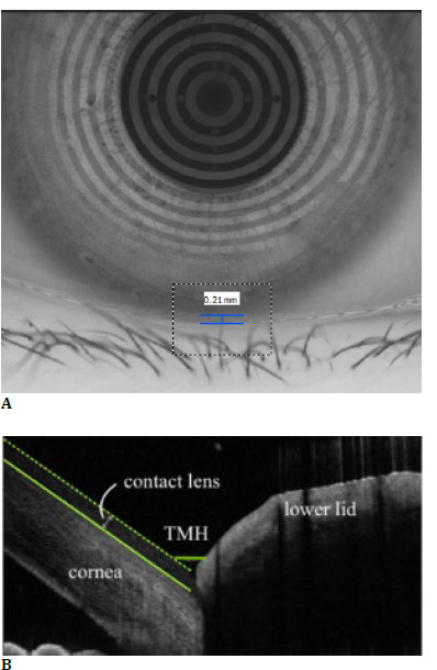 Fig. 1
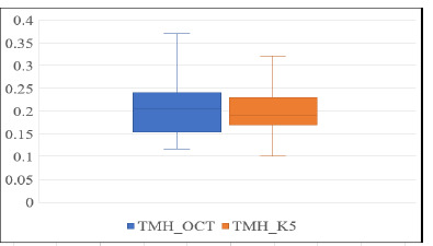 Fig. 2
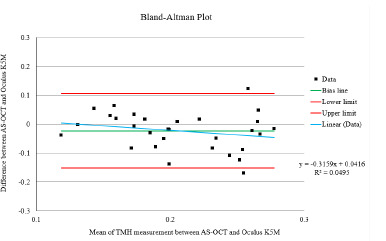 Fig. 3
| TMH | Mean ± Std Deviation (mm) |
|---|---|
| AS-OCT | 0.21 ± 0.06 |
| Oculus K5M | 0.20 ± 0.05 |
| AS-OCT with CL | 0.19 ± 0.06 |
| Oculus K5M with CL | 0.18 ± 0.04 |
Peer Reviews
No public reviews on file for this paper yet. If you reviewed it on a platform where reviews are public (OpenReview, ICLR, NeurIPS, ICML), you can paste yours below so the community can read it here.
Videos
No videos yet. Explain this paper in a talk, walkthrough, or lecture? Add one.
Taxonomy
TopicsCorneal surgery and disorders · Ocular Surface and Contact Lens · Glaucoma and retinal disorders
Introduction
The tear film is essential for maintaining ocular comfort and optimal visual function [1]. The tear meniscus is the collection of tears found at the junction of the bulbar conjunctiva and the margins of both eyelids. Tear meniscus height (TMH) is a valuable biomarker for diagnosing aqueous-deficient dry eye [2]. It holds 75-90% of the total tear volume, and tears are applied to the precorneal tear film [3]. Based on the Tear Film & Ocular Surface Society Disease Early Warning System (TFOS DEWS II), TMH is directly related to the lacrimal secretory rate. A consistently stable precorneal tear film has been considered a fundamental aspect of ocular health [4].
Tear film assessment is crucial for diagnosing dry eye disease (DED). Various methods exist, with meniscometry, focusing on tear meniscus volume, being one approach. However, factors like blinking, measurement location, and environmental conditions influence their accuracy. Quantitative tear meniscus height, curvature, and area assessment offer improved diagnostic precision. Still, it relies on operator expertise and is affected by factors such as fluorescein staining and time since blinking. Slit lamp meniscometry and Schirmer’s test have limitations in terms of repeatability and diagnostic accuracy [5].
Spectral-domain optical coherence tomography (OCT) (3D OCT-1 Maestro (Topcon, Tokyo, Japan)) provides high-resolution, noninvasive imaging of ocular tissues, including the tear meniscus. Its ability to generate three-dimensional images offers a precise alternative to traditional meniscometry for measuring the tear meniscus height (TMH) and is recognized as an accurate alternative [1,6].
The Oculus Keratograph® 5M (manufactured by OCULUS Optikgeräte, Wetzlar, Germany) is a corneal topographer device based on the Placido disc principle. This equipment features a modified tear film scanning function. Despite being an invasive technique, it can accurately represent the actual condition of the ocular surface. It serves as a straightforward approach for screening dry eye [7].
AS-OCT is widely used in clinical practice due to its non-invasive nature and ability to generate detailed, three-dimensional images of ocular structures, including the tear meniscus [3,8]. The Oculus K5M is a valuable tool for assessing the anterior eye and tear film, including tear film quality. Its advantages are that it does not require direct contact with the lower eyelid and can provide a broader view of the entire eye to measure TMH.
This study compares the TMH measurements obtained using AS-OCT and Oculus K5M in subjects with and without contact lens wear. Given the widespread availability of AS-OCT in clinical settings compared to the Oculus K5, this study seeks to determine if AS-OCT can be a reliable alternative for measuring TMH and if the values obtained from both instruments are comparable [8].
Materials and methods
This study included healthy adult participants aged 18-30 without ocular pathologies. Subjects were divided into contact lens wearers and non-contact lens wearers. Contact lens wearers were required to abstain from lens wear for 24 hours before the study.
A cross-sectional study design was employed to compare TMH measurements obtained using AS-OCT and Oculus K5M. Participants were randomly assigned to determine the order of instrument use. TMH measurements were initially taken in both eyes without contact lenses. Subsequently, a Delefilcon A (Dailies Total 1) contact lens was applied to the right eye, and measurements were repeated after a 20-minute adaptation period. All measurements were performed within the same timeframe under standardized conditions to minimize bias.
To minimize bias, TMH measurements were performed in a randomized order on each subject within the same day period [2]. Based on the earlier study, the measurements were made below the corneal vertex at the cornea’s 6 o’clock position with the lower eyelid. The method employed in the present study for measuring TMH using AS-OCT and Oculus K5M was like that described in the previous research, with consistent imaging conditions and standardized measurement procedures.
Imaging scans using AS-OCT and Oculus K5M were conducted under dim lighting and similar humidity. All assessments were consistently performed at the same time of day. Subjects were instructed to begin by measuring tear meniscus height through image scans with AS-OCT and Oculus K5M by the sequences determined by the drawing of lots. In the case of AS-OCT, a vertical scan beam was used to image the tear meniscus positioned between the cornea and the eyelid’s tear boundary line. For the Oculus K5M, lower TMH images were taken and assessed at the central position relative to the pupil centre, using a built-in ruler, as shown in Fig. 1 A. The scanning line’s length was 6 mm, and it was directed towards the 6 o’clock position of the cornea, as shown in Fig. 1 B.
The University’s Ethics Committee approved the study, and informed consent was obtained from all participants.
Tear meniscus height measurement examples using (A) Oculus Keratograph 5M and (B) AS-OCT
Results
This study included 54 healthy right eyes (26 males, 28 females). The mean age of the subjects was 22.87 ± 2.45 years (ranging from 19 to 31 years old). Table 1 presents descriptive statistics for TMH measurements obtained using AS-OCT and Oculus K5M, both with and without contact lenses. Data were typically distributed based on the Shapiro-Wilk test.
The paired t-test showed no significant difference in tear meniscus height (TMH) measurements between AS-OCT and Oculus K5M without contact lenses (t (53) = 0.99, p = 0.33). Furthermore, there were no significant differences in TMH when comparing OCT and Oculus K-5 measurements with contact lenses (t (53) = 1.52, p = 0.13).
Moreover, there were no significant differences in tear meniscus height (TMH) when using OCT with and without contact lenses (t (53) = 1.23, p = 0.22). However, a significant difference in TMH was seen when using Oculus K-5 with and without contact lenses (t (53) = 2.27, p < 0.05).
Boxplot showing TMH measurements with AS-OCT and Oculus K5M
According to the boxplot in Fig. 2 above, no outlier data points were detected within the displayed range for AS-OCT or Oculus K5M measurements with TMH measurements without the contact lenses.
A Bland-Altman plot was constructed to assess the agreement between AS-OCT and Oculus K5M for TMH measurements without contact lenses (Fig. 3). The mean difference (bias) was -0.02 mm, with limits of agreement of -0.15 mm and 0.10 mm. No proportional bias was observed (r = 0.22).
The Bland-Altman plot of the difference between the mean of AS-OCT and Oculus K5M. The Green line indicates bias, the red line indicates the upper and lower limits of agreement (LoA), and the blue line indicates linear regression.
Discussion
The present study investigated the tear meniscus height (TMH) measurements using AS-OCT and Oculus K5M in subjects with and without contact lens wear. The primary objective was to determine the agreement between TMH measurements obtained from these two instruments. It is essential to differentiate between absolute TMH and reflex tear TMH. While slit lamp measurements capture reflex tear TMH, non-invasive tools like AS-OCT and Oculus K5M provide values closer to absolute TMH, which are generally higher [9]. This might explain the tendency of the Oculus Keratograph to record higher TMH values compared to slit lamp measurements. The Oculus Keratograph® 5M (OCULUS Optikgeräte, Wetzlar, Germany) uses infrared imaging. At the same time, the AS-OCT (3D OCT-1 Maestro, Topcon, Tokyo, Japan) employs non-invasive, high-resolution, low-coherence interferometry to image the eye’s anterior surface. The differences in illumination systems may contribute to variability in TMH measurements [2].
A previous study reported that mean TMH values above 0.20 mm were observed in non-clinical participants, and reproducibility and repeatability were demonstrated among different observers [10,11]. The present study showed that the mean TMH value was 0.20 ± 0.05 mm and 0.21 ± 0.06 mm for Oculus K5M and AS-OCT, respectively, without the contact lens. This study showed that TMH measured with Oculus K5M was slightly lower than the AS-OCT, in which both instruments are significantly correlated. A previous study reported that Oculus K5M was lower by 0.01 mm than FD-OCT amongst healthy participants [2].
Another study investigated the mean TMH measurements, which were 0.29 ± 0.16 mm for Oculus K5M and 0.27 ± 0.16 mm for AS-OCT in non-clinical participants [6,12]. The mean TMH value for Medmont Meridia was 0.24 ± 0.09 mm. From the comparison of TMH values, it can be concluded that Medmont Meridia shows good agreement with AS-OCT, as there were no statistically significant differences between them. However, there is a substantial difference between the TMH measurements obtained by Medmont Meridia and Oculus Keratograph 5 M, with Oculus Keratograph 5 M showing higher values [2].
In another study done by Wei et al. (2012), the mean TMH values in the dry eye group, as measured by Oculus Keratograph 4 and AS-OCT, were found to be 0.24 ± 0.07 mm and 0.25 ± 0.08 mm whereas 0.31 ± 0.04 mm and 0.32 ± 0.12 mm in non-dry eye groups respectively [9]. They concluded that the results of Oculus Keratography 4 of TMH values significantly correlated with AS-OCT. Another study reported that the TMH was obtained 3-4 seconds after an average blink to analyze the accuracy of the TMH by using different techniques. The TMH measurement using the slit lamp microscope with reticule was 0.20 ± 0.05 mm and 0.22 ± 0.01 mm, and the OCT measurements were 0.24 ± 0.07 mm. TMH measurements using AS-OCT are higher than those measured using slit-lamp [13].
The TFOS DEWS II Report recommended that the cut-off values for AS-OCT TMH values be 0.22 mm or 0.24 mm to discriminate between normal and abnormal TMH [14]. However, baseline TMH values in regular groups were 0.20 ± 0.05 mm using Oculus K5M [13]. Therefore, based on previous studies’ TMH measurements, the present study indicated that the subjects selected had no signs of dry eye disease.
Prevalence rates of Dry Eye Disease (DED) vary from 5% to 50%, reaching up to 75% in adults over 40, with women most affected [15]. A Japanese study found that contact lens wearers are 2.38 times more likely to be diagnosed with dry eye and 3.61 times more likely to experience severe symptoms than non-wearers. Patients with Contact Lens Associated Dry Eye (CLADE) exhibit distinct ocular characteristics, including Lid Wiper Epitheliopathy (LWE) in 80% of CLADE patients versus 13% of asymptomatic contact lens users [16].
After wearing the contact lenses for 20 minutes, the TMH values returned to baseline [17]. Furthermore, TMH values remained stable for at least 4 hours after wearing the contact lenses. Thus, this suggests that tear film adjusted to the presence of the contact lenses maintained a consistent level of tears around the eyes [17]. In line with the present study, there were no notable differences in TMH measurements obtained using AS-OCT with and without contact lenses, as a 20-minute adaptation period was provided to the subjects before the TMH assessment.
The present study showed that the TMH was reduced by approximately 0.02 mm (20 µm) with contact lens wear. This is supported by another previous study that reported that initially, the pre-contact lens tear film is thicker due to reflex tearing and increased tear production. However, once the reflex tearing subsides, the thickness of the pre-contact lens tear film decreases to approximately 2 micrometers [18].
The Bland-Altman analysis demonstrated good agreement between AS-OCT and Oculus K5M for TMH measurements, with a consistent negative bias of -0.02 mm. While two data points fell outside the limits of agreement, the two instruments exhibited comparable results overall. This is supported by previous research showing a good correlation between Oculus K5M and AS-OCT in dry eye patients. The AS-OCT yielded higher values compared to Oculus K5M. By showing that their differences were only 0.02 mm consistently, the mean difference being close to zero indicated that most data points from the two instruments were similar findings.
Bland-Altman plots were also used to determine the limits of agreement, and most data points fell within the limit of agreement. Two data points fell outside the boundaries of agreement, indicating differences between these measurements. A Bland-Altman plot demonstrates these techniques’ comparability and suggests no evidence of proportional bias.
Considering the two outliers on the Bland-Altman plot, it was found that either the Oculus K5M or the AS-OCT could overestimate the measurements. Instead, these deviations can likely be attributed to random measurement errors or specific variations in the image acquisition process. Despite these two outliers, the Bland-Altman analysis indicated no statistically significant difference between the measurement methods [19].
This previous study investigated dry eye disease subjects and resulted in TMH measurements with Oculus K5M closely correlating with AS-OCT, with good consistency [9,10]. This means that Oculus K5M provides results that agree with the results obtained from AS-OCT, which can also prove that it can be reliably used as an alternative tool for similar measurements. In addition, the Bland-Altman analysis concluded that they can be used interchangeably for TMH measurement.
Conclusion
This study demonstrated a strong agreement between AS-OCT and Oculus K5M in measuring tear meniscus height (TMH) within a controlled study population. These findings suggest that both devices can be reliably used interchangeably for TMH assessment in clinical practice. To further solidify the interchangeability of these instruments, future studies should explore their performance in a broader range of patient populations and clinical settings.
The reference list from the paper itself. Each links out to its DOI / PubMed record.
- 1Canan H Altan-Yaycioglu R Ulas B Sizmaz S Coban-Karatas M Interexaminer reproducibility of optical coherence tomography for measuring the tear film meniscus Curr Eye Res 20143911455010.3109/02713683.2014.89831124749829 · doi ↗ · pubmed ↗
- 2oares I Ramalho E Brardo FM Fernandes A Ramalho E Brardo FM Tear meniscus height agreement and reproducibility between two corneal topographers and spectral-domain optical coherence tomography Clin Exp Optom 2024001710.1080/08164622.2024.234183338627017 · doi ↗ · pubmed ↗
- 3Raj A Dhasmana R Nagpal RC Anterior Segment Optical Coherence Tomography for Tear Meniscus Evaluation and its Correlation with Other Tear Variables in Healthy Individuals J Clin Diagn Res 201610 NC 01410.7860/JCDR/2016/18717.7722 PMC 494842927437253 · doi ↗ · pubmed ↗
- 4Craig JP Nichols KK Akpek EK Caffery B Dua HS Joo C-KTFOS DEWS II Definition and Classification Report Ocul Surf 2017152768310.1016/j.jtos.2017.05.00828736335 · doi ↗ · pubmed ↗
- 5Wolffsohn JS Arita R Chalmers R Djalilian A Dogru M Dumbleton KTFOS DEWS II Diagnostic Methodology report Ocul Surf 2017155397410.1016/j.jtos.2017.05.00128736342 · doi ↗ · pubmed ↗
- 6Eroglu FC Karalezli A Dursun R Is optical coherence tomography an effective device for evaluation of tear film meniscus in patients with acne rosacea?Eye 2016305455210.1038/eye.2015.27726768918 PMC 5108538 · doi ↗ · pubmed ↗
- 7Tian L Qu JH Zhang XY Sun XG Repeatability and Reproducibility of Noninvasive Keratograph 5M Measurements in Patients with Dry Eye Disease J Ophthalmol 201620168013621 Epub 2016 Apr 1210.1155/2016/801362127190639 PMC 4844888 · doi ↗ · pubmed ↗
- 8Wan C Hua R Guo P Lin P Wang J Yang W Measurement method of tear meniscus height based on deep learning Front Med 20231011010.3389/fmed.2023.1126754 PMC 997100036865061 · doi ↗ · pubmed ↗