Factors influencing osteoradionecrosis progression during hyperbaric oxygen therapy: A case study
Sameh Mezri, Chaima Zitouni, Khadija Bahrini, Mounir Haggui, Wiem Boughzala, Hedi Gharsallah, Amaliya Amaliya, Chaima zitouni, Busra Yilmaz, Chaima zitouni, Atsushi Shudo, Chaima zitouni

TL;DR
This study examines how hyperbaric oxygen therapy affects osteoradionecrosis and identifies factors that influence treatment outcomes.
Contribution
The study identifies specific predictive factors influencing ORN progression during HBOT through statistical analysis.
Findings
ORN regressed in 33% of patients after at least 20 HBOT sessions.
Factors like tumor size, radiation dose, and HBOT session count significantly influence ORN progression.
Multivariate analysis confirmed the number of HBOT sessions and dental care timing as key predictors.
Abstract
Although relatively uncommon, osteoradionecrosis (ORN) remains a serious complication following radiotherapy. Various therapeutic approaches, including hyperbaric oxygen therapy (HBOT), are utilized in managing ORN. This study aims to evaluate the role of HBOT in ORN management and to identify predictive factors influencing the evolution of head and neck ORN after HBOT. This retrospective study includes 46 patients who received HBOT for head and neck ORN between 2017 and 2020. The patients were divided into two groups: Group 1 (n=36) included those with regression or stabilization of ORN, while Group 2 (n=10) comprised patients with worsening lesions. We performed a statistical study in order to identify factors influencing ORN progression under treatment. ORN affected the mandible in 93.5% of patients, the maxilla in 2 cases, and the skull base in 4 cases. All patients received HBOT,…
Genes, proteins, chemicals, diseases, species, mutations and cell lines named across the full text — each resolved to its canonical identifier and authoritative record.
Click any figure to enlarge with its caption.
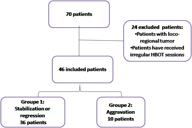 Figure 1
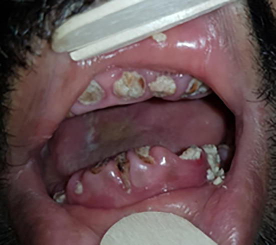 Figure 2
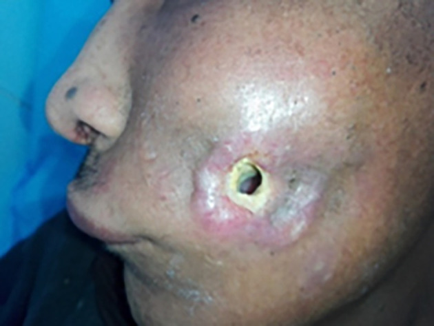 Figure 3
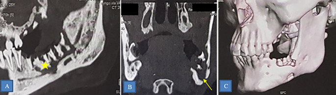 Figure 4
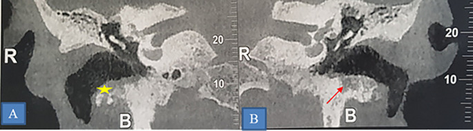 Figure 5
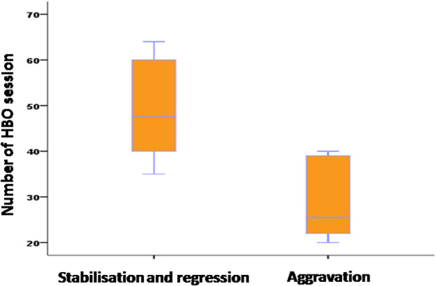 Figure 6
| Tumor histological type and location | Number of patients |
|---|---|
| Undifferentiated carcinoma of nasopharyngeal type (UCNT) | 31 |
| Squamous cell carcinoma of the mandible | 2 |
| Gingival squamous cell carcinoma | 1 |
| Squamous cell carcinoma of the palate | 1 |
| Squamous cell carcinoma of the tonsil | 1 |
| Lymphoma with tonsillar localization | 1 |
| Squamous cell carcinoma of the tongue | 1 |
| Mucoepidermoid carcinoma of the parotid gland | 1 |
| Squamous cell carcinoma of the external auditory canal | 1 |
| Cutaneous squamous cell carcinoma | 1 |
| Breast ductal carcinoma with metastatic cervical lymph nodes | 5 |
| 1st group (%) | 2 nd group (%) | p | ||
|---|---|---|---|---|
| Age groups | < 31 | 1 (3) | 2 (20) | 0.148 |
| [31- 65] | 21 (58) | 5 (50) | ||
| ≥65 | 14 (39) | 3 (30) | ||
| Gender | Male | 19 (53) | 3 (30) | 0.202 |
| Female | 17 (47) | 7 (70) | ||
| Diabetes | Yes | 23 (64) | 7 (70) | 0.720 |
| No | 13 (36) | 3 (30) | ||
| High blood pressure | Yes | 20 (56) | 2 (20) | 0.046 |
| No | 16 (44) | 8 (80) | ||
| Smoking | Yes | 20 (56) | 5 (50) | 0.755 |
| No | 16 (44) | 5 (50) | ||
| Tumor site | Nasopharynx | 22 | 9 | 0.997 |
| Mouth | 8 | 0 | ||
| Breasts | 4 | 1 | ||
| External auditory canal | 1 | 0 | ||
| Skin | 1 | 0 | ||
| Mean tumor size | 27.9 mm | 36 mm | 0.004 | |
| Tumor stage | T2 | 13 (95) | 1 (11) | 0.048 |
| T3 | 8 (36) | 7 (78) | ||
| T4 | 1 (4) | 1 (11) | ||
| Type of RT | Conventional RT | 30 (83) | 10 (100) | 0.590 |
| IMRT | 3 (8) | 0 | ||
| 3D RT | 1 (3) | 0 | ||
| 2D cobalt-60 RT | 2 (6) | 0 | ||
| Mean radiation dose | 42 | 68 | 0.021 | |
| Chemotherapy | Yes | 22 (61) | 8 (80) | 0.267 |
| No | 14 (39) | 2 (20) | ||
| The average interval between dental care (before RT) and RT | 5.46 months | 1.6 months | 0.045 | |
| Dental extraction after RT | Yes | 29 (81) | 9 (90) | 0.486 |
| No | 7 (19) | 1 (10) | ||
| Location of the ORN | Horizontal branch of the mandible | 22 (74) | 5 (50) | 0.049 |
| Mandibular angle | 5 (17) | 5 (50) | ||
| Ascending ramus of the mandible | 3 (10) | |||
| Radiological signs | Bone lysis | 15 (42) | 5 (50) | 0.686 |
| Bone sequestrum | 13 (36) | 4 (40) | ||
| Bone fracture | 8 (22) | 1 (10) | ||
| Antibiotic therapy | Yes | 32(89) | 9(90) | 0.920 |
| No | 4(11) | 1(10) | ||
| Average number of HBOT sessions | 49.11 | 28.6 | 0.001 | |
| Surgery after RT | Yes | 7 (20) | 1 (10) | 0.466 |
| No | 28 (80) | 9 (90) | ||
| Type of surgery | Sequestrectomy | 28 (93) | 6 (100) | 0.515 |
| Mandibular reconstruction with flap | 2 (7) | 0 | ||
| Interval between HBOT and surgery | Before | 4 | 2 | 0.359 |
| Simultaneously | 13 | 2 | ||
| After | 13 | 2 | ||
| P | OR | 95% confidence interval for EXP(B) | ||
|---|---|---|---|---|
| Lower value | Upper value | |||
| High blood pressure | 0.623 | 1.803 | 0.172 | 18.923 |
| Tumor mean size | 0.778 | 1.022 | 0.878 | 1.189 |
| Mean radiation dose | 0.193 | 1.055 | 0.973 | 1.145 |
| The average interval between dental care before RT and RT | 0.043 | 7.055 | 1.067 | 46.649 |
| Dental extraction after RT | 0.121 | 6.159 | 0.618 | 61.371 |
| Location of the ORN | 0.909 | |||
| Average number of HBO sessions | 0.040 | 1.041 | 1.021 | 1.062 |
Peer Reviews
No public reviews on file for this paper yet. If you reviewed it on a platform where reviews are public (OpenReview, ICLR, NeurIPS, ICML), you can paste yours below so the community can read it here.
Videos
No videos yet. Explain this paper in a talk, walkthrough, or lecture? Add one.
Taxonomy
TopicsOral health in cancer treatment · Head and Neck Cancer Studies · Facial Nerve Paralysis Treatment and Research
Introduction
Head and neck cancers rank seventh in the world, with an incidence of 890,000 new cases. ^ 1 ^
Their management typically involves surgery, radiotherapy (RT), and chemotherapy. Despite advancements in techniques, external RT often leads to complications, including osteoradionecrosis (ORN).
ORN is defined as progressive bone destruction, occurring spontaneously or following trauma, with mucosal ulceration exposing irradiated bone, persisting for 3 to 6 months without healing, excluding tumor recurrence. ^ 2 ^
Although relatively uncommon, ORN remains a serious complication, significantly affecting patients’ quality of life.
Since its first description, the pathophysiology of ORN remains unclear, and management strategies are complex and lack consensus.
Various therapeutic approaches are utilized in the treatment of ORN. They include antibiotics, hyperbaric oxygen therapy (HBOT), the PENTOCLO protocol, and conservative surgery.
Our study aims to evaluate the role of HBOT in ORN management and describe predictive factors influencing post-therapeutic evolution of head and neck ORN.
Methods
The cohort study
This is a retrospective study conducted at the HBOT Department of the Military Hospital of Tunis over a period of 4 years, from January 1, 2017, to December 31, 2020. The study included patients who met the following criteria:
- -Treated with HBOT for head and neck ORN
- -Completed at least 20 HBOT sessions
- -Had a complete medical record
- -Were available for follow-up evaluations for at least six months after the end of the therapy
All cases with recurrent loco-regional tumor and who received discontinued or irregular HBOT sessions were excluded from this study.
For this study, we developed a specialized data collection tool called a case report form (CRF). This form was used to document various aspects of each patient’s case, including their demographics, clinical examination, laboratory results, imaging, treatment, and follow-up.
ORN was classified into four grades according to the LENT/SOMA (Late Effects of Normal Tissues/Subjective Objective Management Analytic) classification. ^ 3 ^
Grade 1: Normal bone appearance or debatable modifications
Grade 2: Bone lysis or condensation
Grade 3: Bone sequestration
Grade 4: Bone fracture
OHB protocol
HBOT sessions were conducted in a multiplace hyperbaric chambers type HAUX-STARMED 2400. The HBOT protocol included 3 steps:
- ➢Step 1: Compression phase: Around 0.1 ATA (Atmosphere Absolute) and the Compression rate was adjusted based on patient’s tolerance.
- ➢Step 2: Plateau phase: in this phase patients received 100% oxygen using facial mask for 60 and or 90 minutes with 5-minute air breaks every 25 minutes.
- ➢Step 3: decompression phase: this step is slow typically around 0.1 ATA/min.
Patients were followed up during and following HBOT therapy. Clinical and imaging evolution of injuries was recorded.
Radiological examinations, if needed, were performed after at least 40 HBOT sessions. However, if the hyperbaric medicine specialist noticed any signs of worsening condition after 20 HBOT sessions, CT scans were ordered earlier. In our study, only CT was used for radiological control.
ORN injuries were assessed clinically and by imaging following HBOT treatment. Thereby lesion evolution was classified into three categories:
- -Regression: Clinically and/or radiologically confirmed the disappearance of functional and radiological signs
- -Stabilization: It involves partial healing of the ORN with no progression of bone necrosis. Functional signs are moderate.
- -Worsening or Aggravation: Progressive worsening of the ORN characterized by the persistence of functional and radiological signs. There is no bone healing and the ORN was progressed to a more severe stage.
Statistical analysis
Statistical analyses were performed using SPSS version 22.0. Categorical variables were reported as percentages, and their significance was assessed using Fisher’s exact test or chi-square tests. Quantitative variables were presented as mean (standard deviation) or median ± interquartile range (IQR25%, 75%). Normality was tested using the Kolmogorov-Smirnov test. An unpaired t-test was used to compare two groups when continuous variables were normally distributed. For continuous variables that were not normally distributed, a two-tailed unpaired Mann-Whitney U test was applied. Both univariate and multivariate analyses were conducted to evaluate the effectiveness of HBOT on ORN patients. The cases were divided into two groups: Group 1 (n=36) included patients with regression or stabilization, and Group 2 (n=10) included patients with worsening signs of injury after HBOT ( Figure 1). Differences were considered significant if p ≤ 0.05.
Diagram of patient recruitment, exclusions, and study groups.
Ethics statement
This study was approved by the Ethics Committee of the Military Hospital of Tunis, under decision number 81/2024/CLPP, dated July 22, 2024. The study was conducted in accordance with the ethical principles outlined in the Declaration of Helsinki ( https://www.wma.net/policies-post/wma-declaration-of-helsinki-ethical-principles-for-medical-research-involving-human-subjects/).
Results
Demographic characteristics of the cohort study
This study included 46 patients. The average age of patients was 58 ± 13.9 years, with the majority of patients (61%) aged between 31 and 65 years old. Fifty-two percent were female, and 48% were male. Two-thirds of the patients had diabetes and 59% were smokers.
Radiotherapy treatment
All patients had undergone RT for head and neck malignancies, primarily nasopharyngeal carcinoma (67%) ( Table 1).
The mean tumor size was 29.67 mm.
RT was conventional in 40 cases, cobalt-60 2D dimensional in two cases, three-dimensional in one case, and Intensity-Modulated Radiation Therapy (IMRT) in three cases; with a mean radiation dose of 48.7 Gy. The association with chemotherapy was noted in thirty cases, accounting for 65% of the cases.
Dental care was provided to all patients before RT, with 67% requiring tooth extractions. Twelve patients used the custom fluoride tray correctly. The interval between dental care and the start of RT ranged from one month to 48 months, with a median of two months.
Osteoradionecrosis diagnosis
ORN typically occurred due to dental extractions (83% of cases) or surgical procedures (15%). The median time from RT to ORN onset was 6 years, while from dental care to ORN onset was 36 months.
Common clinical symptoms included pain (100% of patients) and mastication difficulties (98%). Trismus was present in all patients, with varying degrees of severity. Dental mobility was observed in 33% of cases.
We also noted the presence of bone exposure ( Figure 2) in 6 cases, bone sequestration in 15 cases, or bone fracture in 9 cases. The presence of a cutaneous fistula ( Figure 3) was noted in 39% of cases.
Exposure of the mandibular bone.
Cutaneous fistula in osteoradionecrosis.
We performed panoramic radiograph (76%), cone beam imaging (11%), computed tomography (CT scan) (80%), and magnetic resonance imaging (MRI) (28%).
Imaging revealed the following grades of ORN according to the LENT/SOMA classification: Grade 2 (43% of cases), Grade 3 (37%), and Grade 4 (20%).
ORN was predominantly mandibular (40 patients), affecting various regions such as the horizontal branch (27 patients) ( Figure 4), mandibular angle (10 patients), and ascending branch (3 patients). Other locations included the maxilla (2 cases) and the skull base (4 cases) ( Figure 5).
Osteoradionecrosis of the left mandible.CT scan showing osteoradionecrosis of the horizontal branch of the left mandible complicated by sequestra (star) and mandibulo-cutaneous fistula (yellow arrow).A: Sagittal section.B: Frontal section.C: 3D reconstruction.
Osteoradionecrosis of the temporal bone.A and B: Coronal section showing of a CT-scan showing bilateral lytic erosion of the tympanic bone (star) and thickening of the walls of the external auditory canal.
The anatomopathological examination was performed in one case due to delayed alveolar consolidation and diagnostic doubt for differential diagnosis. It concluded with fibro-inflammatory chronic scarring remodeling, displaying features of ORN without specific granuloma or signs of malignancy.
Treatment of osteoradionecrosis
The therapeutic management of ORN included topical antibacterial mouthwash prescribed to all patients. All patients received analgesic treatment based on their pain intensity. Antibiotic therapy was initiated in 89% of cases.
Surgical treatment was performed in 36 patients, predominantly involving sequestrectomy (94% of cases). Mandibulectomy with mandibular reconstruction using flaps was conducted in 6% of cases.
HBOT was provided to all patients, with a mean of 44.65 sessions. HBOT sessions preceded ORN surgery in 17% of cases and were indicated after surgery in 42% of cases.
Complications of HBOT were observed in two patients, including middle ear barotrauma, necessitating transient discontinuation of therapy.
Evolution of ORN lesions was evaluated clinically after 20 HBOT sessions and radiologically after 40 sessions. Results showed regression in 33% of cases, stabilization in 45%, and progression or aggravation in 22% ( Figure 6).
Evolution of osteoradionecrosis based on the number of hyperbaric oxygen therapy sessions.
Risk factors associated with osteoradionecrosis aggravation
In the univariate study ( Table 2), age and sex did not show statistical significance in influencing ORN progression. However, among comorbidities, hypertension was significantly associated with worsened ORN evolution.
Our study also revealed that larger tumor size and advanced tumor stages correlated with worse outcomes, while tumor location did not statistically influence ORN evolution.
The type of radiotherapy did not statistically influence the progression of ORN lesions, nor did its association with chemotherapy. Higher radiation therapy doses statistically influenced ORN evolution.
Delays between dental care (before RT) and RT were associated with worse ORN evolution, while dental care after RT did not significantly influence it.
The location of ORN within the mandible significantly influenced lesion evolution, but radiological signs did not.
The use of antibiotics did not statistically influence the progression of ORN lesions. HBOT significantly influenced ORN evolution, with more sessions correlating with better outcomes. We observed that the progression of lesions in patients who underwent surgery was not influenced by the timing of surgery, whether it was before, concurrent with, or after HBOT therapy. The type of surgical treatment did not statistically influence lesion evolution.
In the multivariate analysis ( Table 3), variables such as the average interval between dental care (before RT) and RT and the number of HBOT sessions emerged as significant influencers of ORN evolution. Specifically, extending the delay between dental care and RT by one month correlated with a sevenfold enhancement in ORN lesion evolution. Furthermore, each additional ten HBOT sessions were linked with a tenfold improvement in ORN lesion evolution.
Discussion
In 1973, Mainous et al. ^ 4 ^ first proposed HBOT as a treatment for ORN. Subsequent studies have highlighted the significant benefits of HBOT in managing ORN, primarily due to its ability to deliver hyper-concentrated oxygen levels, up to 20 times higher than normal conditions. HBOT facilitates increased oxygen diffusion in hypoxic tissues, stimulates osteogenesis and mucosal tissue epithelialization, promotes collagen synthesis and osteoblast proliferation in irradiated tissues, enhances the expression of Vascular Endothelial Growth Factor (VEGF) leading to angiogenesis, improves tissue oxygen perfusion, and exerts a bactericidal and bacteriostatic effect against various pathogens. These mechanisms collectively underscore the therapeutic potential of HBOT in the management of ORN. ^ 5 ^
Several studies have reported healing rates ranging from zero to 100%. ^ 6– 11 ^ However, comparing these results is challenging due to the variability of protocols applied and the variability in case indications (varying severity of ORN cases).
Many authors have demonstrated improvement in ORN with HBOT. ^ 12– 14 ^
Besides its therapeutic effect on ORN lesions, HBOT can help improve patients’ quality of life by reducing pain, promoting wound healing, and lowering the risk of infectious complications. ^ 15 ^
However, Annane et al. ^ 16 ^ halted their trial due to a significantly better healing rate in the placebo group (32%) compared to the HBOT group (19%). The main criticisms of this trial were the monomodal use of HBOT in treating his patients and the lack of strict and clear criteria for defining ORN and its severity.
According to recommendations established by medical organizations such as the Undersea and Hyperbaric Medical Society (UHMS) ^ 15 ^ and the Tenth European Consensus Conference on Hyperbaric Medicine, ^ 17 ^ HBOT is recommended for treating symptomatic ORN cases or as an adjunct treatment to surgical intervention in order to enhance wound healing, reduce infection risk, and promote tissue healing after reconstructive or debridement surgery.
According to the UHMS, to achieve these effects, the recommended pressure should be equal to or greater than 1.4 atmospheres (atm). However, all current indications approved by the UHMS require patients to breathe nearly 100% oxygen in a pressurized chamber at a minimum pressure of 2 ATA. ^ 15 ^ However, results appear controversial and inconclusive. Thus, at the limit of our literature search, only one randomized clinical trial has been published, with other studies mainly consisting of cohort studies of varying quality.
A study by Annane et al. ^ 16 ^ examined the efficacy of HBOT in the treatment of ORN and found that patients who received a higher number of HBOT sessions had higher healing rates.
A meta-analysis by Bennett et al. (2016) ^ 18 ^ also examined the results of several studies on HBOT in the treatment of ORN. They found that healing rates were higher in patients who received a higher number of HBOT sessions, although the results varied depending on the healing criteria used in the different studies.
Marx et al. ^ 19 ^ reported healing rates of up to 85% in patients who received more than 20 HBOT sessions. This was applicable to our study where a minimum of 20 sessions was indicated for our patients.
The combination of HBOT and surgical treatment has been addressed in the literature. Several authors have reported ORN healing rates of 15% to 45% with HBOT alone and 20% to 90% when HBOT was combined with surgery. ^ 20– 22 ^ However, these studies lack precision with somewhat heterogeneous groups.
In a recently published multicenter randomized trial in 2021, Forner et al. ^ 23 ^ reported a significantly better healing rate in the HBOT-surgery group (surgical treatment preceded by 30 HBOT sessions and followed by 10 HBOT sessions) at 70% (21/30) compared to 51% (18/35) in the surgery-only group. HBOT was associated with improved healing rates, regardless of ORN severity. It also reduced the severity of xerostomia and dysphagia and improved total unstimulated salivary flow. However, despite the most supported multimodal approach to ORN management being the combination of HBOT and surgery, due to the divergent conclusions of the literature, establishing standardized protocols for HBOT use in parallel with surgery seems compromised.
The evolution of ORN under treatment can be influenced by several factors.
Oh et al., ^ 11 ^ studied factors influencing ORN evolution, collecting 114 patients treated for ORN over a 16-year period. They were divided into two groups: group 1 of 47 patients treated with conservative treatment (sequestrectomy, debridement, and/or HBOT) and group 2 of 67 patients treated with immediate intervention or after failure of conservative treatment.
Patients whose ORN was associated with an early-stage tumor or extraction before irradiation responded favorably to conservative treatment. However, patients with advanced primary tumors, who continued to smoke and drink after RT, who received palliative RT or a radiation dose exceeding 60Gy, and who had oro-cutaneous fistulas, pathological fractures, swelling, or trismus responded poorly to conservative treatment. In these latter cases, radical resection of the affected tissue proved useful.
The onset time of ORN relative to RT is a factor influencing the evolution of ORN lesions. This notion was supported by Oh et al., ^ 11 ^ who reported that patients whose ORN occurred within 12 months after RT had a higher resolution rate with conservative treatment than patients whose ORN occurred after 12 months.
Beumer et al. ^ 24 ^ reported that ORN occurring after a dose of 70 Gy did not systematically respond to conservative treatment measures, thus requiring non-conservative treatment.
In the same study, the authors reported that ORN occurring due to irritation from dental prostheses or extraction before irradiation respond more effectively to conservative treatment than ORN occurring due to dental disease, either spontaneously or in association with post-irradiation extraction. The latter often requires a radical approach; this result was refuted by Oh et al. ^ 11 ^
In a retrospective study conducted by De Felice ^ 25 ^ published in 2016, comparing resolved ORN and unresolved ORN, no factor was identified as influencing the resolution or progression of ORN during logistic regression.
In our study, the evolution of ORN lesions was influenced by the presence of hypertension, tumor size, T and N stages, RT dose, dental care delays relative to RT, ORN location within the mandible, and HBOT sessions. During logistic regression, only delays in dental care relative to RT would influence the evolution of ORN lesions.
Despite being the only study conducted in Tunisia on this topic, this research has several limitations that should be acknowledged. First, as a retrospective study, it has missing clinical data. Second, evaluating the efficacy of HBOT ideally requires a control group, which could potentially be addressed with a matched sample from another center. Third, the limited number of cases and the heterogeneity among patients in terms of therapeutic modalities prevent definitive conclusions. For these reasons, further research is needed to gain a deeper understanding of the therapeutic strategies for ORN tailored to individual patient profiles.
Despite these limitations, our study offers valuable insights into the management of ORN within the Tunisian context and provides a foundation for future, more comprehensive investigations.
Conclusion
In conclusion, our study yields crucial insights into the management of ORN by identifying significant predictors influencing post-therapeutic evolution of head and neck ORN. These findings underscore the multifactorial nature of ORN progression, implicating patient characteristics, tumor attributes, and treatment modalities.
Notably, advanced T stage, higher RT doses, and shorter delays between dental care and RT initiation were associated with worsened ORN evolution. Conversely, longer delays between dental care and RT initiation, mandibular horizontal branch localization and increased number of HBOT sessions were associated with improved lesion evolution. Logistic regression identified delay between dental care and RT initiation and number of HBOT sessions as independent factors influencing lesion evolution.
These findings highlight the importance of timely intervention and comprehensive treatment strategies in reducing ORN progression and improving patient outcomes. Moreover, our study underscores the potential of HBOT as a valuable adjunctive treatment option in ORN management. However, further research is warranted to validate these findings and develop targeted therapeutic approaches tailored to individual patient profiles.
Consent for publication
All photographs included in this manuscript have been published with the explicit written consent of the individuals depicted. Written consent to publish these images was obtained from the participants prior to submission of the manuscript. Any identifiable features, such as personal details or medical record numbers, have been removed to ensure privacy and confidentiality.
Ethical and consent
This study was approved by the Ethics Committee of the Military Hospital of Tunis, under decision number 81/2024/CLPP, dated July 22, 2024. The study was conducted in accordance with the ethical principles outlined in the Declaration of Helsinki ( https://www.wma.net/policies-post/wma-declaration-of-helsinki-ethical-principles-for-medical-research-involving-human-subjects/).
The research was conducted ethically, with all study procedures being performed in accordance with the requirements of the World Medical Association’s Declaration of Helsinki.
Written informed consent for publication of their clinical details and/or clinical images was obtained from the patient/parent/guardian/relative of the patient.
Author contributions
Dr Sameh Mezri: Conceptualization, Methodology
Dr Chaima Zitouni: Writing – Original Draft Preparation – Review and editing
Dr Khadija Bahrini: Formal Analysis
Dr Mounir Haggui: Supervision
Dr Wiem Boughzala: Investigation
Dr Hedi Gharsallah: Resources
The reference list from the paper itself. Each links out to its DOI / PubMed record.
- 1Mody MD Rocco JW Yom SS : Head and neck cancer. Lancet. 2021 Dec;398(10318):2289–2299. 10.1016/S 0140-6736(21)01550-6 34562395 · doi ↗ · pubmed ↗
- 2Lee IJ Koom WS Lee CG : Risk factors and dose-effect relationship for mandibular osteoradionecrosis in oral and oropharyngeal cancer patients. Int. J. Radiat. Oncol. Biol. Phys. 2009;75(4):1084–1091. 10.1016/j.ijrobp.2008.12.052 19327914 · doi ↗ · pubmed ↗
- 3Nabil S Samman N : Risk factors for osteoradionecrosis after head and neck radiation: a systematic review. Oral Surg. Oral Med. Oral Pathol. Oral Radiol. 2012;113:54–69. 10.1016/j.tripleo.2011.07.042 22669065 · doi ↗ · pubmed ↗
- 4Mainous EG Hart GB : Osteoradionecrosis of the mandible. Treatment with hyperbaric oxygen. Arch. Otolaryngol. Chic. Ill 1960. Mar 1975;101(3):173–177.10.1001/archotol.1975.00780320031007804303 · doi ↗ · pubmed ↗
- 5Dieleman FJ Phan TTT Hoogen FJ Avan den : The efficacy of hyperbaric oxygen therapy related to the clinical stage of osteoradionecrosis of the mandible. Int. J. Oral Maxillofac. Surg. 2017;46(4):428–433.28043745 10.1016/j.ijom.2016.12.004 · doi ↗ · pubmed ↗
- 6Owosho AA Tsai CJ Lee RS : The prevalence and risk factors associated with osteoradionecrosis of the jaw in oral and oropharyngeal cancer patients treated with intensity-modulated radiation therapy (IMRT): The Memorial Sloan Kettering Cancer Center experience. Oral Oncol. 2017;64:44–51. 10.1016/j.oraloncology.2016.11.015 28024723 PMC 5560021 · doi ↗ · pubmed ↗
- 7D’Souza J Goru J Goru S : The influence of hyperbaric oxygen on the outcome of patients treated for osteoradionecrosis: 8 year study. Int. J. Oral Maxillofac. Surg. 2007;36(9):783–787. 10.1016/j.ijom.2007.05.007 17614258 · doi ↗ · pubmed ↗
- 8Chen JA Wang CC Wong YK : Osteoradionecrosis of mandible bone in patients with oral cancer—associated factors and treatment outcomes. Head Neck. 2016;38(5):762–768. 10.1002/hed.23949 25521838 · doi ↗ · pubmed ↗