Spontaneous transomental hernia: a rare cause of closed loop bowel obstruction
Caitlin Zhang, Marie Shella De Robles

TL;DR
A rare case of spontaneous transomental hernia causing bowel obstruction was diagnosed and treated successfully in a patient with no prior abdominal surgery.
Contribution
Highlights the importance of considering transomental hernias in patients without surgical history for timely diagnosis and treatment.
Findings
Spontaneous transomental hernia can present with non-specific obstructive symptoms.
Emergency laparoscopy confirmed the diagnosis and allowed preservation of bowel viability.
Early surgical intervention reduces morbidity in such rare cases.
Abstract
Transomental hernias are the rarest subtype of internal hernias, accounting for 0.5%–3% of bowel obstructions. We report an unusual case of a spontaneous transomental hernia in a 47-year-old male presenting with non-specific obstructive symptoms. A CT scan revealed a closed-loop small bowel obstruction, but the diagnosis of a spontaneous transomental hernia was confirmed during emergency diagnostic laparoscopy. The small bowel remained viable, avoiding the need for resection, and the patient had an uncomplicated postoperative recovery. Clinical suspicion for transomental hernias is crucial, especially in patients with no prior abdominal surgery, to ensure early surgical intervention and reduced morbidity.
Genes, proteins, chemicals, diseases, species, mutations and cell lines named across the full text — each resolved to its canonical identifier and authoritative record.
Click any figure to enlarge with its caption.
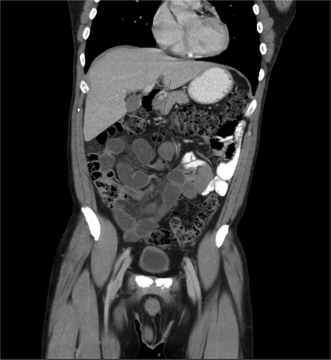 Figure 1
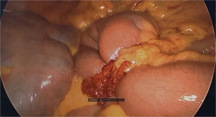 Figure 2
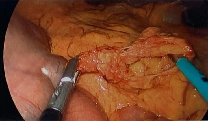 Figure 3
Peer Reviews
No public reviews on file for this paper yet. If you reviewed it on a platform where reviews are public (OpenReview, ICLR, NeurIPS, ICML), you can paste yours below so the community can read it here.
Videos
No videos yet. Explain this paper in a talk, walkthrough, or lecture? Add one.
Taxonomy
TopicsIntestinal and Peritoneal Adhesions · Hernia repair and management · Intestinal Malrotation and Obstruction Disorders
Introduction
Internal hernias are defined as the protrusion of viscera through a mesenteric or peritoneal aperture, a rare occurrence, accounting for only 0.5%–3% of bowel obstructions [9]. The most common subtypes include paraduodenal (53%) and pericaecal (13%), with less common occurrences involving the foramen of Winslow (8%), transmesenteric and transmesocolic (8%), intersigmoid (6%), and retroanastomotic (5%). Transomental hernias, where bowel protrudes through a defect in the omentum, are the rarest subtype, representing 1%–4% of internal hernias [6]. We present the case of a 47-year-old male with a closed-loop bowel obstruction secondary to a spontaneous transomental hernia.
Case report
A 47-year-old Caucasian male presented to the Emergency Department with several hours of worsening upper abdominal pain. The pain was initially colicky but progressed to constant, with associated nausea but no vomiting or fever. He had a normal bowel motion the day prior to presentation. He had no comorbidities and no history of prior abdominal surgeries. All vital signs were within normal limits, and he was afebrile. On examination, his abdomen was mildly distended, soft, and tender to palpation on the left side and centrally. His white cell count was elevated at 13.2 × 10^9^/L, with normal C-reactive protein, liver function tests, lipase, pH, and lactate of 1.2.
A CT abdomen and pelvis with oral and intravenous contrast revealed a closed-loop small bowel obstruction, with dilated loops of small bowel in the right mid to lower abdomen and associated mesenteric fluid (Fig. 1). A nasogastric tube was inserted, and the patient was taken for emergency surgery.
Abdominal CT scan demonstrating closed-loop small bowel obstruction involving jejunal loops, with two distinct transition points indicated by arrows.
During diagnostic laparoscopy, a loop of jejunum was found to be transomentally herniated, congested but viable (Figs 2 and 3). Adhesiolysis was performed, and no small bowel resection was required. The patient was discharged on postoperative day 2 after an uneventful recovery and has not experienced any complications since.
Intraoperative image from diagnostic laparoscopy showing a loop of jejunum herniating through a defect in the greater omentum.
Intraoperative image showing the omental defect after reduction of the herniated loop of jejunum.
Discussion
Transomental hernias are a rare cause of small bowel obstruction, with only 24 cases reported in the English literature over 60 years [5]. They tend to present in a bimodal age distribution, affecting pediatric patients and adults over 50 years of age. Omental defects may be congenital, due to senile atrophy, or acquired from surgery, trauma, or inflammation [9, 12]. Yamaguchi classified transomental hernias into three types: A (through the fused layers of the greater omentum), B (through the omental bursa), and C (into the omental bursa) [11].
Diagnosing transomental hernias preoperatively is difficult [9], as symptoms mimic other intestinal obstructions—abdominal pain, nausea, vomiting, distension, and obstipation [5, 6]. However, they have a higher risk of strangulation due to the small orifice, leading to up to 30% morbidity [8]. CT is the most common imaging modality, often showing dilated bowel loops, collapsed distal bowel segments, and the ‘beak sign,’ indicative of a triangular-shaped transition zone [2]. Other findings include the ‘whirl sign,’ representing a swirling pattern of mesenteric vessels [1]. Localization of dilated small bowel loops in the lesser sac suggests transomental hernia, though definitive diagnosis often requires surgery [4, 6].
Surgical reduction of the herniated bowel is the treatment of choice, performed either laparoscopically or via laparotomy [7]. Laparoscopy is typically possible, given that proximal jejunal loops are usually involved, providing adequate space for pneumoperitoneum [4, 5, 10]. Small bowel resection is required if ischemia or necrosis is present. Repair of the omental defect, often with sutures or partial omentectomy, helps prevent recurrence [3].
Conclusion
Transomental hernias are a rare cause of small bowel obstruction, often presenting with nonspecific clinical and radiological signs. Maintaining a high index of suspicion is essential for early surgical intervention and reducing morbidity.
The reference list from the paper itself. Each links out to its DOI / PubMed record.
- 1Camera L, De Gennaro A, Longobardi M, et al. A spontaneous strangulated transomental hernia: prospective and retrospective multi-detector computed tomography findings. World J Radiol 2014;6:26–30. 10.4329/wjr.v 6.i 2.26.24578790 PMC 3935064 · doi ↗ · pubmed ↗
- 2Delabrousse E, Couvreur M, Saguet O, et al. Strangulated transomental hernia: CT findings. Abdom Imaging 2001;26:86–8. 10.1007/s 002610000135.11116369 · doi ↗ · pubmed ↗
- 3Ergenc M, Uprak TK. A spontaneous transomental hernia: a rare cause of bowel obstruction. Iran J Public Health 2023;52:201–4. 10.18502/ijph.v 52i 1.11683.36824238 PMC 9941434 · doi ↗ · pubmed ↗
- 4Guinier D, Tissot O. Strangulated lesser sac hernia. J Visc Surg 2012;149:e 221–2. 10.1016/j.jviscsurg.2012.02.002.22424797 · doi ↗ · pubmed ↗
- 5Inukai K, Takashima N, Miyai H, et al. Two patients with spontaneous transomental hernia treated with laparoscopic surgery: a review. J Surg Case Rep 2018;2018:rjy 070. 10.1093/jscr/rjy 070.PMC 588883129644047 · doi ↗ · pubmed ↗
- 6Lee SH, Lee SH. Spontaneous transomental hernia. Ann Coloproctol 2016;32:38–41. 10.3393/ac.2016.32.1.38.26962535 PMC 4783511 · doi ↗ · pubmed ↗
- 7Narjis Y, Rabbani K, El Mansouri MN, et al. An unusual cause of small bowel obstruction: “the Swiss cheese transomental hernia”. Indian J Surg 2010;72:369–70. 10.1007/s 12262-010-0106-4.23133305 PMC 3451843 · doi ↗ · pubmed ↗
- 8Newsom BD, Kukora JS. Congenital and acquired internal hernias: unusual causes of small bowel obstruction. Am J Surg 1986;152:279–85. 10.1016/0002-9610(86)90258-8.3752377 · doi ↗ · pubmed ↗