The genome sequence of a braconid wasp, Microplitis deprimator (Fabricius, 1798)
Hannah King, Gavin R. Broad, Cheng Sun, Terrence Sylvester

TL;DR
This paper presents the genome sequence of the braconid wasp Microplitis deprimator, including a detailed assembly of its chromosomes and mitochondrial genome.
Contribution
The study provides the first genome assembly for Microplitis deprimator, including 11 chromosomal pseudomolecules and the mitochondrial genome.
Findings
The genome assembly is 233.20 megabases in total length.
99.12% of the assembly is scaffolded into 11 chromosomal pseudomolecules.
The mitochondrial genome is 19.62 kilobases in length.
Abstract
We present a genome assembly from an individual female braconid wasp, Microplitis deprimator (Arthropoda; Insecta; Hymenoptera; Braconidae). The genome sequence has a total length of 233.20 megabases. Most of the assembly (99.12%) is scaffolded into 11 chromosomal pseudomolecules. The mitochondrial genome has also been assembled and is 19.62 kilobases in length.
Genes, proteins, chemicals, diseases, species, mutations and cell lines named across the full text — each resolved to its canonical identifier and authoritative record.
Click any figure to enlarge with its caption.
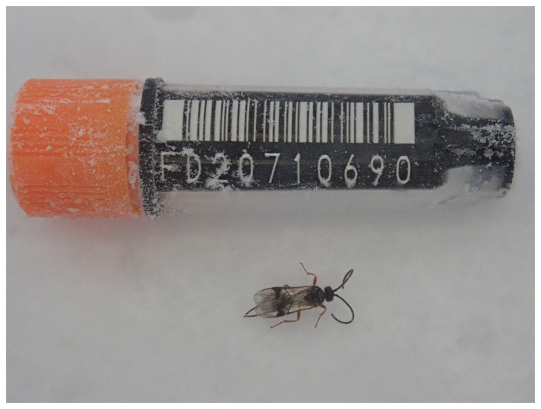 Figure 1
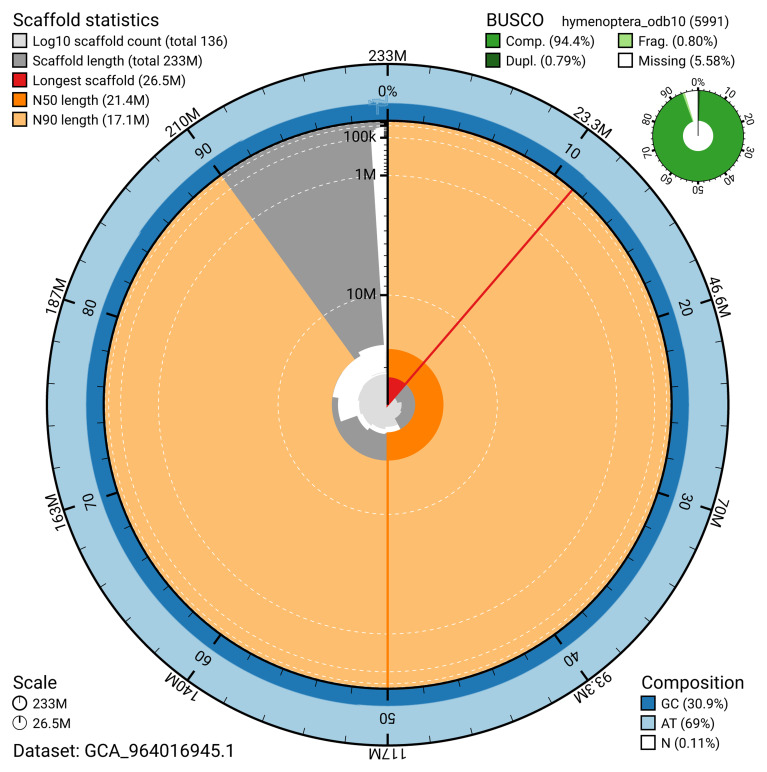 Figure 2
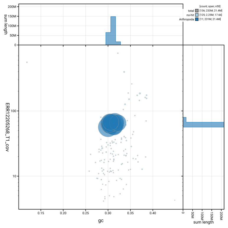 Figure 3
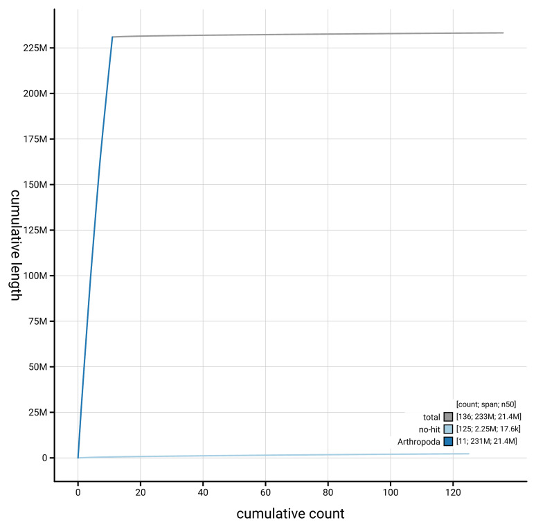 Figure 4
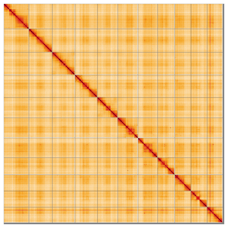 Figure 5
| Project information | |||
|---|---|---|---|
|
|
| ||
|
| PRJEB67617 | ||
|
|
| ||
|
| SAMEA110451492 | ||
|
| 2964682 | ||
| Specimen information | |||
|
|
|
|
|
|
| iyMicDepi1 | SAMEA110451663 | Whole organism |
|
| iyMicDepi1 | SAMEA110451663 | Whole organism |
| Sequencing information | |||
|
|
|
|
|
|
| ERR12144003 | 1.10e+09 | 165.61 |
|
| ERR12205268 | 1.96e+06 | 16.31 |
| Genome assembly | ||
|---|---|---|
| Assembly name | iyMicDepi1.1 | |
| Assembly accession | GCA_964016945.1 | |
|
|
| |
| Span (Mb) | 233.20 | |
| Number of contigs | 1,428 | |
| Number of scaffolds | 135 | |
| Longest scaffold (Mb) | 26.47 | |
| Assembly metrics
|
| |
| Contig N50 length (Mb) | 0.4 |
|
| Scaffold N50 length (Mb) | 21.4 |
|
| Consensus quality (QV) | 54.2 |
|
|
| 99.99% |
|
| BUSCO
| C:94.1%[S:93.3%,D:0.8%],
|
|
| Percentage of assembly
| 99.12% |
|
| Sex chromosomes | None |
|
| Organelles | Mitochondrial genome:
|
|
| INSDC
| Name | Length
| GC% |
|---|---|---|---|
| 1 | 26.47 | 31.0 | |
| 2 | 24.7 | 30.0 | |
| 3 | 23.93 | 30.5 | |
| 4 | 23.69 | 30.5 | |
| 5 | 21.4 | 30.5 | |
| 6 | 21.28 | 31.5 | |
| 7 | 20.78 | 31.5 | |
| 8 | 18.03 | 31.0 | |
| 9 | 17.14 | 30.5 | |
| 10 | 17.1 | 32.0 | |
| 11 | 16.48 | 30.5 | |
| MT | 0.02 | 12.0 |
| Software tool | Version | Source |
|---|---|---|
| BEDTools | 2.30.0 |
|
| BLAST | 2.14.0 |
|
| BlobToolKit | 4.3.7 |
|
| BUSCO | 5.4.3 and 5.5.0 |
|
| bwa-mem2 | 2.2.1 |
|
| Cooler | 0.8.11 |
|
| DIAMOND | 2.1.8 |
|
| fasta_windows | 0.2.4 |
|
| FastK | 427104ea91c78c3b8b8b49f1a7d6bbeaa869ba1c |
|
| Gfastats | 1.3.6 |
|
| GoaT CLI | 0.2.5 |
|
| Hifiasm | 0.19.8-r587 |
|
| HiGlass | 44086069ee7d4d3f6f3f0012569789ec138f42b84
|
|
| Merqury.FK | d00d98157618f4e8d1a9190026b19b471055b22e |
|
| MitoHiFi | 3 |
|
| MultiQC | 1.14, 1.17, and 1.18 |
|
| NCBI Datasets | 15.12.0 |
|
| Nextflow | 23.04.0-5857 |
|
| PretextView | 0.2 |
|
| purge_dups | 1.2.5 |
|
| samtools | 1.16.1, 1.17, and 1.18 |
|
| sanger-tol/ascc | - |
|
| sanger-tol/
| 1.1.1 |
|
| sanger-tol/
| 1.2.1 |
|
| Seqtk | 1.3 |
|
| Singularity | 3.9.0 |
|
| TreeVal | 1.0.0 |
|
| YaHS | 1.2a.2 |
|
- —Wellcome Trust
Peer Reviews
No public reviews on file for this paper yet. If you reviewed it on a platform where reviews are public (OpenReview, ICLR, NeurIPS, ICML), you can paste yours below so the community can read it here.
Videos
No videos yet. Explain this paper in a talk, walkthrough, or lecture? Add one.
Taxonomy
TopicsInsect Resistance and Genetics · Lepidoptera: Biology and Taxonomy · Genomics and Phylogenetic Studies
Species taxonomy
Eukaryota; Opisthokonta; Metazoa; Eumetazoa; Bilateria; Protostomia; Ecdysozoa; Panarthropoda; Arthropoda; Mandibulata; Pancrustacea; Hexapoda; Insecta; Dicondylia; Pterygota; Neoptera; Endopterygota; Hymenoptera; Apocrita; Ichneumonoidea; Braconidae; Microgastrinae; Microplitis; Microplitis deprimator (Fabricius, 1798) (NCBI:txid2964682).
Background
Microplitis deprimator is a relatively large species of Microgastrinae, a very species-rich subfamily of Braconidae, all parasitoids of Lepidoptera larvae. Along with most other microgastrines, M. deprimator is predominantly black with paler legs, but identifiable as a Microplitis mainly by the presence of a second submarginal cell and relatively short hind coxa compared to similar genera, such as Microgaster ( Shaw et al., 2024). Nixon (1970) provides a key to species which is a bit out of date but through which M. deprimator should still key easily (to M. sordipes).
The specimen sequenced was reared from Acronicta psi (Linnaeus), which is a typical host for M. deprimator in the sense that we are using the name. However, there has been confusion over the correct name for this braconid: Nixon (1970) Fernandez-Triana et al. (2020), and other authors have called this species Microplitis sordipes (Ziegler), with M. deprimator implicitly or explicitly treated as a separate species. We follow the latest British checklist ( Broad et al., 2016), in which sordipes is treated as a junior synonym of deprimator.
Regardless of which name is valid, this species is fairly well-known biologically, as a solitary, koinobiont endoparasitoid of hairy Lepidoptera larvae. Oviposition is into small larvae and emergence is from the host larva before it is fully fed. Nixon (1970) recorded several species of, especially, Acronicta (Noctuidae) and Clostera (Notodontidae) as hosts, hosts all belonging to Noctuoidea and being exposed, hairy foliage feeders. The winter is spent in a tough, brown-banded cocoon attached to a twig ( Nixon, 1970). There is apparently a second generation, in the summer, with wasps pupating in a much thinner cocoon and attacking a slightly wider range of hosts, including Euproctis (Erebidae: Lymantriinae). Whether or not these morphologically very similar specimens are conspecific remains to be tested ( Nixon, 1970). Microplitis species are haemolymph feeders, leaving the host to pupate often when the host is still alive ( Shaw & Huddleston, 1991). Microgastrinae are known to inject hosts with bracoviruses along with the egg, which help overcome the host immune system, and the functions of these bracovirus genes have been characterized for another Microplitis species, M. demolitor Wilkinson ( Lorenzi et al., 2023). Genomes of microgastrines, and other braconids, are helping us understand the evolution of the association of bracoviruses with these wasps.
Genome sequence report
The genome of an adult female specimen of Microplitis deprimator ( Figure 1) was sequenced using Pacific Biosciences single-molecule HiFi long reads, generating a total of 16.31 Gb (gigabases) from 1.96 million reads, providing approximately 69-fold coverage. Primary assembly contigs were scaffolded with chromosome conformation Hi-C data, which produced 165.61 Gb from 1,096.75 million reads, yielding an approximate coverage of 710-fold. Specimen and sequencing details are provided in Table 1.
Photograph of the Microplitis deprimator (iyMicDepi1) specimen used for genome sequencing.
Table 1.: Specimen and sequencing data for Microplitis deprimator.
Assembly errors were corrected by manual curation, including 590 missing joins or mis-joins and 15 haplotypic duplications, reducing the scaffold number by 59.04%, and increasing the scaffold N50 by 10.43%. The final assembly has a total length of 233.20 Mb in 135 sequence scaffolds, with 1,292 gaps, and a scaffold N50 of 21.4 Mb ( Table 2). The snail plot in Figure 2 provides a summary of the assembly statistics, while the distribution of assembly scaffolds on GC proportion and coverage is shown in Figure 3. The cumulative assembly plot in Figure 4 shows curves for subsets of scaffolds assigned to different phyla. Most (99.12%) of the assembly sequence was assigned to 11 chromosomal-level scaffolds, which are named in order of size ( Figure 5; Table 3).
Table 2.: Genome assembly data for Microplitis deprimator, iyMicDepi1.1.
Genome assembly of Microplitis deprimator, iyMicDepi1.1: metrics.The BlobToolKit snail plot shows N50 metrics and BUSCO gene completeness. The main plot is divided into 1,000 size-ordered bins around the circumference with each bin representing 0.1% of the 233,239,307 bp assembly. The distribution of scaffold lengths is shown in dark grey with the plot radius scaled to the longest scaffold present in the assembly (26,466,399 bp, shown in red). Orange and pale-orange arcs show the N50 and N90 scaffold lengths (21,403,585 and 17,099,948 bp), respectively. The pale grey spiral shows the cumulative scaffold count on a log scale with white scale lines showing successive orders of magnitude. The blue and pale-blue area around the outside of the plot shows the distribution of GC, AT and N percentages in the same bins as the inner plot. A summary of complete, fragmented, duplicated and missing BUSCO genes in the hymenoptera_odb10 set is shown in the top right. An interactive version of this figure is available at https://blobtoolkit.genomehubs.org/view/GCA_964016945.1/dataset/GCA_964016945.1/snail.
Genome assembly of Microplitis deprimator, iyMicDepi1.1: BlobToolKit GC-coverage plot showing sequence coverage (vertical axis) and GC content (horizontal axis).The circles represent scaffolds, with the size proportional to scaffold length and the colour representing phylum membership. The histograms along the axes display the total length of sequences distributed across different levels of coverage and GC content. An interactive version of this figure is available at https://blobtoolkit.genomehubs.org/view/GCA_964016945.1/dataset/GCA_964016945.1/blob.
Genome assembly of Microplitis deprimator iyMicDepi1.1: BlobToolKit cumulative sequence plot.The grey line shows cumulative length for all scaffolds. Coloured lines show cumulative lengths of scaffolds assigned to each phylum using the buscogenes taxrule. An interactive version of this figure is available at https://blobtoolkit.genomehubs.org/view/GCA_964016945.1/dataset/GCA_964016945.1/cumulative.
Genome assembly of Microplitis deprimator iyMicDepi1.1: Hi-C contact map of the iyMicDepi1.1 assembly, visualised using HiGlass.Chromosomes are shown in order of size from left to right and top to bottom. An interactive version of this figure may be viewed at https://genome-note-higlass.tol.sanger.ac.uk/l/?d=IgLLfDePTRGEl6T0R6kpaw
Table 3.: Chromosomal pseudomolecules in the genome assembly of Microplitis deprimator, iyMicDepi1.
While not fully phased, the assembly deposited is of one haplotype. Contigs corresponding to the second haplotype have also been deposited. The mitochondrial genome was also assembled and can be found as a contig within the multifasta file of the genome submission.
The estimated Quality Value (QV) of the final assembly is 54.2 with k-mer completeness of 99.99%, and the assembly has a BUSCO v5.4.3 completeness of 94.1% (single = 93.3%, duplicated = 0.8%), using the hymenoptera_odb10 reference set ( n = 5,991).
Metadata for specimens, BOLD barcode results, spectra estimates, sequencing runs, contaminants and pre-curation assembly statistics are given at https://links.tol.sanger.ac.uk/species/2964682.
Methods
Sample acquisition and DNA barcoding
An adult female specimen of Microplitis deprimator (specimen ID Ox001837, ToLID iyMicDepi1) was reared from Acronicta psi larva and collected on 2021-08-20. The specimen was collected by Hannah King (University of Oxford), formally identified by Gavin Broad (Natural History Museum) and preserved on dry ice.
The initial identification was verified by an additional DNA barcoding process according to the framework developed by Twyford et al. (2024). A small sample was dissected from the specimens and stored in ethanol, while the remaining parts were shipped on dry ice to the Wellcome Sanger Institute (WSI). The tissue was lysed, the COI marker region was amplified by PCR, and amplicons were sequenced and compared to the BOLD database, confirming the species identification ( Crowley et al., 2023). Following whole genome sequence generation, the relevant DNA barcode region was also used alongside the initial barcoding data for sample tracking at the WSI ( Twyford et al., 2024). The standard operating procedures for Darwin Tree of Life barcoding have been deposited on protocols.io ( Beasley et al., 2023).
Nucleic acid extraction
The workflow for high molecular weight (HMW) DNA extraction at the Wellcome Sanger Institute (WSI) Tree of Life Core Laboratory includes a sequence of core procedures: sample preparation and homogenisation, DNA extraction, fragmentation and purification. Detailed protocols are available on protocols.io ( Denton et al., 2023b). The iyMicDepi1 sample was prepared for DNA extraction by weighing and dissecting it on dry ice ( Jay et al., 2023). Tissue from the whole organism was homogenised using a PowerMasher II tissue disruptor ( Denton et al., 2023a).
HMW DNA was extracted in the WSI Scientific Operations core using the Automated MagAttract v2 protocol ( Oatley et al., 2023). The DNA was sheared into an average fragment size of 12–20 kb in a Megaruptor 3 system ( Bates et al., 2023). Sheared DNA was purified by solid-phase reversible immobilisation, using AMPure PB beads to eliminate shorter fragments and concentrate the DNA ( Strickland et al., 2023). The concentration of the sheared and purified DNA was assessed using a Nanodrop spectrophotometer and Qubit Fluorometer using the Qubit dsDNA High Sensitivity Assay kit. Fragment size distribution was evaluated by running the sample on the FemtoPulse system.
Hi-C preparation
Tissue from the whole organism of the iyMicDepi1 sample was processed at the WSI Scientific Operations core, using the Arima-HiC v2 kit. In brief, frozen tissue (stored at –80 °C) was fixed, and the DNA crosslinked using a TC buffer with 22% formaldehyde. After crosslinking, the tissue was homogenised using the Diagnocine Power Masher-II and BioMasher-II tubes and pestles. Following the kit manufacturer's instructions, crosslinked DNA was digested using a restriction enzyme master mix. The 5’-overhangs were then filled in and labelled with biotinylated nucleotides and proximally ligated. An overnight incubation was carried out for enzymes to digest remaining proteins and for crosslinks to reverse. A clean up was performed with SPRIselect beads prior to library preparation.
Library preparation and sequencing
Library preparation and sequencing were performed at the WSI Scientific Operations core. Pacific Biosciences HiFi circular consensus DNA sequencing libraries were prepared using the PacBio Express Template Preparation Kit v2.0 (Pacific Biosciences, California, USA) as per the manufacturer's instructions. The kit includes the reagents required for removal of single-strand overhangs, DNA damage repair, end repair/A-tailing, adapter ligation, and nuclease treatment. Library preparation also included a library purification step using AMPure PB beads (Pacific Biosciences, California, USA) and size selection step to remove templates shorter than 3 kb using AMPure PB modified SPRI. DNA concentration was quantified using the Qubit Fluorometer v2.0 and Qubit HS Assay Kit and the final library fragment size analysis was carried out using the Agilent Femto Pulse Automated Pulsed Field CE Instrument and gDNA 165kb gDNA and 55kb BAC analysis kit. Samples were sequenced using the Sequel IIe system (Pacific Biosciences, California, USA). The concentration of the library loaded onto the Sequel IIe was between 40–135 pM. The SMRT link software, a PacBio web-based end-to-end workflow manager, was used to set-up and monitor the run, as well as perform primary and secondary analysis of the data upon completion.
For Hi-C library preparation, DNA was fragmented to a size of 400 to 600 bp using a Covaris E220 sonicator. The DNA was then enriched, barcoded, and amplified using the NEBNext Ultra II DNA Library Prep Kit following manufacturers’ instructions. The Hi-C sequencing was performed using paired-end sequencing with a read length of 150 bp on an Illumina NovaSeq 6000 instrument.
Genome assembly, curation and evaluation
** Assembly **
The HiFi reads were first assembled using Hifiasm ( Cheng et al., 2021) with the --primary option. Haplotypic duplications were identified and removed using purge_dups ( Guan et al., 2020). The Hi-C reads were mapped to the primary contigs using bwa-mem2 ( Vasimuddin et al., 2019). The contigs were further scaffolded using the provided Hi-C data ( Rao et al., 2014) in YaHS ( Zhou et al., 2023) using the --break option. The scaffolded assemblies were evaluated using Gfastats ( Formenti et al., 2022), BUSCO ( Manni et al., 2021) and MERQURY.FK ( Rhie et al., 2020).
The mitochondrial genome was assembled using MitoHiFi ( Uliano-Silva et al., 2023), which runs MitoFinder ( Allio et al., 2020) and uses these annotations to select the final mitochondrial contig and to ensure the general quality of the sequence.
** Assembly curation **
The assembly was decontaminated using the Assembly Screen for Cobionts and Contaminants (ASCC) pipeline (article in preparation). Flat files and maps used in curation were generated in TreeVal ( Pointon et al., 2023). Manual curation was primarily conducted using PretextView ( Harry, 2022), with additional insights provided by JBrowse2 ( Diesh et al., 2023) and HiGlass ( Kerpedjiev et al., 2018). Scaffolds were visually inspected and corrected as described by Howe et al. (2021). Any identified contamination, missed joins, and mis-joins were corrected, and duplicate sequences were tagged and removed. The curation process is documented at https://gitlab.com/wtsi-grit/rapid-curation (article in preparation).
** Evaluation of the final assembly **
The final assembly was post-processed and evaluated using the three Nextflow ( Di Tommaso et al., 2017) DSL2 pipelines: sanger-tol/readmapping ( Surana et al., 2023a), sanger-tol/genomenote ( Surana et al., 2023b), and sanger-tol/blobtoolkit ( Muffato et al., 2024). The readmapping pipeline aligns the Hi-C reads using bwa-mem2 ( Vasimuddin et al., 2019) and combines the alignment files with SAMtools ( Danecek et al., 2021). The genomenote pipeline converts the Hi-C alignments into a contact map using BEDTools ( Quinlan & Hall, 2010) and the Cooler tool suite ( Abdennur & Mirny, 2020). The contact map is visualised in HiGlass ( Kerpedjiev et al., 2018). This pipeline also generates assembly statistics using the NCBI datasets report ( Sayers et al., 2024), computes k-mer completeness and QV consensus quality values with FastK and MERQURY.FK, and runs BUSCO ( Manni et al., 2021) to assess completeness.
The blobtoolkit pipeline is a Nextflow port of the previous Snakemake Blobtoolkit pipeline ( Challis et al., 2020). It aligns the PacBio reads in SAMtools and minimap2 ( Li, 2018) and generates coverage tracks for regions of fixed size. In parallel, it queries the GoaT database ( Challis et al., 2023) to identify all matching BUSCO lineages to run BUSCO ( Manni et al., 2021). For the three domain-level BUSCO lineages, the pipeline aligns the BUSCO genes to the UniProt Reference Proteomes database ( Bateman et al., 2023) with DIAMOND ( Buchfink et al., 2021) blastp. The genome is also split into chunks according to the density of the BUSCO genes from the closest taxonomic lineage, and each chunk is aligned to the UniProt Reference Proteomes database with DIAMOND blastx. Genome sequences without a hit are chunked with seqtk and aligned to the NT database with blastn ( Altschul et al., 1990). The blobtools suite combines all these outputs into a blobdir for visualisation.
The genome assembly and evaluation pipelines were developed using nf-core tooling ( Ewels et al., 2020) and MultiQC ( Ewels et al., 2016), relying on the Conda package manager, the Bioconda initiative ( Grüning et al., 2018), the Biocontainers infrastructure ( da Veiga Leprevost et al., 2017), as well as the Docker ( Merkel, 2014) and Singularity ( Kurtzer et al., 2017) containerisation solutions.
Table 4 contains a list of relevant software tool versions and sources.
Wellcome Sanger Institute – Legal and Governance
The materials that have contributed to this genome note have been supplied by a Darwin Tree of Life Partner. The submission of materials by a Darwin Tree of Life Partner is subject to the ‘Darwin Tree of Life Project Sampling Code of Practice’, which can be found in full on the Darwin Tree of Life website here. By agreeing with and signing up to the Sampling Code of Practice, the Darwin Tree of Life Partner agrees they will meet the legal and ethical requirements and standards set out within this document in respect of all samples acquired for, and supplied to, the Darwin Tree of Life Project.
Further, the Wellcome Sanger Institute employs a process whereby due diligence is carried out proportionate to the nature of the materials themselves, and the circumstances under which they have been/are to be collected and provided for use. The purpose of this is to address and mitigate any potential legal and/or ethical implications of receipt and use of the materials as part of the research project, and to ensure that in doing so we align with best practice wherever possible. The overarching areas of consideration are:
• Ethical review of provenance and sourcing of the material
• Legality of collection, transfer and use (national and international)
Each transfer of samples is further undertaken according to a Research Collaboration Agreement or Material Transfer Agreement entered into by the Darwin Tree of Life Partner, Genome Research Limited (operating as the Wellcome Sanger Institute), and in some circumstances other Darwin Tree of Life collaborators.
The reference list from the paper itself. Each links out to its DOI / PubMed record.
- 1Abdennur N Mirny LA : Cooler: scalable storage for Hi-C data and other genomically labeled arrays. Bioinformatics. 2020;36(1):311–316. 10.1093/bioinformatics/btz 540 31290943 PMC 8205516 · doi ↗ · pubmed ↗
- 2Allio R Schomaker-Bastos A Romiguier J : Mito Finder: efficient automated large-scale extraction of mitogenomic data in target enrichment phylogenomics. Mol Ecol Resour. 2020;20(4):892–905. 10.1111/1755-0998.13160 32243090 PMC 7497042 · doi ↗ · pubmed ↗
- 3Altschul SF Gish W Miller W : Basic Local Alignment Search Tool. J Mol Biol. 1990;215(3):403–410. 10.1016/S 0022-2836(05)80360-2 2231712 · doi ↗ · pubmed ↗
- 4Bateman A Martin MJ Orchard S : Uni Prot: the universal protein knowledgebase in 2023. Nucleic Acids Res. 2023;51(D 1):D 523–D 531. 10.1093/nar/gkac 1052 36408920 PMC 9825514 · doi ↗ · pubmed ↗
- 5Bates A Clayton-Lucey I Howard C : Sanger Tree of Life HMW DNA fragmentation: diagenode Megaruptor ®3 for LI Pac Bio. protocols.io. 2023. 10.17504/protocols.io.81wgbxzq 3lpk/v 1 · doi ↗
- 6Beasley J Uhl R Forrest LL : DNA barcoding SO Ps for the Darwin Tree of Life Project. protocols.io. 2023; [Accessed 25 June 2024]. 10.17504/protocols.io.261ged 91jv 47/v 1 · doi ↗
- 7Broad GR Shaw MR Godfray HCJ : Checklist of British and Irish Hymenoptera - Braconidae. Biodivers Data J. 2016;4: e 8151. 10.3897/BDJ.4.e 8151 27226759 PMC 4867695 · doi ↗ · pubmed ↗
- 8Buchfink B Reuter K Drost HG : Sensitive protein alignments at Tree-of-Life scale using DIAMOND. Nat Methods. 2021;18(4):366–368. 10.1038/s 41592-021-01101-x 33828273 PMC 8026399 · doi ↗ · pubmed ↗