Orbital Adipose Tissue: The Optimal Control for Back-Table Fluorescence Imaging of Orbital Tumors
Lan Yao, Wenhua Zhang, Xuedong Wang, Lishuang Guo, Wenlu Liu, Yueyue Li, Rui Ma, Yan Hei, Xinji Yang, Zeyu Zhang, Wei Wu

TL;DR
This study finds that adipose tissue is the best control for fluorescence imaging of orbital tumors due to its consistent low fluorescence and minimal influence from patient factors.
Contribution
The study identifies adipose tissue as the optimal control tissue for back-table fluorescence imaging of orbital tumors.
Findings
Adipose tissue showed consistent hypofluorescence and was not significantly affected by patient clinical variables.
Larger adipose tissues (>1 cm) had a 27% higher signal-to-background ratio compared to smaller ones.
Skin tissue had higher fluorescence than diseased tissue, while muscle tissue showed high variability.
Abstract
Control tissue is essential for ensuring the precision of semiquantitative analysis in back-table fluorescence imaging. However, there remains a lack of agreement on the appropriate selection of control tissues. To evaluate the back-table fluorescence imaging performance of different normal tissues and identify the optimal normal tissue, a cohort of 39 patients with orbital tumors were enrolled in the study. Prior to surgery, these patients received indocyanine green (ICG) and following resection, 43 normal control tissues (34 adipose tissues, 3 skin tissues, 3 periosteal tissues, and 3 muscle tissues) were examined using back-table fluorescence imaging. The skin tissue demonstrated significantly elevated fluorescence intensity in comparison to the diseased tissue, whereas the muscle tissue exhibited a broad range and standard deviation of fluorescence signal intensity. Conversely, the…
Genes, proteins, chemicals, diseases, species, mutations and cell lines named across the full text — each resolved to its canonical identifier and authoritative record.
Click any figure to enlarge with its caption.
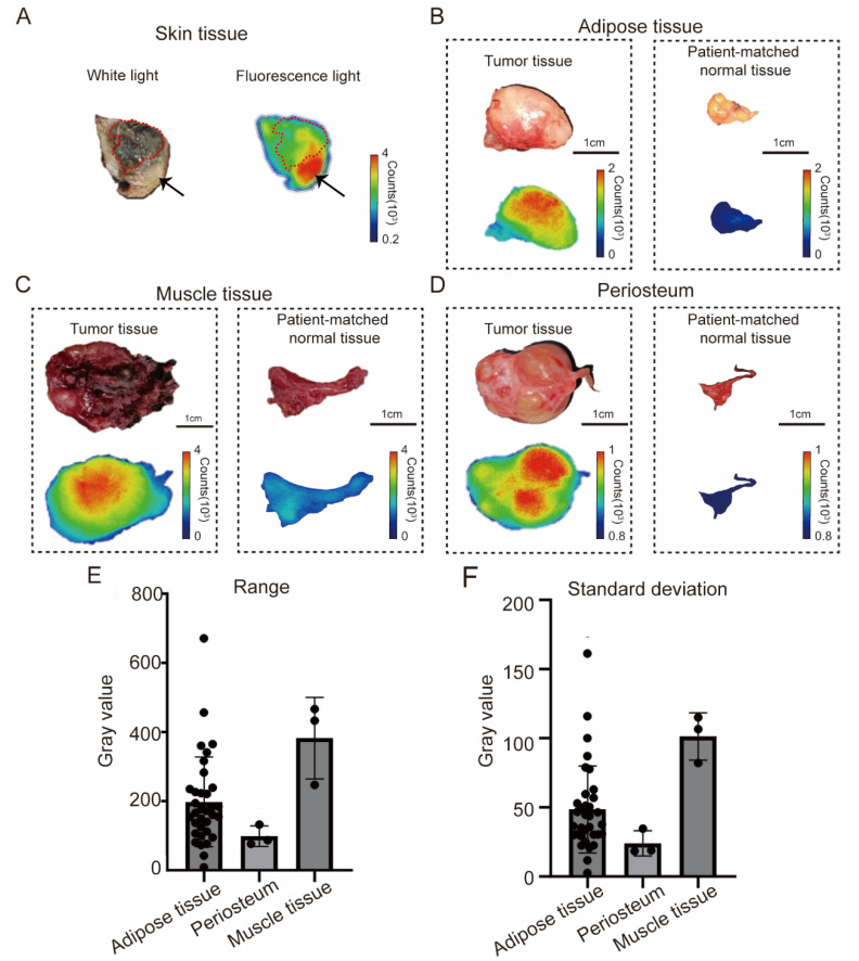 Figure 1
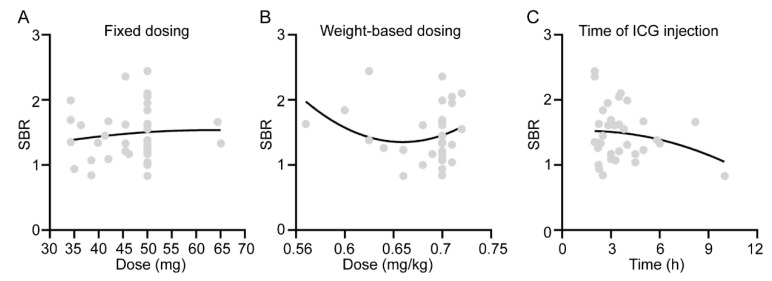 Figure 2
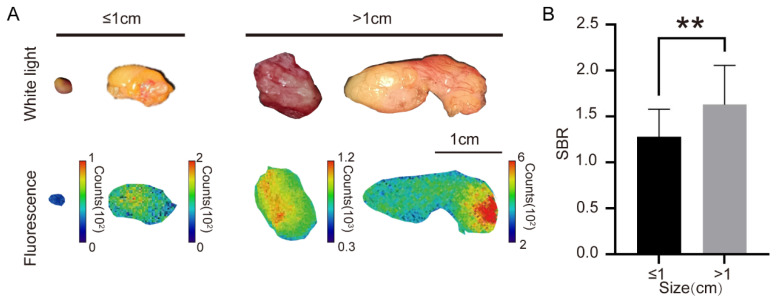 Figure 3
Peer Reviews
No public reviews on file for this paper yet. If you reviewed it on a platform where reviews are public (OpenReview, ICLR, NeurIPS, ICML), you can paste yours below so the community can read it here.
Videos
No videos yet. Explain this paper in a talk, walkthrough, or lecture? Add one.
Taxonomy
TopicsNanoplatforms for cancer theranostics · Photoacoustic and Ultrasonic Imaging · Meningioma and schwannoma management