Magnetic Resonance Imaging With a Novel Hip Flexion Scanning Position for Diagnosing Proximal Hamstring Tendinopathy
Aleksi Jokela, Pekka Niemi, Ilona Koski, Jussi Kosola, Xavier Valle, Ricard Pruna, Sakari Orava, Carles Pedret, Ramon Balius, Giulio Pasta, Juha-Jaakko Sinikumpu, Keijo Mäkelä, Lasse Lempainen

TL;DR
A new MRI scanning position improves the diagnosis of proximal hamstring tendinopathy by revealing more severe injuries than standard scans.
Contribution
A novel hip flexion MRI position is introduced to enhance diagnostic accuracy for proximal hamstring tendinopathy.
Findings
71% of patients showed more severe injury in the hip flexion position compared to the standard position.
16% of tendons showed rupture in the hip flexion position, classified as tendinosis in the standard position.
6% of tendons previously classified as normal in the standard position showed rupture in the hip flexion position.
Abstract
Making a diagnosis of proximal hamstring tendinopathy (PHT) may be challenging, as patients with correlating clinical symptoms may have normal or minimal findings on magnetic resonance imaging (MRI) scans. The purpose of this study was to assess the effect of a novel hip flexion (HF) scanning position on the MRI diagnosis of PHT. It was hypothesized that the HF position, which simulates the symptom-provoking sitting position, would reveal PHT pathology more accurately than the standard scanning position. Cohort study (diagnosis); Level of evidence, 3. Patients with chronic PHT symptoms were included. Chronicity was defined as symptoms that were present for >3 months. Each patient underwent an MRI in 2 parts: (1) the standard pelvic examination in the supine position and (2) the novel HF position in which the patient lays on his or her side with the hip at 90° of flexion. Tendon…
Genes, proteins, chemicals, diseases, species, mutations and cell lines named across the full text — each resolved to its canonical identifier and authoritative record.
Click any figure to enlarge with its caption.
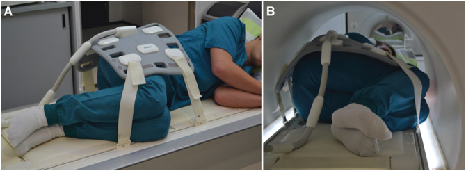 Figure 1
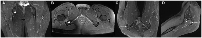 Figure 2
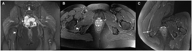 Figure 3
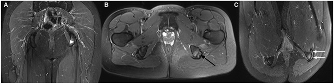 Figure 4
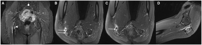 Figure 5
 Figure 6
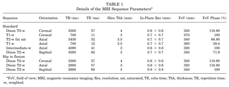 Figure 7
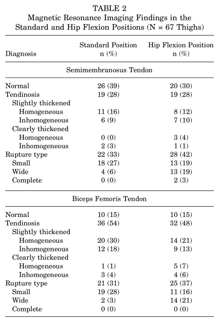 Figure 8
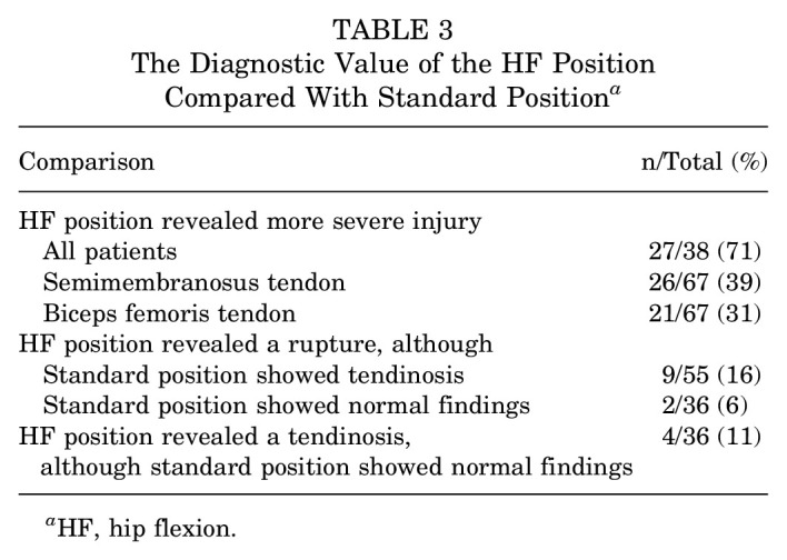 Figure 9
Peer Reviews
No public reviews on file for this paper yet. If you reviewed it on a platform where reviews are public (OpenReview, ICLR, NeurIPS, ICML), you can paste yours below so the community can read it here.
Videos
No videos yet. Explain this paper in a talk, walkthrough, or lecture? Add one.
Taxonomy
TopicsSports injuries and prevention · Tendon Structure and Treatment · Shoulder Injury and Treatment