The genome sequence of a braconid wasp, Aleiodes leptofemur van Achterberg & Shaw, 2016
Gavin R. Broad, Julien Varaldi, Andrew D Austin

TL;DR
This paper presents the genome sequence of a braconid wasp, Aleiodes leptofemur, including a detailed assembly of its chromosomes and mitochondrial DNA.
Contribution
The study provides the first genome assembly for Aleiodes leptofemur, a braconid wasp, with scaffolding into chromosomal pseudomolecules.
Findings
The genome assembly spans 271.20 megabases.
The mitochondrial genome is 32.28 kilobases in length.
Most of the assembly is organized into 15 chromosomal pseudomolecules.
Abstract
We present a genome assembly from an individual male Aleiodes leptofemur (braconid wasp; Arthropoda; Insecta; Hymenoptera; Braconidae). The genome sequence spans 271.20 megabases. Most of the assembly is scaffolded into 15 chromosomal pseudomolecules. The mitochondrial genome has also been assembled and is 32.28 kilobases in length.
Genes, proteins, chemicals, diseases, species, mutations and cell lines named across the full text — each resolved to its canonical identifier and authoritative record.
Click any figure to enlarge with its caption.
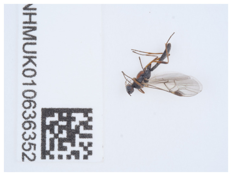 Figure 1
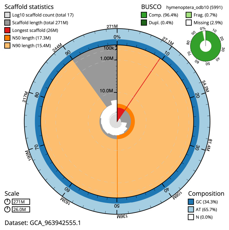 Figure 2
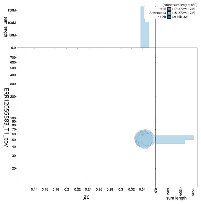 Figure 3
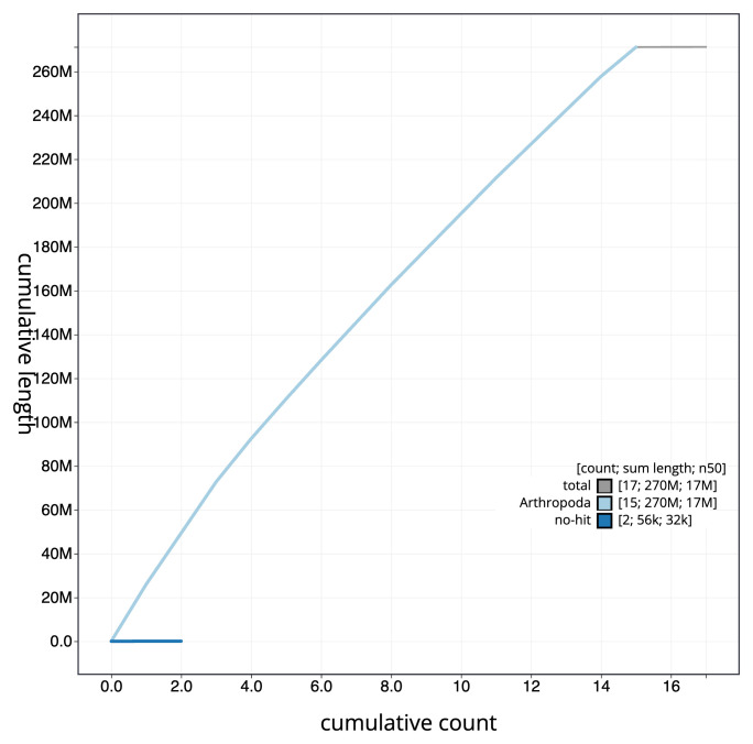 Figure 4
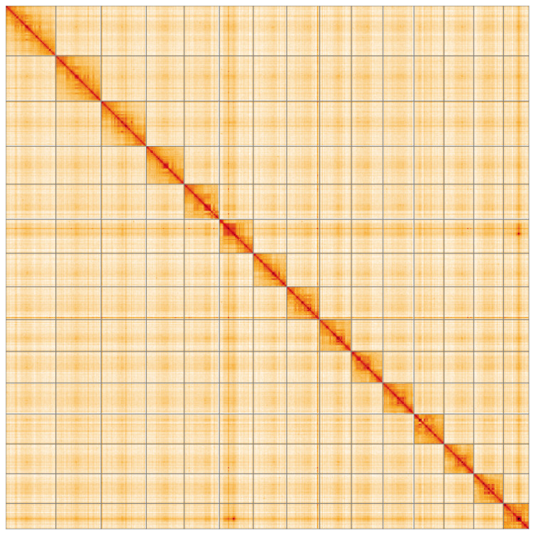 Figure 5
| Project information | |||
|---|---|---|---|
|
| Aleiodes leptofemur | ||
|
| PRJEB66053 | ||
|
|
| ||
|
| SAMEA111458715 | ||
|
| 1844523 | ||
| Specimen information | |||
|
|
|
|
|
|
| iyAleLepo1 | SAMEA111458777 | Whole organism |
|
| iyAleLepo1 | SAMEA111458777 | Whole organism |
| Sequencing information | |||
|
|
|
|
|
|
| ERR12071265 | 7.71e+08 | 116.45 |
|
| ERR12055583 | 1.40e+06 | 14.67 |
| Genome assembly | ||
|---|---|---|
| Assembly name | iyAleLepo1.1 | |
| Assembly accession | GCA_963942555.1 | |
| Span (Mb) | 271.20 | |
| Number of contigs | 280 | |
| Contig N50 length (Mb) | 1.8 | |
| Number of scaffolds | 16 | |
| Scaffold N50 length (Mb) | 17.3 | |
| Longest scaffold (Mb) | 26.01 | |
| Assembly metrics
|
| |
| Consensus quality (QV) | 63.6 |
|
|
| 100.0% |
|
| BUSCO
| C:96.2%[S:95.8%,D:0.4%],
|
|
| Percentage of assembly mapped to chromosomes | 99.97% |
|
| Sex chromosomes | None |
|
| Organelles | Mitochondrial genome: 32.28 kb |
|
| INSDC
| Name | Length
| GC% |
|---|---|---|---|
| 1 | 26.01 | 34.0 | |
| 2 | 23.41 | 34.5 | |
| 3 | 23.36 | 34.0 | |
| 4 | 19.68 | 34.0 | |
| 5 | 18.17 | 34.5 | |
| 6 | 17.64 | 35.0 | |
| 7 | 17.29 | 34.0 | |
| 8 | 17.08 | 34.0 | |
| 9 | 16.46 | 34.0 | |
| 10 | 16.32 | 34.5 | |
| 11 | 16.08 | 34.5 | |
| 12 | 15.52 | 34.5 | |
| 13 | 15.43 | 34.0 | |
| 14 | 15.37 | 34.5 | |
| 15 | 13.37 | 35.0 | |
| MT | 0.03 | 12.5 |
| Software tool | Version | Source |
|---|---|---|
| BEDTools | 2.30.0 |
|
| BLAST | 2.14.0 |
|
| BlobToolKit | 4.3.7 |
|
| BUSCO | 5.4.3 and 5.5.0 |
|
| bwa-mem2 | 2.2.1 |
|
| Cooler | 0.8.11 |
|
| DIAMOND | 2.1.8 |
|
| fasta_windows | 0.2.4 |
|
| FastK | 427104ea91c78c3b8b8b49f1a7d6bbeaa869ba1c |
|
| Gfastats | 1.3.6 |
|
| GoaT CLI | 0.2.5 |
|
| Hifiasm | 0.16.1 |
|
| HiGlass | 44086069ee7d4d3f6f3f0012569789ec138f42b84a
|
|
| Merqury.FK | d00d98157618f4e8d1a9190026b19b471055b22e |
|
| MitoHiFi | 3.2 |
|
| MultiQC | 1.14, 1.17, and 1.18 |
|
| NCBI Datasets | 15.12.0 |
|
| Nextflow | 23.04.0-5857 |
|
| OATK | 1 |
|
| PretextView | 0.2 |
|
| samtools | 1.16.1, 1.17, and 1.18 |
|
| sanger-tol/ascc | - |
|
| sanger-tol/genomenote | 1.1.1 |
|
| sanger-tol/readmapping | 1.2.1 |
|
| Seqtk | 1.3 |
|
| Singularity | 3.9.0 |
|
| TreeVal | 1.0.0 |
|
| YaHS | 1.1a.2 |
|
- —Wellcome Trust
Peer Reviews
No public reviews on file for this paper yet. If you reviewed it on a platform where reviews are public (OpenReview, ICLR, NeurIPS, ICML), you can paste yours below so the community can read it here.
Videos
No videos yet. Explain this paper in a talk, walkthrough, or lecture? Add one.
Taxonomy
TopicsGenomics and Phylogenetic Studies · Genetic diversity and population structure · Identification and Quantification in Food
Species taxonomy
Eukaryota; Opisthokonta; Metazoa; Eumetazoa; Bilateria; Protostomia; Ecdysozoa; Panarthropoda; Arthropoda; Mandibulata; Pancrustacea; Hexapoda; Insecta; Dicondylia; Pterygota; Neoptera; Endopterygota; Hymenoptera; Apocrita; Ichneumonoidea; Braconidae; Rogadinae; Aleiodes; Aleiodes leptofemur van Achterberg & Shaw, 2016 (NCBI:txid1844523).
Background
Aleiodes leptofemur is a small (about 4.5 mm long) braconid wasp of the subfamily Rogadinae. One of many similar species of Aleiodes, A. leptofemur can be identified using van Achterberg and Shaw (2016); useful features for distinguishing A. leptofemur from other dark and reddish-brown Aleiodes include the particularly slender femora and colour details of face, antenna and hind leg.
As with all other Rogadinae, where known, the hosts are Lepidoptera larvae which are mummified by the wasp larva, forming a hardened structure in which the wasp pupates ( Zaldívar-Riverón et al., 2008). A wide range of Noctuidae are attacked, with the common factor being that they feed low down in vegetation. The winter is spent as a larva in the host which is often manipulated to climb to an exposed position before being killed. This habit means that A. leptofemur is frequently reared from the exposed mummies. The adults are partly nocturnal and often light-trapped.
Found across much of Europe, A. leptofemur has multiple generations per year and adults are long-lived ( van Achterberg & Shaw, 2016). Until relatively recently, specimens had been misidentified as other Aleiodes, especially A. borealis (Thomson); however, van Achterberg & Shaw (2016) established that this species had been undescribed and formally described A. leptofemur, including extensive data on the ecology of this widespread species. This genome will help in deciphering the diversification of this very species-rich genus, including testing hypotheses of host range evolution ( Shaw, 2002).
Genome sequence report
The genome of an adult male Aleiodes leptofemur ( Figure 1) was sequenced using Pacific Biosciences single-molecule HiFi long reads, generating a total of 14.67 Gb (gigabases) from 1.40 million reads, providing approximately 59-fold coverage. Primary assembly contigs were scaffolded with chromosome conformation Hi-C data, which produced 116.45 Gbp from 771.22 million reads, yielding an approximate coverage of 429-fold. Specimen and sequencing information is summarised in Table 1.
Photograph of the Aleiodes leptofemur (iyAleLepo1) specimen used for genome sequencing.
Table 1.: Specimen and sequencing data for Aleiodes leptofemur.
Manual assembly curation corrected 87 missing joins or mis-joins, reducing the scaffold number by 66.67%. The final assembly has a total length of 271.20 Mb in 16 sequence scaffolds with a scaffold N50 of 17.3 Mb ( Table 2). The total count of gaps in the scaffolds is 263. The snail plot in Figure 2 provides a summary of the assembly statistics, while the distribution of assembly scaffolds on GC proportion and coverage is shown in Figure 3. The cumulative assembly plot in Figure 4 shows curves for subsets of scaffolds assigned to different phyla. Most (99.97%) of the assembly sequence was assigned to 15 chromosomal-level scaffolds. Chromosome-scale scaffolds confirmed by the Hi-C data are named in order of size ( Figure 5; Table 3). The specimen was a haploid male. The mitochondrial genome was also assembled and can be found as a contig within the multifasta file of the genome submission.
Table 2.: Genome assembly data for Aleiodes leptofemur, iyAleLepo1.1.
Genome assembly of Aleiodes leptofemur, iyAleLepo1.1: metrics.The BlobToolKit snail plot shows N50 metrics and BUSCO gene completeness. The main plot is divided into 1,000 size-ordered bins around the circumference with each bin representing 0.1% of the 271,235,516 bp assembly. The distribution of scaffold lengths is shown in dark grey with the plot radius scaled to the longest scaffold present in the assembly (26,012,348 bp, shown in red). Orange and pale-orange arcs show the N50 and N90 scaffold lengths (17,287,625 and 15,370,087 bp), respectively. The pale grey spiral shows the cumulative scaffold count on a log scale with white scale lines showing successive orders of magnitude. The blue and pale-blue area around the outside of the plot shows the distribution of GC, AT and N percentages in the same bins as the inner plot. A summary of complete, fragmented, duplicated and missing BUSCO genes in the hymenoptera_odb10 set is shown in the top right. An interactive version of this figure is available at https://blobtoolkit.genomehubs.org/view/Aleiodes_leptofemur/dataset/GCA_963942555.1/snail.
Genome assembly of Aleiodes leptofemur, iyAleLepo1.1: BlobToolKit GC-coverage plot.Sequences are coloured by phylum. Circles are sized in proportion to sequence length. Histograms show the distribution of sequence length sum along each axis. An interactive version of this figure is available at https://blobtoolkit.genomehubs.org/view/Aleiodes_leptofemur/dataset/GCA_963942555.1/blob.
Genome assembly of Aleiodes leptofemur iyAleLepo1.1: BlobToolKit cumulative sequence plot.The grey line shows cumulative length for all sequences. Coloured lines show cumulative lengths of sequences assigned to each phylum using the buscogenes taxrule. An interactive version of this figure is available at https://blobtoolkit.genomehubs.org/view/Aleiodes_leptofemur/dataset/GCA_963942555.1/cumulative.
Genome assembly of Aleiodes leptofemur iyAleLepo1.1: Hi-C contact map of the iyAleLepo1.1 assembly, visualised using HiGlass.Chromosomes are shown in order of size from left to right and top to bottom. An interactive version of this figure may be viewed at https://genome-note-higlass.tol.sanger.ac.uk/l/?d=ci1GSod4RD-2U_QcFn3hKA.
Table 3.: Chromosomal pseudomolecules in the genome assembly of Aleiodes leptofemur, iyAleLepo1.
The estimated Quality Value (QV) of the final assembly is 63.6 with k-mer completeness of 100.0%, and the assembly has a BUSCO v5.4.3 completeness of 96.2% (single = 95.8%, duplicated = 0.4%), using the hymenoptera_odb10 reference set ( n = 5,991).
Metadata for specimens, BOLD barcode results, spectra estimates, sequencing runs, contaminants and pre-curation assembly statistics are given at https://links.tol.sanger.ac.uk/species/1844523.
Methods
Sample acquisition
An adult male Aleiodes leptofemur (specimen ID NHMUK010636352, ToLID iyAleLepo1) was collected from Tonbridge, England, UK (latitude 51.19, longitude 0.26) on 2021-05-28 using a light trap. The specimen was collected and identified by Gavin Broad (Natural History Museum), and was preserved by dry freezing at –80 °C.
Following morphological identification, the species taxonomy is verified by DNA barcoding according to the framework developed by Twyford et al. (2024). Briefly, legs from the specimen are taken and stored in ethanol. The tissue is lysed, the CO1 gene is amplified by PCR, and amplicons are sequenced and compared to the BOLD database ( Crowley et al., 2023). A DNA barcode is also generated from the PacBio sequencing data at a later stage for sample tracking through the genome production pipeline at the Wellcome Sanger Institute ( Twyford et al., 2024). The standard operating procedures for the Darwin Tree of Life barcoding have been deposited on protocols.io ( Beasley et al., 2023).
Nucleic acid extraction
The workflow for high molecular weight (HMW) DNA extraction at the Wellcome Sanger Institute (WSI) Tree of Life Core Laboratory includes a sequence of core procedures: sample preparation; sample homogenisation, DNA extraction, fragmentation, and clean-up. In sample preparation, the iyAleLepo1 sample was weighed and dissected on dry ice ( Jay et al., 2023). Tissue from the whole organism was homogenised using a PowerMasher II tissue disruptor ( Denton et al., 2023a).
HMW DNA was extracted at the WSI Scientific Operations core using the Automated MagAttract v2 protocol ( Oatley et al., 2023). The DNA was sheared into an average fragment size of 12–20 kb in a Megaruptor 3 system with speed setting 31 ( Bates et al., 2023). Sheared DNA was purified by solid-phase reversible immobilisation ( Strickland et al., 2023): in brief, the method employs AMPure PB beads to eliminate shorter fragments and concentrate the DNA. The concentration of the sheared and purified DNA was assessed using a Nanodrop spectrophotometer and Qubit Fluorometer using the Qubit dsDNA High Sensitivity Assay kit. Fragment size distribution was evaluated by running the sample on the FemtoPulse system.
Protocols developed by the WSI Tree of Life laboratory are publicly available on protocols.io ( Denton et al., 2023b).
Sequencing
Pacific Biosciences HiFi circular consensus DNA sequencing libraries were constructed according to the manufacturers’ instructions. DNA sequencing was performed by the Scientific Operations core at the WSI on a Pacific Biosciences Sequel IIe instrument. Hi-C data were also generated from remaining whole organism tissue of iyAleLepo1 using the Arima-HiC v2 kit. The Hi-C sequencing was performed using paired-end sequencing with a read length of 150 bp on the Illumina NovaSeq 6000 instrument.
Genome assembly, curation and evaluation
** Assembly **
Original assembly of HiFi reads is performed using Hifiasm ( Cheng et al., 2021) with the --primary option. Hi-C reads are further mapped with bwa-mem2 ( Vasimuddin et al., 2019) to the primary contigs, which are further scaffolded using the provided Hi-C data ( Rao et al., 2014) in YaHS ( Zhou et al., 2023) using the --break option. Scaffolded assemblies are evaluated using Gfastats ( Formenti et al., 2022), BUSCO ( Manni et al., 2021) and MERQURY.FK ( Rhie et al., 2020). The mitochondrial genome was assembled using MitoHiFi ( Uliano-Silva et al., 2023) and OATK ( Zhou, 2023).
** Assembly curation **
The assembly was decontaminated using the Assembly Screen for Cobionts and Contaminants (ASCC) pipeline (article in preparation). Flat files and maps used in curation were generated in TreeVal ( Pointon et al., 2023). Manual curation was primarily conducted using PretextView ( Harry, 2022), with additional insights provided by JBrowse2 ( Diesh et al., 2023) and HiGlass ( Kerpedjiev et al., 2018). Scaffolds were visually inspected and corrected as described by Howe et al. (2021). Any identified contamination, missed joins, and mis-joins were corrected, and duplicate sequences were tagged and removed. The entire process is documented at https://gitlab.com/wtsi-grit/rapid-curation (article in preparation).
** Evaluation of the final assembly **
The final assembly was post-processed and evaluated with the three Nextflow ( Di Tommaso et al., 2017) DSL2 pipelines “sanger-tol/readmapping” ( Surana et al., 2023a), “sanger-tol/genomenote” ( Surana et al., 2023b), and “sanger-tol/blobtoolkit” ( Muffato et al., 2024). The pipeline sanger-tol/readmapping aligns the Hi-C reads with bwa-mem2 ( Vasimuddin et al., 2019) and combines the alignment files with SAMtools ( Danecek et al., 2021). The sanger-tol/genomenote pipeline transforms the Hi-C alignments into a contact map with BEDTools ( Quinlan & Hall, 2010) and the Cooler tool suite ( Abdennur & Mirny, 2020), which is then visualised with HiGlass ( Kerpedjiev et al., 2018). It also provides statistics about the assembly with the NCBI datasets ( Sayers et al., 2024) report, computes k-mer completeness and QV consensus quality values with FastK and MERQURY.FK, and a completeness assessment with BUSCO ( Manni et al., 2021).
The sanger-tol/blobtoolkit pipeline is a Nextflow port of the previous Snakemake Blobtoolkit pipeline ( Challis et al., 2020). It aligns the PacBio reads with SAMtools and minimap2 ( Li, 2018) and generates coverage tracks for regions of fixed size. In parallel, it queries the GoaT database ( Challis et al., 2023) to identify all matching BUSCO lineages to run BUSCO ( Manni et al., 2021). For the three domain-level BUSCO lineage, the pipeline aligns the BUSCO genes to the Uniprot Reference Proteomes database ( Bateman et al., 2023) with DIAMOND ( Buchfink et al., 2021) blastp. The genome is also split into chunks according to the density of the BUSCO genes from the closest taxonomically lineage, and each chunk is aligned to the Uniprot Reference Proteomes database with DIAMOND blastx. Genome sequences that have no hit are then chunked with seqtk and aligned to the NT database with blastn ( Altschul et al., 1990). All those outputs are combined with the blobtools suite into a blobdir for visualisation.
The genome assembly and evaluation pipelines were developed using the nf-core tooling ( Ewels et al., 2020), use MultiQC ( Ewels et al., 2016), and make extensive use of the Conda package manager, the Bioconda initiative ( Grüning et al., 2018), the Biocontainers infrastructure ( da Veiga Leprevost et al., 2017), and the Docker ( Merkel, 2014) and Singularity ( Kurtzer et al., 2017) containerisation solutions.
Table 4 contains a list of relevant software tool versions and sources.
Wellcome Sanger Institute – Legal and Governance
The materials that have contributed to this genome note have been supplied by a Darwin Tree of Life Partner. The submission of materials by a Darwin Tree of Life Partner is subject to the ‘Darwin Tree of Life Project Sampling Code of Practice’, which can be found in full on the Darwin Tree of Life website here. By agreeing with and signing up to the Sampling Code of Practice, the Darwin Tree of Life Partner agrees they will meet the legal and ethical requirements and standards set out within this document in respect of all samples acquired for, and supplied to, the Darwin Tree of Life Project.
Further, the Wellcome Sanger Institute employs a process whereby due diligence is carried out proportionate to the nature of the materials themselves, and the circumstances under which they have been/are to be collected and provided for use. The purpose of this is to address and mitigate any potential legal and/or ethical implications of receipt and use of the materials as part of the research project, and to ensure that in doing so we align with best practice wherever possible. The overarching areas of consideration are:
• Ethical review of provenance and sourcing of the material
• Legality of collection, transfer and use (national and international)
Each transfer of samples is further undertaken according to a Research Collaboration Agreement or Material Transfer Agreement entered into by the Darwin Tree of Life Partner, Genome Research Limited (operating as the Wellcome Sanger Institute), and in some circumstances other Darwin Tree of Life collaborators.
The reference list from the paper itself. Each links out to its DOI / PubMed record.
- 1Abdennur N Mirny LA : Cooler: scalable storage for Hi-C data and other genomically labeled arrays. Bioinformatics. 2020;36(1):311–316. 10.1093/bioinformatics/btz 540 31290943 PMC 8205516 · doi ↗ · pubmed ↗
- 2Altschul SF Gish W Miller W : Basic Local Alignment Search Tool. J Mol Biol. 1990;215(3):403–410. 10.1016/S 0022-2836(05)80360-2 2231712 · doi ↗ · pubmed ↗
- 3Bateman A Martin MJ Orchard S : Uni Prot: the universal protein knowledgebase in 2023. Nucleic Acids Res. 2023;51(D 1):D 523–D 531. 10.1093/nar/gkac 1052 36408920 PMC 9825514 · doi ↗ · pubmed ↗
- 4Bates A Clayton-Lucey I Howard C : Sanger Tree of Life HMW DNA fragmentation: diagenode Megaruptor ®3 for LI Pac Bio. protocols.io. 2023. 10.17504/protocols.io.81wgbxzq 3lpk/v 1 · doi ↗
- 5Beasley J Uhl R Forrest LL : DNA barcoding sops for the Darwin Tree of Life project. protocols.io. 2023; [Accessed 25 June 2024]. 10.17504/protocols.io.261ged 91jv 47/v 1 · doi ↗
- 6Buchfink B Reuter K Drost HG : Sensitive protein alignments at Tree-of-Life scale using DIAMOND. Nat Methods. 2021;18(4):366–368. 10.1038/s 41592-021-01101-x 33828273 PMC 8026399 · doi ↗ · pubmed ↗
- 7Challis R Kumar S Sotero-Caio C : Genomes on a Tree (Goa T): a versatile, scalable search engine for genomic and sequencing project metadata across the eukaryotic tree of life [version 1; peer review: 2 approved]. Wellcome Open Res. 2023;8:24. 10.12688/wellcomeopenres.18658.1 36864925 PMC 9971660 · doi ↗ · pubmed ↗
- 8Challis R Richards E Rajan J : Blob Tool Kit – interactive quality assessment of genome assemblies. G 3 (Bethesda). 2020;10(4):1361–1374. 10.1534/g 3.119.400908 32071071 PMC 7144090 · doi ↗ · pubmed ↗